1
Phyllotaxis Turns over a New Leaf – A Review
1 2
Derek T. A. Lamport 1,*, Li Tan 2, Michael Held 3 and Marcia J. Kieliszewski 3 3
1 School of Life Sciences, University of Sussex, Falmer, Brighton BN1 9QG, UK
4
2 Complex Carbohydrate Research Center, University of Georgia, Athens, GA 30602, USA;
5
6
3 Department of Chemistry and Biochemistry, Ohio University, Athens, OH 45701, USA;
7
[email protected] (M.H.); [email protected] (M.J.K.)
8
* Correspondence: [email protected]
9 10
Abstract: Phyllotaxis describes the periodic arrangement of plant organs most 11
conspicuously floral. Oscillators generally underlie periodic phenomena. A 12
hypothetical algorithm generates phyllotaxis regulated by the Hechtian growth 13
Oscillator of the stem apical meristem (SAM) protoderm. The oscillator integrates 14
biochemical and mechanical force that regulate morphogenetic gradients of three 15
ionic species, auxin, protons and Ca2+. Hechtian adhesion between cell wall and 16
plasma membrane transduces wall stress that opens Ca2+ channels and reorients auxin 17
efflux “PIN” proteins; they control the auxin-activated proton pump that dissociates 18
Ca2+ bound by periplasmic arabinogalactan proteins (AGP-Ca2+) hence the source of 19
cytosolic Ca2+ waves that activate exocytosis of wall precursors, AGPs and PIN 20
proteins essential for morphogenesis. This novel approach identifies the critical 21
determinants of an algorithm that generates phyllotaxis spiral and Fibonaccian 22
symmetry: These determinants in order of their relative contribution are: (1) size of 23
the apical meristem and the AGP-Ca2+ capacitor; (2) proton pump activity (3) auxin 24
efflux proteins (4) Ca2+ channel activity (5) Hechtian adhesion that mediates the cell 25
wall stress vector. Arguably, AGPs and the AGP-Ca2+ capacitor play a decisive role 26
in phyllotaxis periodicity and its evolutionary origins. 27
28
Keywords: arabinogalactan proteins, phyllotaxis, Hechtian Oscillator, calcium
29
homeostasis, auxin
30 31 32
Introduction: 33
Agnes Arber [1] in “The Natural Philosophy of Plant Form” comprehensively 34
described the development of plant morphology from the ancient philosophers, Plato, 35
Aristotle and Theophrastus, to the more recent Cambridge botanical tradition that 36
extends from William Turner, Nehemiah Grew and “Robin” Hill to the present. 37
William Turner (1508-68) father of English botany published the first herbal in 38
English (1551) as a Fellow of Pembroke College; Nehemiah Grew (1641-1712) 39
another Pembroke graduate, father of plant anatomy published “The Anatomy of 40
Plants” (1682) depicted in the exquisite stained glass windows of the college library. 41
Finally, the Hill reaction demonstrated the photolysis of water as the source of 42
atmospheric oxygen and established molecular botany as a new level of scientific 43
enquiry. Arber’s historical perspective may help resolve some long standing problems 44
2
of plant morphogenesis. Chapter 10 of Arber “The mechanism of plant morphology”45
presented an insightful approach to the pivotal role of the cell wall and the stress-46
strain of cell expansion that results in “form conditioned by pressure” where “even a 47
minor [cell wall] alteration may be associated with striking changes in the external
48
form.” In D.H.Northcote’s laboratory those ideas catalysed the first Ph.D. dissertation 49
devoted to the primary cell wall and the discovery of cell wall proteins as a new field 50
of study. These hydroxyproline-rich glycoproteins, especially the arabinogalactan 51
proteins (AGPs) are involved in a hypothetical Hechtian growth oscillator; it involves 52
transduction of the wall stress-strain to the plasma membrane where an auxin-53
activated proton pump dissociates AGP-Ca2+. Elevated cytosolic Ca2+activates 54
exocytosis thus regulating plant growth. Discussion of the Hechtian oscillator
vis-a-55
vis the role of the primary cell wall in plant morphogenesis [2] suggests extrapolating 56
the oscillator to phyllotaxis based on the premise that presence of the oscillator 57
components implies presence of a functional Hechtian oscillator. Indeed, recent work 58
suggests mechanotransduction of stress relocates auxin efflux PIN proteins that 59
generate new protoderm primordia. However, the precise biochemical mechanisms 60
involved in stress transduction and the role of auxin and calcium homeostasis remain 61
to be elucidated. Here we invoke Hechtian adhesion and AGPs as essential 62
components that lead us to propose a novel biochemical algorithm for floral 63
phyllotaxis and an explanation of its strong tendency towards Fibonacci periodicity. 64
This approach contrasts with many previous studies with an overwhelming 65
mathematical bias. Indeed, many observations in Nature involve periodicity and the 66
probable underlying oscillations. 67
Oscillatory plant growth, known since (Darwin [3] was subsequently confirmed by 68
rapid tip growth of pollen tubes and root hairs [4]. Plant morphogenesis also involves 69
periodicity strikingly displayed by the pattern of leaves and floral organs[5] that often 70
appear as Fibonacci spirals typified by whorls of 3, 5, 8, 13, 21 and 34 petals [6]. 71
Hypothetically, such periodicity depends on an underlying oscillator such as the 72
recently formulated Hechtian growth oscillator [2,7] that involves auxin-driven Ca2+ 73
release from arabinogalactan proteins (AGPs) of the cell surface; it accounts for the 74
origin of oscillations in molecular detail absent from previous models of tip growth 75
[8]. Here we extrapolate the Hechtian Oscillator to the challenging problem of 76
phyllotaxis and the generation of primordia in the protoderm, outermost cell layer of 77
3
mathematical approach comprehensively reviewed in [6,9,10] . However, more recent79
work emphasizes a cell wall stress vector generated by rapid cell expansion in the 80
protoderm that re-orientates auxin efflux PIN proteins of neighbouring cells and thus 81
directs auxin transport (and the inferred generation of Ca2+ waves) that regulate 82
growth and differentiation (e.g. [11-14]). The present paper complements these and 83
more recent models of [15] but with the notable exception of [16] none consider a 84
possible role for cell surface AGPs. However, “Nature keeps some of her secrets 85
longer than others” [17]. That includes the elusive molecular function of classical 86
AGPs [18-20]. Identified some fifty years ago [21-23]. AGPs remained “A Great 87
Puzzle” until the recent demonstration that AGP glycomodules bind Ca2+ specifically 88
[24]. They form a cell surface AGP-Ca2+ capacitor that involves the interaction of 89
three essential ions auxin, H+ and Ca2+. These “morphogens” of the Ca2+ signal 90
transduction pathway, (Figure 1.) interact and thus regulate cell expansion and 91
growth: 92
The pathway begins with transduction of the cell wall stress vector to the plasma 93
membrane, via AGP57C [25] as the likely molecular basis of Hechtian adhesion 94
between the cell wall and the plasma membrane. Further transmission of a 95
biochemical signal to the cytoplasm involves stretch-activated proton and Ca2+ ion 96
fluxes of the plasma membrane generated by the Hechtian growth oscillator [7]. The 97
cytoplasmic response to Ca2+ influx presumably involves exocytosis of wall 98
plasticizers and precursors including redirection/reorientation of auxin efflux PIN 99
proteins, eponymously named after their mutant pin-shaped phenotype. These auxin 100
transport proteins channel auxin flow away from slow expansion towards rapid 101
expansion thus generating auxin waves with maxima corresponding to the periodicity 102
of nascent primordia. Alan Turing’s classic paper [26] postulated only two 103
morphogens sufficed to generate spiral phyllotactic periodicity. The sections below 104
expand on Turing’s original suggestion with recent experimental evidence. Turing’s 105
insight was much closer to reality than the “two interacting morphogens” he 106
envisaged. 107
The ingenuity of Mother Nature exceeds our human imagination by involving three 108
interacting ions, auxin, protons and Ca2+ (Figure 1.) as the master regulator of plant 109
growth. Although ion accumulation studied for more than 80 years [27] has generally 110
assumed the relative immobility of Ca2+ ionically bound to the cell wall; non-111
4
dynamic cytosolic Ca2+. Counter-intuitively the mechanism for the release of dynamic113
Ca2+ from ionically bound AGP-Ca2+ is not obvious. However, the paired glucuronic 114
carboxyls of AGP glycomodules explain the remarkable stoichiometric Ca2+-binding 115
properties of periplasmic AGP-Ca2+; its dissociation by an auxin-activated proton 116
pump predicts an essential role of AGPs in Ca2+ homeostasis. 117
118
(a)
119
120
(b)
121 122 123 124 125 126 127 128
129
130
131
132
133
134
135
136
I. Quiescent phase II. Activated
phase
III. Fully Active phase - A
III. Fully Active phase - B
Cell wall
Periplasm
Plasma membrane
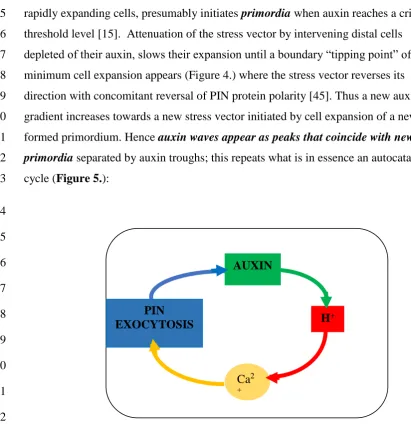
5
Figure 1. The Hechtian oscillator ion fluxes regulate growth137
(a) Depicts a simplified version of the Hechtian oscillator in [2] 138
(b) This figure shows stills from the animation in supplement1: 139
Membrane and ion fluxes are analogous to a molecular “pinball machine”. 140
KEY: protons: red Ca2+ ions: yellow auxin: green 141
Stretch-activates Ca2+ channels; Ca2+ trickle initiates proton pump activity: 142
Phase I. Quiescent: [7s] Proton pump minimally active; 143
Ca2+ channels closed with minimal Ca2+ influx 144
Phase II. Activation: [6s] Turgor increases cell expansion and thus wall stress that 145
increases demand for auxin and opens stretch-activated Ca2+ channels; Ca2+ trickle 146
initiates auxin binding by the proton pump, initiating low level oscillator activity 147
leading to Phase III. 148
Phase III. Fully Activated: [12s] high auxin levels fully activate proton pump. 149
Proton extrusion dissociates periplasmic glycomodule AGP-Ca2+. 150
Entry via Ca2+ channels generates cytosolic Ca2+ waves that activate: 151
exocytosis of: cell wall precursors, wall plasticisers and redirect 152
auxin efflux “PIN” proteins. 153
Phase IV: [9s] Returns to Quiescent state: Stress relaxation closes Ca2+ channels 154
Auxin dissociates from proton pump; Cytosolic Ca2+ recycles to recharge 155
glycomodules. 156
157
Molecular determinants of phyllotaxis periodicity 158
1. Hechtian adhesion. 159
The Profound implicationsof Hecht [28] and many other’s observations are 160
becoming clear. Numerous papers emphasise Hechtian strands of plasmolysed 161
cells but ignore the corollary, strong adhesion between the wall and plasma 162
membrane of turgid cells which until recently has remained a scientific 163
mystery. However, in plasmolysed pollen tubes [7] and root hair tips [29] 164
(Figure 2.) a high density of Hechtian strands correlates rapid tip growth with 165
Hechtian adhesion arguably mediated by AGP57C [25]. This suggests its 166
essential role in transduction of the wall stress vector that initiates 167
oscillations and cytosolic Ca2+ waves as hypothesised by the Hechtian 168
Oscillator (Figure 1.) [7]. 169
170
6
Figure 2. Hechtian strands in root hairs (arrow head) towards the very tip after 172
labelling wheat root hairs with a membrane selective non-permeable fluorescent styryl 173
dye, FM1-43. Reprinted from [29] 174
175
2. Is the Hechtian Oscillator just an Hypothesis?Direct evidence? 176
The correlation between Hechtian adhesion and tip growth also implies that 177
transduction of the wall stress vector with concomitant activation of the proton pump 178
releases Ca2+ from a tip-localised AGP-Ca2+ capacitor, hence a source of the tip-179
focussed Ca2+ influx. Although initially an inference, direct experimental evidence 180
was described most recently by De Vriese et al.[30]: Tobacco BY-2 cells expressing 181
the bioluminescent Ca2+ sensor aequorin responded immediately to addition of the 182
auxin analogue 2,4-D, “the luminescent signal rapidly increased and 183
reached a maximum after 90 s. Thus direct evidence confirms a major prediction of 184
the Hechtian oscillator hypothesis that connects activation of the proton pump and 185
proton extrusion with rapidly increased cytosolic Ca2+. 186
(Figure 1.) 187
The Hechtian oscillator exemplifies the pollen tube paradigm of rapid tip growth in 188
particular [2,7] The rapidly growing cell wall transmits its stress-strain status via 189
Hechtian adhesion to the plasma membrane, The role of Hechtian adhesion in stress 190
transduction, inexplicably overlooked for more than a hundred years also explains 191
how a periplasmic AGP-Ca2+capacitor, as a major component of the oscillator and its 192
auxin-activated proton pump, can regulate plant growth in general. The biochemical 193
physiological and ecological properties of the Hechtian oscillator also avoid the 194
vagaries of a variable external Ca2+ supply; it guarantees immediate access to Ca2+ 195
while recycling cytosolic Ca2+ replenishes the AGP-Ca2+ capacitor. Such efficient use 196
of Ca2+ may ensure the survival of calcifuge species in Ca2+-deficient habitats where 197
over-expression of AGPs also observed under salt stress [31] may enhance the ability 198
to scavenge Ca2+. Marine plants such as Zostera (Eelgrass) support that hypothesis; 199
recent characterisation of their AGPs shows an elevated glucuronic acid content 200
suggestive of enhanced Ca2+ binding in high salt [32]. 201
202
3. Auxin activity is a proxy for the Hechtian Oscillator. 203
(Heisler et al., [13] concluded that in the shoot apical meristem of Arabidopsis 204
“cycles of auxin build-up and depletion accompany, and may direct, different
205
7
of Arabidopsis embryogenesis [33]. Auxin waves indicate the presence of an207
auxin-activated proton pump an essential component of the Hechtian 208
oscillator. Therefore auxin activity itself can be taken as a proxy for an active 209
Hechtian oscillator, consistent with the well- known association of auxin 210
with cell expansion. 211
Thus, H+ dissociation of periplasmic AGP-Ca2+ [24] is the inferred source of 212
cytosolic Ca2+ that activates exocytosis in the AGP-rich protoderm. Indeed 213
ubiquitous distribution of AGPs throughout the Plant Kingdom [34,35] 214
implies an absolute requirement for AGPs. Lethal knockouts of pollen AGPs 215
[36] confirm their essential global role. Indeed, AGPs are closely associated 216
with morphogenesis even at the very earliest stages such as microspore 217
embryogenesis [37]. Therefore we hypothesize that a biological oscillator 218
generates oscillatory growth and contributes to primordia periodicity; 219
phyllotaxis is a test case of the Hechtian oscillator and its general 220
applicability developed in the following sections: 221
222
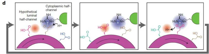
4. A molecular pin-ball machine regulates ion fluxes at the plasma 223
membrane 224
Auxinactivates plasma membrane H+-ATPase proton pump by increasing its 225
phosphorylation which increases the rate of proton extrusion [38]. The extent of 226
ATPase phosphorylation [39] exerts fine gain control of the proton pump over a wide 227
range. Hydrolysis of a single ATP molecule fuels the extrusion of about three protons 228
by an H+-ATPase “turbo-molecular” proton pump. The molecular pathway involves 229
successive glutamate protonation and deprotonation from the cytoplasmic side to the 230
periplasm and cell wall (Figure 3.) [39] and initial extremely fast lateral proton 231
diffusion on the plasma membrane surface [40]. 232
233
234
Figure 3. The proton pump pathway Sequential protonation and deprotonation of 235
8
residue at the cytoplasmic half-channel with subsequent deprotonation of a Glu237
residue at a luminal half-channel. Reprinted from [39] 238
239
Proton extrusion and a concomitantly low wall pH associated with cell extension so-240
called “acid growth” [41] exemplifies Lord Rutherford’s dictum that “No
241
experimental result is ever wrong.” A widely accepted (textbook) explanation 242
invokes low pH-dependent wall loosening “enzymes” and expansins all of unknown 243
specificity [42] but ignore dissociation of AGP-Ca2+ that provides an alternative 244
reinterpretation of “acid growth” based on a visual analogy of the plasma membrane 245
depicted as a metaphorical “molecular pin-ball machine” in (Supplement S1) that 246
regulates three ion fluxes, H+, Ca2+ and auxin (anions at neutral pH, neutral at low 247
pH). When activated by auxin the proton pump shoots fast protons into the periplasm 248
where they dislodge Ca2+ ions from the periplasmic AGP glycomodules; i.e. proton 249
efflux generates Ca2+influx. Thus free Ca2+ ions then enter the cytosol via stretch-250
activated Ca2+channels: Rapid increase of cytosolic Ca2+ [30] activates exocytosis of 251
putative wall plasticisers, namely AGPs and AGP peptides [7] (Figure 1.). 252
Regulation of Ca2+ homeostasis is thus the major function of the proton pump 253
rather than the regulation of wall pH. 254
5. Transduction of the stress vector through the protoderm 255
Primordia are initiated in a “generative annulus” [6]. This thin band of protodermal 256
cells encircles the outermost cell layer of the stem apical meristem (SAM) protoderm 257
where its powerful morphogenetic properties are triggered by the incipient primordia. 258
These most rapidly expanding cells transmit the stress vector to neighbouring cells via 259
their anticlinal walls[14,43-45]. Such stress relocates the auxin efflux “PIN” proteins 260
so their polarisation in the protoderm results in auxin transport towards incipient 261
primordia. The physical basis of stress transduction depends on Hechtian adhesion 262
while its co-localised auxin efflux “PIN” proteins was shown by 263
immunocytochemistry [45]. Such polarised localisation of auxin efflux proteins e.g. 264
PIN1in the protoderm [33] suggested a crucial role for auxin transport in the 265
generation of primordia [46] whoconcluded that “PIN directs auxin to the sites 266
where young primordia are being formed.” Rapid relocation of PIN1 is evidently of
267
huge significance; although the precise mechanism remains obscure. Two 268
9
Hechtian adhesion and AGPs. They mediate transduction of the cell wall stress270
vector, as follows: 271
272
6. Stress, PIN protein redirection and auxin waves 273
“Symmetries control distribution in space” [47] begs the question: What is the origin 274
of symmetry? And how is it broken? A centuries’ old debate gradually developed 275
from “vital force” and the equally unfalsifiable “morphic resonance” to Spemann’s 276
organiser, morphogenetic fields, Waddington’s “evocators” and Turing’s morphogen 277
gradients to current concepts of homeobox genes and a plethora of cognate 278
transcription factors. They illustrate the complexity of animal morphogenesis 279
compared with the sublime sessile simplicity of plants and the view here that auxin 280
gradients control proton and Ca2+ fluxespredominant regulators of growth and 281
differentiation. 282
Auxin transport involves diffusion facilitated by auxin efflux, eponymous “PIN” 283
proteins; they evoke the idea of the membrane as a “molecular pin-ball machine” 284
generating auxin waves that break the perfect symmetry of the protoderm as 285
follows: 286
Morphogenesis frequently involves auxin waves [48]. That includes phyllotaxis [5] 287
where a recent theoretical biophysical model involving complex linear wave 288
equations predict auxin waves that specify the site of new primordia [16]. Those 289
authors noted that “The role of auxin transport in phyllotaxis must be universal” and 290
also inferred that “electromechanical feedbacks apparently involve the Ca2+ and H+
291
ions.” Recent direct experimental evidence [7,24] explains how the cell wall stress 292
vector and transcytosis relocate PIN proteins and thus together with AGP-Ca2+ 293
generate the auxin and Ca2+ waves that initiate primordia formation as follows: 294
Rapidly expanding cells of the protoderm transmit the stress vector via anticlinal 295
walls towards slower cell expansion Figure 4.). The biophysical basis of stress 296
transduction arguably involves Hechtian adhesion between the wall and plasma 297
membrane as described for growth of pollen tubes and epidermal cells of root tips 298
[45]. Hechtian adhesion is also evident in the protoderm: For example, Figure 1A of 299
[49] shows Hechtian strand formation in “protodermal areas. Hechtian adhesion is 300
10
Furthermore, auxin waves are also evidence of an active Hechtian oscillator based on302
Hechtian adhesion. 303
Transmission of the stress vector relocates PIN auxin efflux proteins to the stressed 304
anticlinal walls of stressed cells [45]. Thus auxin moves against its concentration 305
306
307
308
Figure 4. Stress in AGP-rich Protoderm generates primordia. This hypothetical 309
scheme illustrates the likely origin of auxin waves in phyllotaxis [6]. Five-fold 310
rotational symmetry predominates as the archaetype in dicot floral phyllotaxis: A 311
plausible biochemical algorithm generates auxin waves and new primordia: Rapid cell 312
expansion creates the stress vector [red arrows] that orientates PIN proteins; these 313
channel auxin [green arrows] towards rapidly expanding cells to form incipient 314
primordia and deplete auxin from sites of slow expansion, until reaching a “tipping 315
point” (lowest auxin level, slowest cell expansion, minimal stress) where PIN proteins 316
reverse their orientation. Auxin maxima and minima generate regions of rapid 317
expansion at auxin peaks corresponding to incipient primordia P1 to P5 separated by 318
slowest growth at auxin troughs or “tipping points”. Precise spacing of growth peaks 319
corresponds to the frequency of auxin waves controlled by three primary 320
determinants, proton pump, auxin flux and AGP-Ca2+ capacitor size. 321
322
gradient towards cells with highest auxin levels therefore depleting the auxin of less 323
11
rapidly expanding cells, presumably initiates primordia when auxin reaches a critical325
threshold level [15]. Attenuation of the stress vector by intervening distal cells 326
depleted of their auxin, slows their expansion until a boundary “tipping point” of 327
minimum cell expansion appears (Figure 4.) where the stress vector reverses its 328
direction with concomitant reversal of PIN protein polarity [45]. Thus a new auxin 329
gradient increases towards a new stress vector initiated by cell expansion of a newly 330
formed primordium. Hence auxin waves appear as peaks that coincide with new 331
primordia separated by auxin troughs; this repeats what is in essence an autocatalytic 332
cycle (Figure 5.): 333
334
335
336
337
338
339
340
341
342
Figure 5. The auxin autocatalytic cycle While Turing proposed a model based on 343
the interaction between simple diffusion gradients of two morphogens, Nature 344
exploits the interaction of three ions: PIN proteins boost the uphill diffusion of auxin 345
against the concentration gradient while protons the fastest diffusing ions, dissociate 346
AGP-Ca2+ thus enhancing exocytosis of PIN proteins (and Ca2+channels) that 347
propagate the Ca2+ message. This is summarised by the canalization theory [12] in 348
which “small local differences in auxin concentration are amplified by a
self-349
reinforcing accumulation mechanism, resulting in local auxin elevation and auxin
350
depletion in the surrounding tissue.”
351 352
We conclude that the generation of successive auxin waves varying in amplitude and 353
frequency depends on the response of the proton pump and cell surface AGP. Indeed, 354
there is an increasingly clear correlation between enhanced AGP expression and 355
tissue morphogenesis: 356
Membrane-bound PIN proteins recycle rapidly via transcytosis; for example, 357
relocalization of PIN7 occurs within two minutes after the gravity stimulus [50]. 358
AUXIN
H+
PIN EXOCYTOSIS
12
Arguably the mechanism involves Hechtian adhesion that transmits wall stress359
directly to the plasma membrane rather than indirect transmission via “statoliths”. 360
During rapid tip growth of pollen tubes and root hairs, Hechtian adhesion 361
predominates at the growing tip where wall stress-strain is most apparent and 362
exocytosis maximal. This correlation implies that the stress vector relocates Hechtian 363
adhesion sites at a malleable cell wall and this in turn directs the exocytosis of wall 364
precursors including auxin-efflux PIN proteins. (cf. Figure 4.] Thus, auxin waves 365
generated by transmission of the cell wall stress vector depend on two additional 366
factors, transcytosis and cell wall rheology: 367
368
7. Transcytosis rules the waves 369
Although electron microscopy depicts a static plasma membrane it is in fact hugely 370
dynamic involved in secretion, recycling, ion homeostasis, dynamic rearrangement of 371
PIN proteins and Hechtian adhesion! While for simplicity sake we view 372
endo/exocytosis or transcytosis as a single mechanism, of course it involves a hugely 373
complex network of interactions controlled by Ca2+ levels [51] ultimately leading to 374
the control of cell wall rheology and cell expansion. 375
376
8. Cell wall rheology 377
Transmission of the stress vector from rapidly expanding cells of incipient primordia 378
involves plasticity of the anticlinal cell walls. Although Anton Heyn [52] identified 379
wall plasticity as a crucial determinant of cell expansion, even after eighty years the 380
biochemical basis of the Heyn paradigm remains elusive. Despite Heyn’s emphasis on 381
plasticity, cleavage of covalent cell wall crosslinks remain the predominant but 382
elusive explanation [53]. Most synthetic plastics depend on plasticisers like 383
phthalates, small molecules that disrupt the alignment of linear polymer chains but do 384
not cleave covalent bonds. Analogous plasticisers of pectin include classical AGPs 385
but their molecular size precludes simple diffusion through a pectic matrix. However, 386
the much smaller diffusible AGP peptides upregulated by auxin [54] are also potential 387
plasticisers; significantly their glucuronic acid content [55] indicates Ca2+-binding 388
capacity similar to the much larger non-diffusible classical AGPs. Thus small AGP 389
peptides diffusing though the wall can compete for Ca2+ crosslinks and thus favour a 390
pectic gel-sol transition with a concomitant increased wall plasticity: Cosgrove [56] 391
13
auxin-induced patterning of the shoot apical meristem… this correlation of
de-393
esterified pectin with softer meristem regions is perplexing” but consistent with 394
electrostatic repulsion of ionised pectic carboxylates their Ca2+ depleted and 395
scavenged by AGPs and AGP peptides with a higher affinity for Ca2+. However, 396
Altartouri et al. [57] represent the prevailing view that Ca2+ crosslinkage of de-397
esterified pectin decreases wall plasticity. This implicitly assumes sufficient free Ca2+ 398
for pectin crosslinking but ignores AGP-Ca2+ homeostasis that determines the 399
availability of both free and bound apoplastic Ca2+. 400
Fine control of pectin rheology by small diffusible AGP peptides has not previously 401
been considered. Similar reasoning may apply to some monocots where 402
glucuronoxylans largely replace pectin [58]. Finally, we can only agree that: “Cell
403
expansion thus appears to be intimately linked to these wall sensor pathways in ways
404
we are only beginning to fathom.”[56]. 405
406
9. A phyllotaxis algorithm 407
“While progress has been made, there are many fascinating challenges in phyllotaxis
408
still open for the curious mind to explore. The story is far from over. While careful
409
experiments are crucial to continued progress, it does not require elaborate
410
experiments for ordinary folk to enjoy the wonderful architectures seen near the
411
meristems of plants.” [6] also summarising much recent work: “Key results stem from
412
the observed facts that phyllotactic patterns are naturally produced by instabilities,
413
connected to both the distribution of the growth hormone auxin and to the local
414
stress–strain fields.” Although those “instabilities” remain undefined, phyllotaxis per
415
se is remarkably stable but with exceptions described by Arber [1] in several species: 416
For example, a completely dimerous flower of Iris on a shoot also bearing a normal 417
trimerous flower; and Potentilla flowers with their parts in three, four, five, or six 418
(p.165), and concluded that “phyllotaxis “depends upon the rhythmic development of 419
primordia at the growing apex.” And somewhat ahead of its time eighty years ago the
420
insightful observation: “it seems reasonable to suppose that these variations are 421
associated with internal chemical oscillations.” (p.195), with a final intuitive leap to 422
“Physico-chemical factors...one such factor has been so universal as to affect the
423
whole of the vegetable kingdom; this is the development of a cell wall encasing each
424
unit of the plant body.” “The challenge now is to describe how the stem apical 425
14
mathematical approaches based on optimal packing shows that Fibonacci patterns can427
arise naturally in many pattern-forming systems but this is not obviously connected 428
with the biochemical mechanisms involved in patterning. Both approaches achieve 429
optimal packing but in quite different ways. All the components of the Hechtian 430
oscillator are present. Thus, a dynamic algorithm involving protoderm biochemistry 431
and mechanotransduction is now feasible as a working hypothesis: 432
Protoderm cell expansion generates new primordia N. For example in a floral 433
phyllotaxis N is a function of the stem apical meristem (SAM) size and the magnitude 434
of major variables that define the symmetry and periodicity of new primordia. To 435
sustain their growth, rapidly expanding cells demand auxin by generating the cell wall 436
stress vector (CWsv) that redirects PIN proteins thus channelling auxin towards these 437
incipient primordia (Figure 4.). A resulting auxin gradient then appears as waves in 438
the annulus a narrow band of morphogenetic cells surrounding the outer protoderm 439
(SAMPa) with auxin maxima and minima corresponding to future primordia and 440
boundary tipping points respectively: Generally increasing the magnitude of a 441
variable increases auxin transport towards a primordium hence rapidly depleting distal 442
cells. Reversal of PIN protein orientation then generates a new primordium. Thus, an 443
increased auxin depletion rate increases the number of new primordia. However, they 444
also depend on Ca2+ availability determined by the expression of AGPs. Strong 445
expression of AGPs in the protoderm predicts increased periodicity of primordia 446
while weaker expression will decrease it. Thus to that extent the algorithm is 447
semiquantitative and dependent on strong expression of AGPs in the protoderm of 448
Arabidopsis meristems [59] Euphorbia embryonic cultures [60] and during somatic 449
embryogenesis of Arabidopsis [61]. The novel conclusion that AGPs play a decisive 450
role as crucial determinants of phyllotaxis periodicity (figure 6.) is a complex 451
function of a hypothetical algorithm derived from the foregoing considerations: 452
Stem apical meristem protoderm SAMPa generates N new primordia as a function of 453
an equation comprised of the following variables: 454
1. AGP-Ca2+ capacitor: AGPc 455
2. Stem apical meristem protoderm annulus radius: SAMPa 456
3. PP proton pump activity: PP 457
4. Auxin efflux activity: Aefflux [hence auxin levels: Aux] 458
5. Ca2+ channels: Cch 459
15
6. Hechtian adhesion: Had461
7. Cell wall stress vector: CWsv 462
463
N = SAMPa ___________ 464
AGP-1 + (PP . Aux)-1 + (Cch-1 + CWsv)+ Had-1 465
466
The cell wall stress CWsv vector determines auxin, proton and Ca2+ ion fluxes 467
involving four phases of the Hechtian Oscillator proton pump that regulate cell 468
expansion. 469
Phase I. Quiescent: Minimal cell wall stress corresponds to minimal Ca2+ influx and 470
minimal activity of the oscillator. 471
Phase II. Activation: Cell expansion increases wall stress, demand for auxin and 472
opens Ca2+ channels; entry of Ca2+ initiates auxin binding to the proton pump and 473
initial oscillator activation leading to Phase III. 474
Phase III. Maximum Activation: occurs at the high auxin levels supplied by 475
redirected auxin efflux PIN proteins; accelerated proton extrusion dissociates 476
glycomodule AGP-Ca2+and supplies the Ca2+channels thus generating cytosolic 477
Ca2+waves that activate exocytosis, notably of wall precursors and plasticisers but 478
also enabling dynamic redirection of PIN proteins. 479
Addition of precursors reinforce wall and slow its expansion, leading to Phase IV: 480
Phase IV. Return to quiescent phase: 481
Stress relaxation of reinforced wall closes Ca2+ channels. 482
Cytosolic Ca2+ recycles via AGP precursors and Ca2+transporters replenish 483
periplasmic AGP-Ca2+ 484
When attenuation of the stress vector reaches a tipping point of minimal oscillator 485
activity and least rapid cell expansion, distant cells expand more rapidly and now 486
exert a new stress vector in an opposing direction thus generating a new primordium 487
16
489490
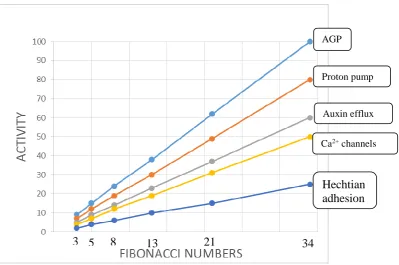
Figure 6. Major variables of the phyllotaxis algorithm 491
SAMPa stem apical meristem protoderm generates new primordia as a 492
function of the activity of five major variables or determinants shown in the 493
speculative graph taking petal phyllotaxis as an example ranging from 3 to 34 petals. 494
It assumes that the cell wall stress vector CWsv is essentially constant for a given 495
SAMPa. 496
Protoderm AGP content [activity] is the primary determinant based on the 497
simple hypothesis that size of the AGP-Ca2+ capacitor determines the periodicity of 498
primordia initiation (see Fig 4. ) where a large capacitor generates more primordia. 499
The graph correlates the Fibonacci series with AGP expression and activity of other 500
factors particularly the proton pump not previously connected with phyllotaxis. Other 501
determinants illustrate an inferred heirarchy based on the size of their relative input to 502
the Hechtian growth oscillator. 503
The beguiling simplicity of the above plot inferred largely from biochemical 504
observations, is in strong contrast to previous complex formulations based largely on 505
mathematical/geometrical logic. 506
507
10. Evolutionary origin of angiosperm phyllotaxis 508
The evolutionary history of the stem apical meristem from a relatively simple 509
arrangement of apical cells in the Bryophytes and ferns culminates in the 510
morphogenetic protoderm of the angiosperms. Here we conjecture that hybridisation 511
may solve the riddle of Fibonacci phyllotaxis and its evolutionary origins. 512
Strong AGP expression predicts numerous closely spaced primordia (Figure 4.) that 513
generate floral phyllotaxis, arguably determined by the amplitude of the Ca2+ signal 514
that depends on the AGP-Ca2+ capacitor size, proton flux and Ca2+ channel status. 515
Thus cells of the protoderm with a large AGP-Ca2+ capacitor will increase cytosolic 516
AGP
Proton pump
Auxin efflux
Ca2+ channels
Hechtian adhesion
3 5 8 13 3
21 3
17
Ca2+ rapidly and therefore generate numerous primordia within a shorter range than in517
a protoderm with weaker AGP expression and therefore with more cells between 518
primordia. For example, lower activity of the proton pump and a smaller AGP-Ca2+ 519
capacitor increase spacing between primordia. Unlike the animal kingdom plants have 520
an enormous a propensity for polyploidy and hybridisation that we suggest provides a 521
simple biochemical explanation for an evolutionary origin of the well-known 522
Fibonacci series of floral organs exemplified by the 3, 5, 8, 13, 21 and 34 petals 523
(Figure 6.) e.g. Ranunculus ficaria (13), Erigeron canadensis (21): Can hybridisation 524
between contiguous members of the series generate a Fibonacci sequence? If so, how? 525
Consider a hybrid expressing the sum of AGPs from both parents! If AGPs play a 526
dominant role in defining phylotaxis then a hybrid of two-fold and threefold 527
symmetry, with a corresponding increased size of the AGP-Ca2+ capacitor, would 528
generate the most common five-fold symmetry And so on for subsequent members of 529
the series (Figure 6). D’Arcy Thompson [17] viewed Fibonacci (1170-1240) not as 530
the cause but merely “a consequence of optimal space filling in systems adding new 531
units at a pole.”Thus, we infer that hybridisation generates floral Fibonacci 532
phyllotaxis and accounts for the evolutionary origin of a discrete series rather than a 533
smooth arithmetic progression. This suggestion has the merit of simplicity based on 534
Occam’s Razor in contrast to all preceding mathematical conjectures [9] and is 535
supported by the Hechtian Oscillator as a predictive paradigm. 536
537
538
11. D’Arcy Thompson, Alan Turing and Peter Mitchell revisited 539
D’Arcy Thompson’s classical “Growth and Form” [17] exemplified a purely 540
descriptive mathematical approach that collated a huge corpus of biophysical 541
observations rather than hypothesis-driven experiment. On the other hand, Turing [26] 542
combined a mathematical with a physical chemical approach. Thus, while a Turing 543
self-replicating machine aptly fits cell replication, Turing postulated that ontogeny 544
based on biological parsimony might involve the diffusion of only two chemical 545
morphogens that would suffice to create morphogenetic gradients. Those ideas 546
preceded the biochemical insights of Mitchell (Mitchell, [62] experimentalist par
547
excellence who questioned conventional wisdom and proposed the versatile 548
chemiosmotic proton pump. A universal energy transduction machine couples proton 549
18
consumes ATP and pumps protons. This vectorial chemical system differs551
fundamentally from conventionally scalar chemical ones as explained by Mitchell 552
[63]: “It was obviously my hope that the chemiosmotic rationale of vectorial 553
metabolism and biological energy transfer might one day come to be generally
554
accepted, and I have done my best to argue in favour of that state of affairs for more
555
than twenty years…was it not the great Max Planck who remarked that a new
556
scientific idea does not triumph by convincing its opponents, but rather because its
557
opponents eventually die?”. Although Mitchell’s unconventional ideas were initially 558
rejected they were finally recognised. Their universal applicability has become 559
apparent more recently. In simple photoautotrophs, light-driven proton gradients 560
involve bacteriorhodopsin while in more advanced eukaryotes an electron transport 561
chain generates mitochondrial proton gradients. Proton pumps and their regulation are 562
thus at the epicentre of plant growth that, stripped to its bare essentials, depends on 563
three morphogen gradients, auxin, protons and Ca2+ rather than just two. 564
However, these gradients do not arise by simple diffusion but are regulated by auxin 565
efflux “PIN” proteins whose discovery began with Rubery and Sheldrake’s [64] 566
classic experiments in the laboratory of D.H. Northcote [65]. PIN proteins control 567
auxin gradients and auxin levels that activate the proton pump while the cell wall 568
stress vector opens Ca2+ channels that generate cytosolic Ca2+ gradients. Thus the 569
Hechtian growth oscillator is an extrapolation of Mitchell’s chemiosmosis that unifies 570
physics and chemistry in a minimalist approach to regulating plant growth. Indeed 571
precursors to life surely involve proton gradients as a basis of prebiotic energy 572
transduction and the universal proton pump of exoplanet life in the habitable zone. 573
574
Acknowledgements: 575
We gratefully acknowledge our home Academic institutions for the past many years 576
of support that has made this and previous work possible. 577
578
We are indeed greatly indebted to our colleague Mr. Ben Coleman for the 579
supplementary information S1 animation of the Hechtian Oscillator ion fluxes. 580
19
Reference List583 584
1.Arber, A. The Natural Philosophy of Plant Form; Cambridge University Press: 585
1950. 586
2.Lamport, D. T. A.; Tan, L.; Held, M. A.; Kieliszewksi, M. J. The Role of the 587
Primary Cell Wall in Plant Morphogenesis. Int. J. Mol. Sci. 2018, 19, 2674. 588
3.Darwin, C. The power of movement in plants; John Murray: London, 1880. 589
4.Pierson, E. S.; Li, Y. Q.; Zhang, H. Q.; Willemse, M. T. M.; Linskens, H. F.; 590
Cresti, M. Pulsatory growth of pollen tubes: investigation of a possible 591
relationship with the periodic distribution of cell wall components. Acta Bot.
592
Neerl. 1995, 44 (2), 121-128. 593
5.Zoulias, N.; Duttke, S. H. C.; Garces, H.; Spencer, V.; Kim, M. The Role of 594
Auxin in the Pattern Formation of the Asteraceae Flower Head (Capitulum). 595
Plant Physiol. 2019, 179, 391-401. 596
6.Pennybacker, M. F.; Shipman, P. D.; Newell, A. C. Phyllotaxis: Some progress, 597
but a story far from over. Physica D 2015, 306, 48-81. 598
7.Lamport, D. T. A.; Tan, L.; Held, M. A.; Kieliszewksi, M. J. Pollen tube growth 599
and guidance: Occam's razor sharpened on a molecular arabinogalactan 600
glycoprotein Rosetta Stone. New Phytologist 2018, 217, 491-500. 601
8.Chebli, Y.; Geitmann, A. Mechanical principles governing pollen tube growth. 602
Functional Plant Science and Biotechnology 2007, 1 (2), 232-245. 603
9.Adler, I.; Barabe, D.; Jean, R. V. A history of the study of Phyllotaxis. Annals of
604
Botany 1997, 80, 231-244. 605
10.Okabe, T. The riddle of phyllotaxis: exquisite control of divergence angle. 606
Acta Soc BotPol. 2016, 85 (4), 3527. 607
11.Sachs, T. The Control of the Patterned Differentiation of Vascular Tissues. 608
Advances in Botanical Research 1981, 9, 151-262. 609
12.Reinhardt, D.; Pesce, E.-R.; Stieger, P.; Mandel, T.; Baltensperger, K.; 610
Bennett, M.; Traas, J.; Friml, J. Regulation of phyllotaxis by polar auxin 611
transport. Nature 2003, 426, 255-260. 612
13.Heisler, G.; Ohno, C.; Das, P.; Sieber, P.; Reddy, G. V.; Long, J. A.; 613
Meyerowitz, E. M. Patterns of Auxin Transport and Gene Expression during 614
Primordium Development Revealed by Live Imaging of the Arabidopsis
615
Inflorescence Meristem. Current Biology 2005, 15, 1899-1911. 616
14.Heisler, M. G.; Hamant, O.; Krupinski, P.; Uyttewaal, M.; Ohno, C.; Jonsson, 617
H.; Traas, J.; Meyerowitz, E. M. Alignment between PIN1 Polarity and 618
Microtubule Orientation in the Shoot Apical Meristem Reveals a Tight 619
Coupling between Morphogenesis and Auxin Transport. PLoS Biology 2010, 8
620
20
15.Bhatia, N.; Heisler, M. G. Self-organising periodicity in development: organ622
positioning in plants. Development 2018, 145 (149336). 623
16.Abraham-Shrauner, B.; Pickard, B. G. A model for leaf initiation 624
Determination of phyllotaxis by waves in the generative circle. Plant
625
Signaling & Behavior 2011, 6 (11), 1755-1768. 626
17.Thompson, D. On Growth and Form; Abridged ed.; 1961. 627
18.Clarke, A. E.; Anderson, R. L.; Stone, B. A. Form and function of 628
arabinogalactans and arabinogalactan-proteins. Phytochem. 1979, 18, 521-540. 629
19.Gaspar, Y. M.; Johnson, K. L.; McKenna, J. A.; Bacic, A.; Schultz, C. J. The 630
complex structures of arabinogalactan-proteins and the journey towards 631
understanding. Plant Mol. Biol. 2001, 47, 161-176. 632
20.Ellis, M.; Egelund, J.; Schultz, C. J.; Bacic, A. Arabinogalactan-proteins: Key 633
regulators at the cell surface? Plant Physiol. 2010, 153, 403-419. 634
21.Aspinall, G. O.; Malloy, J. A.; Craig, J. W. T. Extracellular polysaccharides 635
from suspension-cultured sycamore cells. Can. J. Biochem. 1969, 47, 1063-636
1070. 637
22.Lamport, D. T. A. Cell wall metabolism. Ann. Rev. Plant Physiol. 1970, 21, 638
235-270. 639
23.Jermyn, M. A.; Yeow, Y. M. A class of lectins present in the tissues of seed 640
plants. Aust. J. Plant Physiol. 1975, 2, 501-531. 641
24.Lamport, D. T. A.; Varnai, P. Periplasmic arabinogalactan glycoproteins act as 642
a calcium capacitor that regulates plant growth and development. New
643
Phytologist 2013, 197, 58-64. 644
25.Tan, Li.; Eberhard, S.; Pattathil, S.; Warder, C.; Glushka, J.; Yuan, C.; Hao, 645
Z.; Zhu, X.; Avci, U.; Miller, J. S.; Baldwin, D.; Pham, C.; Orlando, R.; 646
Darvill, A.; Hahn, M. G.; Kieliszewksi, M. J.; Mohnen, D. An Arabidopsis 647
Cell Wall Proteoglycan Consists of Pectin and Arabinoxylan Covalently 648
Linked to an Arabinogalactan Protein. Plant Cell 2013, 25, 270-287. 649
26.Turing, A. M. The chemical basis of morphogenesis. Phil. Trans. R. Soc.
650
Lond. B 1952, 237, 37-72. 651
27.Briggs, G. E. The Absorption of Salts by Plant Tissues, considered as Ionic 652
Interchange. Annals of Botany 1933, 46 (182), 301-322. 653
28.Hecht, K. Studien uber den Vorgang der Plasmophysiologie der 654
Grenzschichten lebender Pflanzenzellen. Cohns Beitrage zur Biologie der
655
Pflanzen 1912, 11, 137-189. 656
29.Volgger, M.; Lang, I.; Ovecka, M.; Lichtscheidl, I. Plasmolysis and cell wall 657
deposition in wheat root hairs under osmotic stress. Protoplasma 2010, 243, 658
21
30.De Vriese, K.; Himschoot, E.; Dunser, K.; Nguyen, L.; Drozdzecki, A.; Costa,660
A.; Nowack, M. K.; Kleine-Vehn, J.; Audenaert, D.; Beeckman, T.; Vanneste, 661
S. Identification of Novel Inhibitors of Auxin-Induced Ca2+ Signaling via a 662
Plant-Based Chemical Screen. Plant Physiology 2019, 180, 480-496. 663
31.Lamport, D. T. A.; Kieliszewksi, M. J.; Showalter, A. M. Salt-stress 664
upregulates periplasmic arabinogalactan-proteins: using salt-stress to analyse 665
AGP function. New Phytologist 2006, 169 (3), 479-492. 666
32.Pfeifer, L.; Classen, B. First structural characterisation of seagrass 667
arabinogalactan-proteins reveals habitat-driven adaptation to marine 668
environment. Cell Wall Meeting 2019, 15, P003. 669
33.Moller, B.; Weijers, D. Auxin Control of Embryo Patterning. Cold Spring
670
Harb Perspect Biol 2009, 1, 1-13. 671
34.Showalter, A. M. Arabinogalactan-proteins: structure, expression and 672
function. Cell. Mol. Life Sci. 2001, 58, 1399-1417. 673
35.Palacio-Lopez, K.; Tinaz, B.; Holzinger, A.; Domozych, D. S. 674
Arabinogalactan proteins and the extracellular matrix od Charaophytes: a 675
sticky business. Front. Plant Sci. 2019, 10 (447). 676
36.Coimbra, S.; Costa, M.; Jones, B. J.; Mendes, M. A.; Pereira, L. G. Pollen 677
grain development is compromised in Arabidopsis agp6 agp11 null mutants. 678
Journal of Experimental Botany 2009, 60, 3133-3142. 679
37.Corral-Martinez, P.; Driouich, A.; Segui-Simarro, J. M. Dynamic Changes in 680
Arabinogalactan-Protein, Pectin, Xyloglucan and Xylan Composition of the 681
Cell Wall During Microspore Embryogenesis in Brassica napus. Front. Plant
682
Sci. 2019, 10, 332. 683
38.Koji, T.; Hayashi, K.; Kinoshita, T. Auxin Activates the Plasma Membrane 684
H+-ATPase by Phosphorylation during Hypocotyl Elongation in Arabidopsis. 685
Plant Physiol. 2012, 159, 632-641. 686
39.Mazhab-Jafari, M. T.; Rohou, A.; Schmidt, C.; Bueler, S. A.; Benlekbir, S.; 687
Robinson, C. V.; Rubinstein, J. L. Atomic model for the membrane-embedded 688
VO motor of a eukaryotic V-ATPase. Nature 2016, 539, 118-130. 689
40.Amdursky, N.; Lin, Y.; Aho, N.; Groenhof, G. Exploring fast proton transfer 690
events associated with lateral proton diffusion on the surface of membranes. 691
Proc. Natl. Acad, Sci. 2019, 116 (7), 2443-2451. 692
41.Rayle, D. L.; Cleland, R. E. The acid growth theory of auxin-induced cell 693
elongation is alive and well. Plant Physiol. 1992, 99, 1271-1274. 694
42.Ramakrishna, P.; Duarte, P. R.; Rance, G. A.; Schubert, A.; Vordermaier, V.; 695
Vu, L. D.; Murphy, E.; Barro, A. V.; Swarup, K.; Moirangthem, K.; 696
Jorgensen, B.; Moirangthem, K.; van de Cotte, B.; Goh, T.; Lin, Z.; Voss, U.; 697
Beeckman, T.; Bennett, M. J.; Gevaert, K.; Maizel, A.; De Smet, I. 698
22
cell divisions during lateral root initiation. Proc. Natl. Acad, Sci. 2019, 116,700
8597-8602. 701
43.Hamant, O.; Heisler, M. G.; Jonnson, H.; Krupinski, P.; Uyttewaal, M.; 702
Bokov, P.; Corson, F.; Sahlin, P.; Boudaoud, A.; Meyerowitz, E. M.; Couder, 703
Y.; Traas, J. Developmental patterning by mechanical signals in Arabidopsis. 704
Science 2008, 322, 1650-1654. 705
44.Sampathkumar, A.; Yan, A.; Krupinski, P.; Meyerowitz, E. M. Physical 706
Forces Regulate Plant Development and Morphogenesis. Current Biology
707
2014, 24 (10), 475-483. 708
45.Feraru, E.; Feraru, M. I.; Kleine-Vehn, J.; Martinie, A.; Mouille, G.; Vanneste, 709
S.; Vernhettes, S.; Runions, J.; Friml, J. PIN Polarity Maintenance by the Cell 710
Wall in Arabidopsis. Current Biology 2011, 21, 338-343. 711
46.Vernoux, T.; Besnard, F.; Traas, J. Auxin at the Shoot Apical Meristem. Cold
712
Spring Harb Perspect Biol 2010, 2 (a001487), 1-14. 713
47.Critchlow, K. The hidden geometry of Flowers; Floris Books: Edinburgh, 714
2011. 715
48.Zajaczkowski, S.; Wodzicki, T. J.; Romberger, J. A. Auxin Waves and Plant 716
Morphogenesis. In Hormonal Regulation of Development II: The Functions of
717
Hormones from the Level of the Cell to the Whole Plant, Scott, T. K., Ed.; 718
Springer Berlin Heidelberg: Berlin, Heidelberg, 1984; pp 244-262. 719
49.Komis, G.; Apostolakos, P.; Galatis, B. Altered patterns of tubulin 720
polymerization in dividing leaf cells of Chlorophyton comosum after a
721
hyperosmotic treatment. New Phytologist 2001, 149, 193-207.
722
50.Sato, E. M.; Hijazi, H.; Bennett, M. J.; Vissenjberg, K.; Swarup, R. New 723
insights into root gravitropic signalling. J. Exp. Bot. 2015, 66 (8), 2155-2165. 724
51.Roy, S.; Holdaway-Clarke, T. L.; Hackett, G. R.; Kunkel, J. G.; Lord, E. M.; 725
Hepler, P. K. Uncoupling secretion and tip growth in lily pollen tubes: 726
evidence for the role of calcium in exocytosis. Plant J. 1999, 19 (4), 379-386. 727
52.Heyn, A. N. J. The physiology of cell elongation. Botanical Review 1940, 6
728
(10), 515-574. 729
53.Dyson, R. J.; Band, L. R.; Jensen, O. E. A model of crosslink kinetics in the 730
expanding plant cell wall: Yield stress and enzyme action. J. Theor. Biol.
731
2012, 307, 125-136. 732
54.Pacheco-Villalobos, D.; Diaz-Moeeno, M.; van der Schuren, A.; Tamaki, T.; 733
Kang, Y. H.; Gujas, B.; Novak, O.; Jaspert, N.; Li, Z.; Wolf, S.; Oecking, C.; 734
Ljung, K.; Bulone, V.; Hardtke, S. The Effects of High Steady State Auxin 735
Levels on Root Cell Elongation in Brachypodium. Plant Cell 2016, 28, 1009-736
23
55.Tryfona, T.; Liang, H.-C.; Kotake, T.; Kaneko, S.; Marsh, J.; Ichinose, H.;738
Lovegrove, A.; Tsumaraya, Y.; Shewry, P. R.; Dupree, P. Carbohydrate 739
structural analysis of wheat flour arabinogalactan protein. Carbohydrate
740
Research 2010, 345 (2648), 2656. 741
56.Cosgrove, D. J. Diffuse Growth of Plant Cell Walls. Plant Physiology 2018, 742
176, 17-27. 743
57.Altartouri, B.; Bidhendi, A.; Tani, T.; Suzuki, J.; Conrad, C.; Chebli, Y.; Liu, 744
N.; Karunakaran, C.; Scacelli, G.; Geitmann, A. Pectin chemistry and 745
cellulose crystallinity govern pavement cell morphogenesis in a multi-step 746
mechanism. Plant Physiology 2019, 181, 127-141. 747
58.Zelaya, V. M.; Fernandez, P. V.; Vega, A. S.; Mantese, A. I.; Federico, A. A.; 748
Ciancia, M. Glucuronoarabinoxylans as major cell walls polymers from young 749
shoots of the woody bamboo Phyllostachys aurea. Carbohydrate Polymers
750
2017, 167, 240-249. 751
59.Abe, M.; Takahashi, T.; Komeda, Y. Cloning and characterization of an LI 752
Layer-specific gene in Arabidopsis thaliana. Plant Cell Physiol. 1999, 40 (6), 753
571-580. 754
60.Saare-Surminski, K.; Preil, W.; Knox, J. P.; Lieberei, R. Arabinogalactan 755
proteins in embryogenic and non-embryogenic callus cultures of Euphorbia 756
pulcherrima. Physiol. Plant. (2000) 2000, 108 (2), 180-187. 757
61.Potocka, I.; Godel, K.; Dobrowolska, I.; Kurczyñska, E. U. Spatio-temporal 758
localization of selected pectic and arabinogalactan protein epitopes and the 759
ultrastructural characteristics of explant cells that accompany the changes in 760
the cell fate during somatic embryogenesis in Arabidopsis thaliana. Plant
761
Physiology and Biochemistry 2018, 127, 573-589. 762
62.Mitchell, P. Coupling of phosphorylation to electron and hydrogen transfer by 763
a chemi-osmotic type of mechanism. Nature 1961, 191, 144-148. 764
63.Mitchell, P. David Keilin's Respiratory Chain Concept and Its Chemiosmotic 765
Consequences. Nobel Lectures, Chemistry 1971-1980, 1978, 295-330. 766
64.Rubery, P. H.; Sheldrake, A. R. Carrier-mediated Auxin Transport. Planta
767
1974, 118, 101-121. 768
65.Crompton, D.W.T. Donald Henry Northcote (1921-2004). Biogr. Mems. Fell.
769
R. Soc. 2019, 67, 359-370 770
771 772
![Figure 4. Stress in AGP-rich Protoderm generates primordia. This hypothetical scheme illustrates the likely origin of auxin waves in phyllotaxis [6]](https://thumb-us.123doks.com/thumbv2/123dok_us/7883323.1308013/10.595.64.544.180.467/figure-stress-protoderm-generates-primordia-hypothetical-illustrates-phyllotaxis.webp)