Article
Key Steps in the Early Evolution of Life from the
Origin of Protein Synthesis to Modern Cellular Life
Sankar Chatterjee*
Department of Geosciences, Museum of Texas Tech University, Box 4319, 3301 4th Street, Lubbock, Texas 79409, USA
* Correspondence: [email protected]; Tel: +1-806-787-4332
Abstract: The emergence of proteins in the prebiotic world was a watershed event at the origin of life. With their astonishing versatility, the protein enzymes catalyzed crucial biochemical reactions within protocells into more complex biomolecules in diverse metabolic pathways, whereas structural proteins provided strength and permeability in the cell membrane. Five major biochemical innovations followed in succession after availability of various kinds of protein molecules during decoding and translation of mRNAs. These are: (1) the modification of the phospholipid membrane into the plasma membrane; (2) the origin of primitive cytoplasm; (3) primitive gene regulation; (4) the beginnings of the virus world; and (5) the advent of DNA. The creative role of viruses during prebiotic synthesis led to the origin of the DNA world, when DNA replaced mRNA as the major genome of the protocells. With the advent of DNA, replication of information was entirely dissociated from its expression. Because DNA is much more stable than mRNA with more storage capacity, it is a superb archive for information systems in the form of base sequences. DNA progressively took over the replicative storage function of mRNA, leaving the latter for protein synthesis. Genetic information began to flow from DNA to mRNA to protein in a two-step process involving transcription and translation. In the biological stage, DNA replication was central to the binary fission of the first cell, orchestrated by the duplication of genomes and then the division of the parent cell into two identical daughter cells. With the onset of binary fission, the population of primitive cells grew rapidly in the hydrothermal vent environment, undergoing Darwinian evolution and diversification by mutation. The habitat of the earliest fossil record (≥ 3.5 Ga) from the Archean sedimentary rocks of Canada, Greenland, Australia, South Africa, and India offers a new window on the early radiation of microbial life. The development of anoxygenic and then oxygenic photosynthesis from early hyperthermophiles would have allowed life to escape the hydrothermal setting to the mesophilic global ocean.
Keywords: protein/RNA world: plasma membrane; cytoplasm; gene regulation; virus world; pre-retro virus; emergence of DNA; transcription and replication; first cells; hyperthermophiles; LUCA; Bacteria and Archaea; anoxygenic bacteria; oxygenic bacteria; global distribution of cyanobacteria
1. Introduction
The origin of life is one of the great unsolved scientific problems of our age. While the details of this process of origin are still shrouded in mystery, the prevailing scientific hypothesis is that transition from non-living matter, such as simple organic compounds to living cell was not a single event, but a gradual process of increasing complexity. The past decade has seen a resurgence of interest in the origin of life, in part because of new evidence from astrobiology. To understand how life could begin on young Earth, it is essential to know what organic compounds–the essential building blocks of life–were likely to have been available and how they interacted in a suitable environment. Using the early history of the young Earth, I have suggested previously that life probably arose through five hierarchical stages of increasing molecular complexity in a steaming hot environment of hydrothermal crater basins about 4 billion years ago—cosmic, geologic, chemical,
information, and biological. Each stage gave birth to a new sequence of biomolecules with new characteristics and increasing complexity that led to the emergence of the first cells. The building blocks of life had their beginnings in the interstellar space during the explosion of a supernova. Most likely, carbonaceous chondrites delivered both building blocks of life and water to early Earth during recurrent impacts on the early Archean crust. Asteroid collisions also created innumerable hydrothermal crater basins that became the perfect cradle for prebiotic chemistry. The hydrothermal crater vents provided both chemicals and energy sources, making them feasible as a niche for the emergence of life [1-3].
The RNA world model has become the main paradigm in the current origin of life research in which RNA assumed informational and functional roles [4-8]. In our previous paper [9], we have argued that the peptide/RNA world is more parsimonious than the RNA world for biogenesis. This view was supported by several authors in recent times [9-15]. We suggested that the demand for a wide range of protein enzymes over peptides in the prebiotic reactions was the main selective pressure for the origin of information-directed protein synthesis [9]. Once the mRNA-directed protein synthesis became established, various enzymes were created to meet the demand of catalysis and metabolism. The next step involved encapsulation of a more complex enzyme system capable of catalyzing fatty acids to form phospholipids. The transition from single-chain lipids to phospholipids was triggered by the availability of a wide range of enzymes in the protein/RNA world that catalyzed the conversion of lipid membranes into phospholipid membranes. In this paper, we discuss the evolution and functional repertoire of translation proteins that led to hierarchical emergences of plasma membranes, cytoplasm, gene regulation, viruses, DNA, and finally the first cells.
2. Protein/RNA World
Proteins were assembled from amino acids using information encoded in mRNA and translated to protein by tRNA. The ensuing peptide elongation was catalyzed by rRNA in the ribosome. Each protein had its own unique amino acid sequence of the mRNA gene. Proteins control most of the functions of a cell, breaking down nutrients, assembling cellular components, copying DNA, and so on. They truly occupy a central position in the organization of the first cell. They act as enzymes that permit only a few of the many possible reactions among cellular components to take place. Enzymes play an essential role in prebiotic synthesis. They are catalysts that greatly accelerate reactions by providing an alternate reaction pathway with a much lower energy barrier. Thus, although they do not create new reactions, they greatly enhance the rate at which a particular substrate is changed into a particular product. Without them, the translation of genetic material into proteins would be impossible. Proteins form channels in plasma membranes, allowing specific substances to enter and leave, while excluding others. They form channels in plasma membranes, allowing specific substances to enter and leave, while excluding others. Protein molecules owe their properties to their three-dimensional shapes, which are themselves determined by their amino acid sequences of their constituent chains. These properties in turn determine how a protein biologically functions: whether it will bind certain organic molecules and catalyze their reactions or form regular structures, such as a helix, and act as a building material.
protocells at the time [16]. These newly formed enzymes carried out hundreds of chemical reactions that took place in the protocell. Structural proteins, on the other hand, provided structure and support for the cell membrane. Ancient enzymes, tryptophan synthases, resurrected from the common ancestor of all bacteria indicate that such sophistication of primordial enzyme complexes existed more than 3.4 billion years ago [17].
Proteins mediate most functions of modern cells. Four major events followed after the availability of template-directed proteins but with considerable overlap. These affected, first, the efficiency of the translation machinery, then, resilience of the coding system, and finally, the quality of the synthesized proteins. These protein-mediated events are: (1) the transition from the phospholipid membrane to the plasma membrane; (2) the origin of prebiotic cytoplasm; (3) the beginnings of the virus world; and (4) the advent of DNA. The newly synthesized protein enzymes helped to catalyze and mediate these critical molecular evolutions, favored by strong selective forces.
3. The Evolution of Phospholipid Membranes
Self-assembly of cell membranes that encapsulated the polymers into populations of protocells were essential to the development of life forms in the prebiotic Earth. The primitive cell membranes enclosed the life-building molecules, defined the boundaries from environments, and enhanced chemical reactions inside protocells. Each cell membrane was a very thin film of lipid molecules, about 5 nm thick and was a dynamic fluid structure. The origin of cellular life presumably occurred by self-assembly of cosmic organic compounds into encapsulated systems capable of catalyzed polymer synthesis [18,19]. Thus, the availability of a primitive cell membrane component in a hydrothermal vent environment was a prerequisite for biogenesis. The cell membranes are built of components that have a remarkable capability of spontaneous self-assembly. The chemical requirements for formation of cell membranes from individual molecules are remarkably simple. The lipids that form the lipid bilayer common to all biological membranes are not gene products. Therefore, the development of genetic material need not have to precede the development of the membrane. However, the development of membranes could have facilitated the development of self-replicating genes by providing them a protected space in which to evolve and eventually function. Most likely, the phospholipids did not become available in the prebiotic environment until various protein enzymes were available and metabolic pathways for their catalyzed synthesis evolved. The phospholipid bilayers were impermeable to most water-soluble molecules. This property made bilayers excellent boundaries that allowed for protocells to maintain an internal composition different from that of the surrounding medium. But protocells could not survive and evolve sealed off from the outside. They must be able to take up nutrients, get rid of waste products, and respond to environmental signals. These functions were carried out by proteins inserted into phospholipid bilayers to form a plasma membrane, improving the bilayer’s permeability.
3.1. The Origin of the Phospholipid Membrane
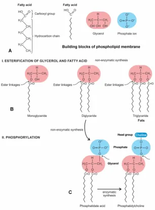
The synthesis of the phospholipids requires an activated intermediate, phosphatidate acid (diaglycerol 3-phosphate). The prebiotic pathway from fatty acids to the simplest phospholipid, phosphatidate acid, occurs via successive acyl- and phosphotransfer reactions. The first step in the synthesis of the phospholipids is the synthesis of phosphatidate acid, which is formed by the addition of two fatty acids to glycerol 3-phosphate. Here we explore the non-enzymatic pathways of the emergence of the phosphatidate in the peptide/RNA world. From three building blocks—fatty acids, glycerol, and a phosphate ion, which were available in the prebiotic environment [28], we show the gradual evolution of monoglyceride, diglyceride, and triglyceride by the condensation reaction (Fig. 1B). When glycerol and fatty acid react, a water molecule is expelled, forming an ester linkage. The production of diglyceride is considered first, since it will lead directly to the biosynthesis of the phosphatidate. Diglyceride is formed when the glycerol and fatty acid chains become joined by two ester linkages. Non-enzymatic synthesis of ester bonds to produce the diglycerides might have been the first step toward glycerolipids [16,22,26]. The phosphorylation of diglyceride, in turn, would give rise to the phosphatidate acid, which is essentially a diglyceride in which a phosphate group has been added to a single glycerol molecule (Fig. 1C).
The non-enzymatic synthesis of the activated phosphatidate acid was at a pivotal point in the lipid biosynthetic pathways. It served as the precursor for the formation of the glycerophospholipid (commonly called phospholipid) membrane by enzymatic synthesis. Glycerophospholipids are the main constituents of membrane bilayers. Enzymatic synthesis pathways evolved over time when RNA-directed protein enzymes were available in the protein/RNA world. The phosphate group of the activated phosphatidate acid is esterified to an alcohol to produce a variety of phospholipids including the attachment of choline, ethanolamine, serine, and inositol to the phosphate group of phosphatidic acid. Names of phospholipids then include phosphatidylcholine (phosphate + cholin), phosphatidylethanolamine (phosphate + ethanolamine), phosphatidylserine (phosphate + serine), and phosphatidylinositol (phosphate + inositol). If the alcohol is choline, the product is phosphatidylcholine. Of these, phosphatidylethanolamine is the most common phospholipid in bacterial cell membranes. Different modifiers give the phospholipids different properties and roles in a cell. Three successive enzymatic methylation could convert the phosphatidate to phospholipid. A phospholipid consists of a polar headgroup on one end of the molecule and fatty acid chains on the end. These chemical structures create an amphipathic liquid. In solution, they instantly form bilayers that are selectively permeable. The phospholipids are composed of a polar head group (usually a negatively charged phosphate group and glycerol); it is hydrophilic. Phosphate is a primary anionic component of most phospholipid membrane lipids (Fig. 1A). The phospholipid tails consist of two long fatty acid chains, which are hydrophobic and avoid interactions with water. Two fatty acids are attached to a glycerol by ester or other bonds. The polar head group and fatty acid chains are attached by a 3-carbon glycerol unit [28].
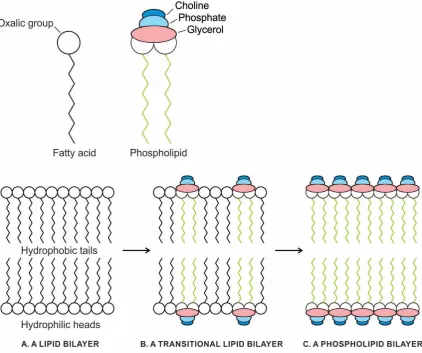
The phospholipid molecules have a hydrophilic head end and two hydrophobic tails that will not mix with water and will avoid being surrounded by it (Fig. 2A). Because these amphipathic molecules have both a hydrophilic and hydrophobic group on the same molecule, they can undergo self-assembly into a cell. In an oil slick, the hydrophobic tails mix with oil while the heads stay close to the water in a monolayer cell (or micelle). When placed in water, the phospholipids will orient themselves in a bilayer in which non-polar tail regions face the inner layer of the bilayer (Fig. 2B). Being cylindrical, the phospholipid molecules contribute structural stability and create a semipermeable environment. The same forces that drive the phospholipids to form bilayers also provide a self-healing property. Admixture of cholesterol helps to stabilize the bilayer.
Figure 1. Origin of the phospholipid membrane from simple fatty acids by an intermediate, the phosphatidate acid by a series of non-enzymatic synthesis. A, fatty acid, glycerol, and phosphate ion are the building blocks of phospholipid. B, in the first stage of phospholipid formation, several glycerides such as monoglyceride, diglyceride, and triglyceride (fat) are formed by esterification of glycerol and fatty acid, with the loss of a water molecule; the covalent bond, an ester linkage results from this reaction. C, the next stage of synthesis of phosphatidate acid is by phosphorylation of a diglyceride molecule, when a phosphate ion is joined. Phosphatidate acid, in turn, would give rise to phospholipid by attaching to an alcohol molecule, such as choline, ethanolamine, serine or inositol. Of these various combinations, phosphatidylcholine (shown in the figure) is the most common phospholipid in the cell membrane.
Figure 2. The self-assembly of a phospholipid membrane in a hydrothermal crater lake in the peptide/RNA world. A, a generalized phospholipid molecule has a hydrophilic (‘water-loving’) head and two hydrophobic (‘water-hating’) tails that do not mix with water and will avoid being
surrounded by it. B, in an oil slick on the surface of a crater lake, the hydrophobic tails mix with oil. whereas the heads stay close to water. During turbulence, phospholipids form two kinds of membranes: a monolayer, which can only capture a drop of oil (left), or a bilayer, which can capture a group of water molecules (right). The bilayer allows the hydrophobic tails to associate with one another, whereas the heads associate with water molecules, on both the inside and outside surfaces of the membrane. A bilayer vesicle is stabilized when it encapsulates protein molecules that interact with the bilayer surface.
Figure 3. The gradual transition from single-chain, highly permeable fatty acid membrane (A) to selective permeable phospholipid membrane with the increase of phospholipid content (C) via a transitional stage (B). Increasing phospholipid content inhibits the permeability of fatty acid membranes through changes in bilayer fluidity.
3.2. The Origin of the Plasma Membrane
Here, we suggest the likely scenario for the origin of the plasma membrane from the phospholipid membrane (Fig. 4). A new class of proteins emerged in the protein/RNA world that played critical roles for the conversion of the phospholipid membrane to the plasma membrane. Proteins could be amphipathic because they are made of amino acids, and amino acids have R-groups that range from highly nonpolar to highly polar. To incorporate in the phospholipid membrane, the nonpolar amino acids would be selected in the interior of the lipid bilayer, while the polar would be selected alongside the polar heads of the surrounding water.
substances from crossing while permitting other substances to enter and leave the protocell. The selective permeability of the plasma membrane and the specificity of transport proteins made it possible to create an environment inside the protocell that was radically different from the prebiotic soup and amenable to biogenesis.
The first step of survival in confinement was the possibility for the protocells to take in nutrients and energy from the outside environment and get rid of waste material. The simplest way in which fully enveloped protocells could fulfill this condition was by means of pores, mere holes kept open in the phospholipid bilayers by some kind of inserted protein framework. The insertion of proteins in bilayer lipid vesicles was an essential first step that affected the phospholipid bilayer’s permeability, facilitating transport and other molecules into the protocells. In primitive membrane transport, passive transport was used when molecules were moved across the plasma membrane of the protocells along, down, or with their concentration gradients. The molecule would flow from where it was at higher concentrations to where it was at a lower concentration.
Next came transport facilitators, which were transmembrane proteins that act as restricted passages for certain specific substances. A more sophisticated kind of molecular passage was the gated channel-like facilitators that let certain substances of a given chemical specificity to move through passively but they were unidirectional and regulated by a gate that needed to be unlocked by some channels or electrical signal.
The next improvement in the building of molecular transport systems was active transport, where molecules were moved against their concentration gradient in energy-requiring processes and the machinery involved was correspondingly more complex. The systems that carry out such active transport are called pumps. The energy used was derived from ATP, the universal currency of energy. There were various ATP-powered pumps that were used to transport ions and molecules against their concentration gradients. The plasma membrane would have played an essential role in the generation of metabolic energy, and transformed it into useful ATP.
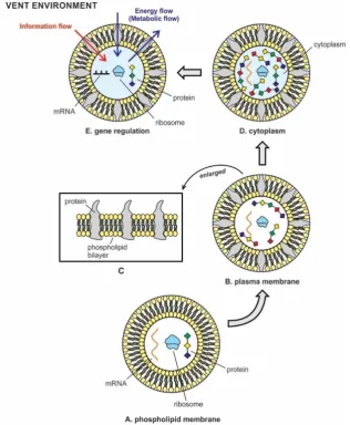
The classical fluid mosaic model of the structure of the cell membrane distinguishes between two types of membrane proteins: peripheral proteins and integral proteins; the former occurs only outside the lipid bilayer, while the latter spans the entire membrane for transport of ions and molecules [29]. The phospholipids and the plasma membranes make back and forth movements within the plasma membrane, making the plasma membrane a fluid structure. The key point is that the arrangement of proteins makes the interior and exterior surfaces of the plasma membrane very different (Fig. 4C). Thus, the plasma membranes are a mosaic of the phospholipids and the different types of proteins, and the overall structure is dynamic and fluid. In protocells, the membrane proteins were responsible for the passage of ions, polar molecules, and large molecules that did not readily cross the phospholipid bilayers on their own.
Figure 4. The transition of phospholipid membrane to plasma membrane. A, phospholipid membrane; B, plasma membrane; C, phospholipid membrane evolved into plasma membrane by inserting protein molecules into bilayers that made the cell membrane selective permeable so that certain ions like potassium and sodium can cross the bilayer barrier; D, cytoplasm inside protocell formed when newly synthesized protein molecules by a translation machine began to concentrate inside the protocell, turning internal water into a gel-like substance; E, gene regulation in RNA protocell; energy flows and information flows are represented as the basic exchanges of the protocell with outer/inner environment. RNA protocell began to respond to environmental cues and target mRNA for regulating gene expression.
Proteins are responsible for most of the dynamic processes carried out by the cell membrane, including the transport of molecules into and out of the cell (Fig. 4B,4C). The plasma membrane separates the cell from its environment and is selectively permeable: it chooses what enters and exists in the cell. Receptor proteins are the gatekeepers; they detect signals from the environment of the cells; the transport proteins help some molecules get across the membrane. Certain membrane proteins act as enzymes. The plasma membrane was the ideal microenvironment to experiment with synthesis of more complex nucleic acid such as DNA for the permanent storage of the information system.
4. The Origin of Cytoplasm
synthesized by translation machinery and available for prebiotic synthesis, they began to accumulate in the primitive cytoplasm inside protocells. This primordial cytoplasm became the ready source of a variety of proteins, when needed. The simple aqueous solution (prebiotic soup) inside the protocell was gradually converted to a viscous, gel-like cytoplasm that increased the protocell volume and provided some rigidity of its spherical shape. The first cytoplasm in protocells and the semipermeable plasma membrane from the phospholipid membrane most likely organized at the same time with the availability of proteins (Fig. 4D). The prebiotic cytoplasm provided a stable microenvironment for the organization of all nucleic acids, lipids, enzymes, proteins, other macromolecules, molecules, ions and ribosomes, as well as water and salts that were all encased in the plasma membrane. This primitive cytoplasm became a complex, crowded system containing a wide range of molecules – from ions and small molecules, to macromolecules like proteins, nucleic acids, and ribosomes. Many metabolic reactions, including protein synthesis, and the transition from RNA to DNA began to take place in this primordial cytoplasm. Over half of the molecules were actively involved in the synthesis of proteins. Some of these proteins were used in the synthesis of viruses and DNA molecules, while others were engaged in energy production. The constituents of the cytoplasm were moved across the protocell depending on their requirements. Primitive cytoplasm supported and suspended these molecules in its gel-like substance. This primordial cytoplasm became the site for most of the enzymatic reactions and metabolic activity of the protocell. The primitive cytoplasm was confined to the outside by the plasma membrane, the latter began to regulate the passage of some substances, such as organic molecules, ions and water, preventing the passage of some other substances to maintain the content of the primitive cytoplasm. Other compounds moved passively across the membrane.
Three major innovations took place in succession, exploiting these new protein reserves in the prebiotic cytoplasm: (1) gene regulation and information flow; (2) the origin of the virus world; and (3) the advent of the DNA world. Here we discuss the origin of gene regulation, followed by the primordial viruses in the vent environment that gave rise to the DNA world.
5. mRNA Gene Regulation and Information Flow
The following steps probably occurred as information flowed from environment to mRNA to proteins represented by arrows in the following expression:
Environmental signal —> receptor proteins —> second messengers —> mRNA —> protein —> activated protein.
6. The Beginnings of the Virus World
Viruses straddle the line between living and nonliving. They are tiny, noncellular, microscopic parasites that infect virtually every type of known cell. Even simpler and smaller than a bacterium, a virus has a diameter of 20–400 nm. Because they are not living organisms in a true sense, they require the biochemical machinery of a cell to reproduce. A virus is nothing more than a few strands of genetic material wrapped in a package of protein—a parasite, unable to function on its own. In order to survive, it must find a cell to infect. Only then can the virus take control of the host’s cellular machinery and use it to churn out thousands of copies of itself. These viruses then move from one cell to the next, transforming each new host into a factory that makes even more viruses.
Their hallmark characteristics, namely their small size, tiny genomes, and parasitic dependence on cellular hosts for reproduction, set them apart from all other living things despite their animation. However, the discovery of the giant viruses (> 400 nm), called the Mimivirus, with massive genomes and the most complete resources for building proteins further blurs the established boundaries between viruses and the smallest parasitic cellular organisms. The simple size-based distinction between viruses and cells is no longer tenable. However, its icosahedral ultrastructure of capsid coat, and its typical eclipse phase in its life cycle, support the viral nature of the Mimivirus. Furthermore, the Mimivirus lacks universal bacterial genes, such as encoding ribosomal RNA and proteins [30].
Viruses can be defined as capsid-encoding organisms as opposed to ribosome-encoding cellular organisms [31,32]. Viral particles are by far the most abundant biological entities on our planet, greatly outnumbering all their cellular hosts put together; most of the biomass in the ocean is made up of viruses [33]. The genetic diversity of viruses is enormous as well, in part because they can acquire genomes from their hosts and they can later paste these genes into new hosts. The viruses are agents for gene dissemination, evolution, and biodiversity.
The simplest viruses have just two components: a nucleic acid core and an outer protein capsid shell. The genome, which may be DNA or RNA, contains the instructions for taking over cells, making capsids, and creating more virions, or viral particles. There are many types of viruses, classified by their size and shape, by their genetic material (RNA or DNA), and by their host organisms. The majority of viruses have a genome based on DNA, although a significant minority has RNA genes. Viruses come in three common shapes: helical, polyhedral (such as icosahedral), and complex viruses, the latter often possess a unique structure or an extension on virions. Viruses are highly diverse in their morphology and in the nature of their genetic material. The genomes may be single-stranded RNA (ssRNA), double-stranded RNA (dsRNA), single-stranded DNA (ssDNA), or double-stranded DNA (dsDNA). Encapsidation of viral genomes constitutes a virus particle.
In their overall structure, viruses fall into two categories: enveloped and non-enveloped. Non-enveloped viruses have an extremely simple structure. They consist of genetic material and possibly one or more enzymes that are encased inside the capsid. Enveloped viruses are more derived and complex, where the capsid is surrounded by an envelope. The envelope consists of a phospholipid bilayer with a mixture of viral proteins and proteins derived from the plasma membrane of the host cell [34,35].
chromosome. In this condition, the host cell continues to live and reproduce normally. Thus, viruses only need to do two things: they need to have a mechanism for reproduction within host cells, and they need a way to get out of their target cells [37]. Viruses develop a simple way of creating new viruses that require only a minimal investment of molecular machinery.
Viral polymerases play a central role in the viral genome replication and transcription. Several steps in the virus life cycle require the activity of a polymerase. Based on the genome type and specific needs of a particular virus, a variety of enzymes are contributed by viral hallmark genes encoding proteins. These are RNA-dependent RNA polymerase (RdRp), RNA-dependent DNA polymerases (RdDp), DNA-dependent RNA polymerases (DdRp), and DNA-dependent DNA polymerase (DdDp). Viral polymerases slowly transformed RNA viruses to DNA viruses step-by-step during a recurrent infection. The evolutionary networks of primordial viruses, their recurrent infections of protocells, and their polymerases accelerated the origin of the first cells. Two capsid proteins that are most widely distributed among viruses are the jelly-roll capsid proteins (JRC) and the superfamily 3 helicase (S3H) [36].
6.1. Viruses and Evolution
Viruses are not self-sustaining and need to enter a cell in order to complete their life cycle. Viruses emphasize parasitic roles far more than cooperation in the evolutionary process. Therefore, we tend to regard viruses only as pathogens and thereby dismiss their crucial importance for the evolution of life. Viruses were not only the probable precursors of the first cells, but they have helped to shape and build genomes of all species including humans. The impact of viruses on life is dramatic. The symbiotic relationship of viruses and cells is not always restricted to parasitism, but extends to a wide range of mutualism. The majority of known viruses are in fact persistent and inapparent, not pathogenic (toxic). Many viruses are beneficial to their hosts, providing essential functions in others. Viruses are major drivers of evolutionary transitions [34-36].
The history of life is a story of coevolution of viruses and their cellular hosts. All cellular life harbors diverse genetic parasites including transposons, plasmids, viruses and other selfish elements. The parasite-host coevolution is a major aspect of the evolution of life [36,37]. The coevolution is often described as an incessant arms race. The billion-year war between viruses and cells is the major source of evolutionary novelties. Viruses evolve, the host adapts, proteins change, and viruses evade them. It never ends. Many novelties first selected in the viral world might have been transferred to cells as a consequence of the continuous flow of viral genes into cellular genomes. The war has driven a dramatic diversification of viruses and of the host defense system. Viruses have a remarkable capacity to invade, replicate, and evolve within living cells. In response, cells developed an array of defense systems. Viruses and protocells were intertwined since the protein/RNA world. Viral reproduction within a living cell always produces changes in the host cell, sometimes resulting in cell death and sometimes slowly killing the infected cells.
The creative role of viruses in the origin and evolution of life has been known for a century. Viruses are truly nature’s genomic laboratory, and they help accelerate evolution of the host in a fast lane. Felix D’Herelle, the discoverer of bacteriophages and one of the founders of virology, proposed as early as 1922 that phages or bacteriophages might be the evolutionary precursors of cells [38]. Similarly, J.B.S. Haldane in his 1928 classic The Origin of Life suggested an early ‘viral’ stage of evolution as an integral part of the proposed scenario for the emergence of life from the prebiotic soup [39]. In our discussion of the origin of life, we have followed Haldane’s insight.
The great billion-year war between viruses and cells are the major source of evolutionary novelties [32]. Host cells are under immense evolutionary pressure from their viral invaders. They have evolved numerous immune systems to cope with this pressure. Viruses use an extensive battery of counter-defense strategies to exist in presence of host cells. Without a cellular victim, viruses cannot function. A prominent group of viruses are bacteriophages or phages. The coevolution of viruses and bacteria has a complex and prolonged interaction. CRISPR arrays and associated cas genes are a family of DNA sequences found within the genomes of bacterial cells. These sequences are derived from DNA fragments of bacteriophages that have previously infected bacteria and are used to detect and destroy DNA from similar phages during subsequent infections. Hence these sequences play a key role in the anti-phage defense system of bacteria. The discovery and exploitation of CRISPR-Cas systems have stimulated a resurgence in the identification and characterization of anti-phage mechanisms and gene editing.
The arms race between protocells and viruses led to the advent of DNA from mRNA precursors. The reverse transcriptase paved the way to generate DNA; they still generate DNA from RNA in retroviruses, cancer cells, and HIV. Viruses donated DNA and their replicating genes to protocells [36,40]; they might have played a central role in the emergence of eukaryotes and their nuclei [36,40,41]; they might have been the cause of the partitioning of biological organisms into three domains of life by horizontal gene transfer [43,44]. Their role in information transfer between extant prokaryotes by horizontal gene transfer complicates efforts to build evolutionary trees depicting early life on Earth and to unravel the origin of particular metabolic pathways.
Many viruses have their own, ancient evolutionary history, dating to the protein/RNA world. They are the relics of the protein/RNA world. Viruses possess genes, replicate, evolve, and adapt to particular hosts, biotic habitats, and ecological niches. From prebiotic protocells to unicellular life to human populations, viruses affect life’s outcomes and give an ever-changing shape to the fitness landscape, often determining which organisms will survive [34]. Since the beginnings of life, viruses have been the major drivers of macroevolution in all branches of life by horizontal gene transfer across three cellular domains—Bacteria, Archaea, and Eukaryotes [41,43,44]. They comprise the principal source of novel genes in the biosphere [34,45]. Some viruses have the ability to become dormant inside of a host cell. The genetic material of dormant viruses may remain in the host cell for long periods of time and is copied as the cell reproduces. Other viruses (e.g., retroviruses) integrate their genetic material into the cell they infect, and if this happens to be a germ line, the viral genome (or its relict) can be maintained essentially forever. About 8% of human genetic material originated from RNA viruses rather than from our vertebrate ancestors [46]. Similarly, retroviruses facilitate the rapid evolution of the mammalian placenta [47]. Virus-host interaction is an important evolutionary force and played a crucial role in the origin and evolution of life.
6.2. The Prebiotic Origin of RNA Viruses
The origin of viruses is shrouded in mystery, but recent advances in genomics shed light on their ancient ancestry. Viruses have never been detected in fossils, probably because they are too small and too fragile for fossilization. Therefore, the evolutionary history of viruses is difficult to reconstruct. For many years, the central debating point in discussions of the origin of viruses is whether they are ancient, first appearing before the Last Universal Common Ancestor (LUCA), or evolved more recently, such that their ancestry lies with genes that ‘escaped’ from the genomes of their cellular host organisms and subsequently evolved through independent reproduction [48].
cellular homologues or only distantly related ones. All these combined evidences argue that viruses did not evolve from free-living cells, but arose independently in the prebiotic world before the first cells. The existence of hallmark genes seems to falsify both the cell degeneration and the escaped genes concepts of viral infection [34,36,49-52]. This primordial origin theory is supported by the strongly inverse relationship between genome size mutation rate across all replications systems, such that pre-LUCA genomes were probably both small and highly error-prone and hence RNA-virus like [48].
In the ancient viral world, the flow of virus-specific genes has gone uninterrupted from the precellular stage of life’s evolution to the present day. In our view, three major classes of viruses originated in the prebiotic world: (1) positive-stranded mRNA viruses in the primordial protein/RNA world, (2) retroid viruses in the RNA–DNA transition world, and (3) DNA viruses in the DNA world. These ancient viruses emerged in a hydrothermal vent environment in which the mixing and matching of diverse genetic elements was more extensive than it is in any modern biological community. Phylogenetic analyses have shown that that the RNA polymerase, DNA polymerase, and DNA helicase that transcribe and replicate DNA in modern cells were recruited from the viral world [48,49,54].
The idea that viruses are very ancient and have co-evolved with the protocells has recently led to several hypotheses stating that viruses have played a major role in several critical evolutionary transitions [31-34]. Viruses ubiquitously infect all members of the three cellular domains of life, strongly suggesting that protocells with RNA genomes were already the victims of a viral attack. Moreover, viruses infecting cells from the three domains of life—Bacteria, Archaea, and Eukaryotes— suggests that viruses emerged very early in the prebiotic world before the first cells [37,48,49].
The notion of viral antiquity seems easier to accept for mRNA viruses in the hydrothermal vent environment. High temperatures in the vent environment favored high diversity of virus-like particles [34]. The only organisms with RNA-coded genomes today are RNA-based viruses, which may shed helpful insight into the protein/RNA world. The virus world retained a distinct flow of genes from the repeated infection of protocells containing RNAs, ribosomes, and proteins. Viruses have maintained their identities and unique parasitic lifestyle ever since, notwithstanding the transfer of many genes between viral and cellular genomes. Several genes that are central to viral replication and structures are shared by all viruses but absent from cellular genomes. In this scenario, the principal lineages of viruses emerged from the primordial pool of primitive genetic elements with a distinct suite of viral genes that retained their identity throughout the entire history of life. Viruses enhanced gene mixing in the prebiotic world by infecting protocells containing RNA and protein molecules, and their subsequent endogenization of them. Therefore, the viral evolution in the prebiotic world is closely intertwined with the origin and early evolution of the cell [51,52,55].
6.3. Pre-virus World
Viruses can be viewed as mobile genetic elements (MGE) [36]. The principal lineages of viruses and related selfish agents emerged from the primordial genetic pool of primitive genetic elements in the hydrothermal vent environment, the ancestors of both cellular and genetic elements. The primordial gene pool was crucible for the major virus lineages, where mixing and matching of diverse genetic elements was extensive. Viruses reflect their origin from capsidless selfish replicons, such as plasmids, transposons, and viroids. In this scenario, viruses are direct descendants of primordial genetic elements [32]. These selfish replicons, called viral hallmark genes, encoded capsid proteins with key roles in genome replication, expression, and encapsidation. These selfish replicons are shared by a broad variety of viruses but are missing from cellular genomes suggesting a flow of virus-specific genes that went uninterrupted from the protein/RNA world of biogenesis to this day. These viral genes are genuine viral hallmarks and can originate either through modification of existing genes or de novo. Eventually, diverse protein-coding RNA elements would develop a capsid coat to give rise to the first viruses [32,52].
available in the prebiotic soup in the hydrothermal crater vent environment. As more and more proteins were manufactured inside protocells, these densely packed biomolecules would exert an osmotic pressure on the phospholipid membrane, occasionally resulting in the rupture of the protocells. The biomolecules would then be dispersed in the prebiotic soup, making it an ideal Nature’s genomic laboratory. Different kinds of genetic innovations took place in the genetic pool of the hydrothermal vent that mixed, matched, and evolved new, increasingly complex gene ensembles.
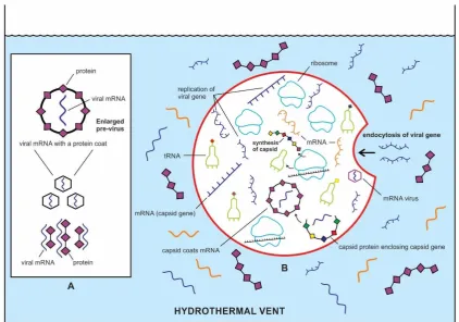
The two important capsid proteins are the jelly-roll capsid proteins (JRC) and the superfamily 3 helicase (S3H), the former is more widespread [32]. We speculate that primordial mRNA gene encoding capsid protein JRC was present in the vent environment (Fig. 5A). New JRC protein genes can originate either through modification of existing genes or de novo. Viruses contain many de novo genes, namely those in which an existing gene has been ‘overprinted’ by a new open reading frame; mutations of the mRNA gene led to the expression of a second reading frame, overlapping the first. Overlapping genes are very common in viral genomes [56].
The origin and evolution of viruses might have occurred on the mineral substrate of the crater floor, where RNAs and proteins were accumulated side by side. During this prebiotic genomic experiment, naked and fragile mRNA genes might have capped occasionally by proteins that offered protection for stability and durability in the vent environment [1-3]. This protein coat of mRNA molecules was prelude to the evolution of a viral structure. Perhaps, in this milieu of different kinds of mRNAs in the prebiotic soup, JRC capsid genes originated de novo within genomes of nonviral mRNAs by overprinting. These capsid genes were capped by proteins on the mineral substrate, transforming them into ancestral viral particles (Fig. 5A). These ancestral pre-viruses had some survival advantages over naked viral genes in the vent environment because of the protective protein coat. Initially, protein coats were random and were not encoded by the enclosed mRNA genes. Perhaps the ancestral pre-viral genes, when engulfed by protocells, could translate it into custom-made capsid protein using ribosomes of the host (Fig. 5B). Primordial viruses could have evolved by encapsidation of these viral genomes.
Some of the ancestral mRNA viruses were accidentally ingested by the protocells by infolding of their membranes while searching for food in the vent environment. The engulfed viruses shed their capsid coat, which might be used by protocells for protein storage. This might have been the beginning of endocytosis. The viral genes, on the other hand began to exploit the translation machinery of the protocell to make the custom-made capsid protein.
6.4. The Reproduction Cycle of mRNA Viruses
The emergence of ancestral virus with capsid-coding sequence of proteins was a big evolutionary step and was mediated by ribosome-coding protocells (Fig. 5B). Here I propose a model for parallel evolution of protocells and the emerging viruses. As more and more proteins were synthesized, various kinds of protocells dominated in the hydrothermal vent environment. Some of these protocells were densely packed with diverse populations of genetic elements, including self-replicating mRNAs, various protein-coding mRNAs, and translation machinery.
Figure 5. Beginnings of the ancient viral world in the prebiotic soup in the hydrothermal vent
environment, which was an ideal Nature’s genomic laboratory. A, in my model, viral selfish genes modified from mRNA molecules appeared de novo by overprinting in the prebiotic soup; these capsid genes were encased by a protein shell for stability and durability. However, these proteins were random, easily available in the prebiotic soup, and were not encoded by viral gene. This initial stage
of viral structure that afforded protection of fragile mRNA is called ‘pre-virus’. B, In the next stage of
evolution, some viral genes entered the protocells by endocytosis, utilizing their ribosomes for synthesis of capsid proteins. Once the capsid protein began to coat the viral gene, the first mRNA virus appeared.
Once some mRNA molecules developed the capsid coat, the first mRNA viruses originated. The capsid affords protection of the viral gene and allows viral genes to gain access to appropriate host cells. As more and more mRNA viruses were created, they exerted osmotic pressure on the phospholipid membrane causing a burst of the protocells (Fig. 6A). Slowly, these newly released viruses learned by trial and error how to infect protocells and swap genes with them without inventing their own translation machinery. This innovative short-cut strategy for virus reproduction inside protocells worked efficiently, hijacking the translation machinery of the host. Viruses preferred this parasitic existence from the beginning of biosynthesis, and it has continued to proliferate throughout the geologic ages.
We speculate that primordial viruses were single-stranded mRNA viruses that could function both as a genome and as a messenger RNA. It could be directly translated into capsid protein in the host cell by host ribosomes. Like living counterparts, the genome contains relatively few genes, usually between three and ten, including as an RNA-dependent RNA polymerase (RdRp) or RNA replicase, a viral protein enzyme that synthesizes mRNA from mRNA template. RdRp is an essential protein-encoded in the genomes of all RNA-containing viruses prior to the DNA stage. Pre-viruses would donate an RdRp gene to the host cell facilitating replication of its mRNA. RNA replication before the emergence of RdRp is difficult to comprehend.
Figure 6. The origin of mRNA viruses and their reproduction cycle. A, synthesis of first generation
of mRNA viruses inside protocells hijacking hosts’ translation machinery. mRNA viral genes
We speculate that primordial virions initially didn’t kill or loose their host protocells, but utilized their translation machinery for reproduction of genomes. Some of the likely stages of the cycle of infection were (Fig. 6B):
1. Attachment. Capsids on the surface of the mRNA virus attach to the surface of the host protocell (Fig. 6B, cycle 1).
2. Entry via endocytosis. The virus enters the interior of the host protocell through the process of endocytosis.
3. Uncoating of capsid. Inside the protocell, the viral genome emerged from the protein capsid; the capsid in turn destroyed the host mRNA so that viral mRNA occupied its place (Fig. 10B, cycle 2, inset).
4. mRNA copying and protein synthesis. RdRp enzymes copy the viral genome. Energy and ribosome from the host protocell are used to build viral proteins. (Fig. 6B, cycle 2 and cycle 3).
5. Assembly of viral progeny. The viral particles assemble by encapsidation to form progeny virions (Fig. 6B, cycle 4).
6. Release via exocytosis. The virus exits in the host protocell by exocytosis (Fig. 6B, cycle 4, inset).
The progeny virions began to infect other protocells to begin the next cycle of infection (Fig. 10B, cycle 5). The life cycle of most viruses is designed to maximize the production of progeny virus particles. Often, the burden of producing a large number of virus particles causes the infected cells to die, the lysis of the host cell. In early stages, primordial viruses probably established a long-term association with the protocell, in which the protocell released a steady stream of viral particles over an extended period of time, benefiting both host and parasite in symbiosis. These ancient RNA viruses had a high mutation rate and underwent evolution and natural selection, just like cellular life, and most of them evolved rapidly. When two viruses infected a protocell at the same time, they might swap genetic material to make new ‘mixed’ viruses with unique properties. The viral infection of a protocell is a prelude to a modern bacteriophage that infects and replicates within Bacteria and Archaea.
This is the beginning of mRNA viruses and their spread in the vent environment. Today, mRNA viruses amount to a large fraction of known viruses including many pathogens, such as the hepatitis C virus, West Nile Virus, dengue virus, and SARS and MERS coronaviruses, as well as less clinically serious pathogens, such as rhinoviruses that cause the common cold [57].
6.5. Retroviruses
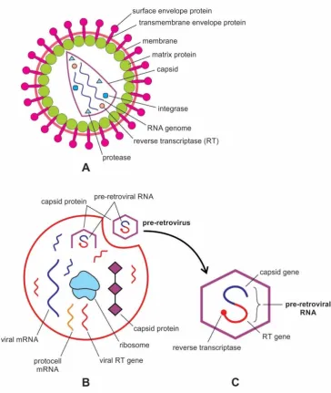
A retrovirus is a highly derived enveloped particle in which the capsid core contains two identical single-stranded RNA molecules, each RNA carries its genetic and structural blueprint. The virion is 80-100 nm in diameter, and its lipid envelope incorporates and displays the viral phospholipids. The hallmark of a retrovirus is its replicative strategy in the sense that it can reverse-transcribe its RNA into DNA using its own reverse transcriptase enzyme. This catalyzed transcription is the reverse flow of information of central dogma, hence the name reverse transcriptase and retrovirus. The new DNA is then integrated into the host cell genome by an integrase enzyme. The host cell treats the viral DNA as part of its own genome, transcribing and translating the viral genes, producing the proteins required to assemble new copies of a retrovirus [34,35].
frameshifting mechanism makes the retroviral genome more compact (Fig. 7A). Retroviruses have evolved to exploit this translational plasticity in order to regulate their own expressions [58].
Figure 7. Retroviruses. A, structure of a modern retrovirus, an enveloped particle in which the capsid core contains two identical single-stranded mRNA molecules. Each mRNA is made up of three genes: integrase, reverse transcriptase, and protease. Once inside the host cell cytoplasm, the virus uses its own reverse transcriptase enzyme to produce DNA from its RNA genome. B, likely origin of pre-retrovirus inside infected protocell, where the viral RT gene was linked to the mRNA viral gene, and was encased by a capsid coat. Once two genes were fused into a single gene for close packing, pre-retrovirus released from the protocell via exocytosis. It was a non-enveloped particle; C, structure of a pre-retrovirus showing how two genes—capsid gene and RT gene were fused into a single gene, and were encased by a capsid protein.
6.6. Origin of Retroviruses
The known virosphere consists of three principal viral types: the RNA viruses, retroid viruses, and DNA viruses. Horizontal gene transfer (HGT) is rampant among viruses within each of these principal types, but is generally confined to closely related viruses, or viruses (and plasmids) with similar replication mechanisms [59]. There are many examples of mixing and matching in the virus world, but somehow, they so far have been confined to the same type of nucleic acid. A novel virus genome discovered in an extreme hot spring environment suggests recombination of two unrelated groups— between a ssDNA virus and an RNA virus—a natural chimera not seen before [60]. In this hybrid genome, alongside the RNA-derived gene, it contained a gene for DNA replication typical of a DNA virus. Surprisingly, these hybrid viruses are present not just in the acidic lake, but more widespread in a couple of oceanic samples. This find proves that modern viruses can combine information in the two normally separate genetic molecules. And it lends support to the idea that it was viruses that performed the upgrade from RNA and effectively gave rise to DNA. These authors suggest that the hybrid virus may have formed when an mRNA virus, retrovirus, and DNA virus all infected a cell at the same time. The retrovirus used its reverse transcriptase enzymes to make a DNA copy of an RNA virus gene, which combined with the DNA genome to yield this hybrid. In our view, this hybrid virus provides a first glimpse at the ancient viral birth of DNA in the hydrothermal vent environment by the mixing and matching of mRNA virus, pre-retrovirus, and DNA virus.
Retroviruses infect a wide range of animals from fish to humans, and can occasionally leave genomic fossils within their host genome, known as endogenous retroviruses (ERVs). ERVs consist of the genetic material of extinct, or ‘fossil’ viruses. Our bodies are littered with the shards of retroviruses. Eight percent of our genome is composed of broken and disabled retroviruses, which, millions of years ago, managed to embed themselves in the DNA of our ancestors. Because they no longer seem to serve a purpose or cause harm, these remains have often been referred as ‘junk DNA’ [34]. Recent phylogenetic study of ERVs placed the time of their most recent common ancestor in the Early Paleozoic [61]. The origin of retroviruses in the Devonian presents an important framework to investigate evolutionary transitions that led to the emergence of the retroviruses. Since vertebrates originated during the Cambrian evolution, ERVs in vertebrate hosts represent the upper limit of the retroviral origin. Molecular precursors of retrovirus probably began in the prebiotic environment billions of years ago in the RNA-DNA Retro world [36]. Retrovirus-like entities are older than the first cells.
Retroviruses bear much similarity to capsidless selfish genetic elements, such as plasmids and various types of transposons because they have close evolutionary connections, both share hallmark genes. These hallmark genes encode key components of the viral replication apparatus (such as polymerases and helicases). These retroelements—capsidless genetic parasites—are key to understanding the origin of viral genes [52]. We speculate that these retroelements were self-assembled to a viral gene such as a positive-strand mRNA virus step-by-step. Positive-sense RNA are particularly suitable for reverse genetics because their genomes are typically infectious in protocells and can be immediately translated by the host’s translation machinery. In our model, ancestral retroviruses could emerge only when various retroelements and protein enzymes were available inside protocells.
One of the critical enzymes synthesized inside protocells was the reverse transcriptase (RT) enzyme. RT is an RNA-dependent DNA polymerase. Due to the limitation of the genome size that can be packaged in the virus shell, viral polymerases are generally active as a single protein capable of carrying out multiple functions related to the viral genome synthesis. Here we propose a simple evolutionary scenario for the origin of retroviruses. The vast class of retroelements is united by a single conserved gene, the RT gene, which also defines the key feature of their reproduction cycle, reverse transcription [52].
mRNA and was enclosed in a capsid shell. In this fused viral gene, one gene was used for synthesis of structural capsid protein, the other for the RT gene for synthesis of reverse transcriptase enzyme (Fig. 7B, C). The RdRp enzyme could be used by pre-retroviruses to replicate their genomes.
The ability to make structural protein and enzyme from the same mRNA gene had distinctly selective advantages over two separate genes performing similar functions in a double-stranded RNA virus. Most likely, protocells, by that time, had developed several weapons to ward off viral attacks by destroying viral genomes. In response, the pre-retrovirus altered their RNAs in such a way as to thwart attacks from the protocells. Moreover, this linking of two genes, capsid gene and RT gene, into one mRNA strand allowed a novel compact, space-saving mechanism of the genome. This ability of linking two genes into one was achieved by the ribosomal frameshifting mechanism as discussed earlier [58]. A pre-retrovirus had now become a single-stranded mRNA virus with a capsid coat to play a formidable parasite during protocell infection (Fig. 7B, C). With the emergence of retroviruses, the protein/RNA world transformed itself into the ‘retro world’ [52].
6.7. The Origin of DNA Viruses from mRNA Viruses
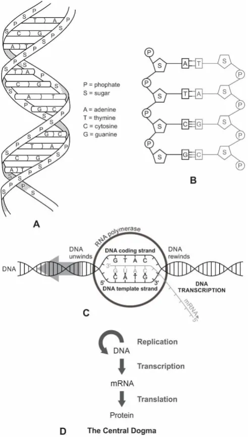
DNA can be considered modified RNA because there are only two chemical differences between RNA and DNA molecules [16]. The first is the removal of a single oxygen atom from RNA (ribonucleic acid) to generate deoxyribonucleic acid, or DNA. The second difference is the addition of a methyl (CH3) group to the nucleotide base uracil (U) to generate thymine (T).
Forterre [40,49] suggests three stages in the evolution of DNA from RNA through viral infection: the RNA world, the RNA-to-DNA transition, and the DNA world. In the RNA world, RNA viruses emerged from the RNA cell. In this model, LUCA is considered as the primitive RNA cell from which the RNA virus emerged. The RNA virus gave rise to three lineages of the DNA viruses in the RNA-DNA transition. These three lineages of RNA-DNA virus evolved in parallel into three domains of life— Bacteria, Archaea, and Eukaryotes.
Forterre [40,49,62] proposes that DNA viruses evolved directly from RNA viruses in two steps during the RNA-to-DNA transition. The critical enzymes for RNA to DNA conversion was supplied by the viral world. In the first step of the RNA-to-DNA transition, the deoxyribose in DNA was generated from the ribose in RNA by enzymes called ribonucleotide reductases, which converted RNA to U-DNA in the genome. In the second step in the evolution of DNA was the conversion of the uracil base to thymine by thymidylate synthases, forming T-DNA (DNA containing thymidine). The emergence of thymidylate synthase activity in some U-DNA virus lineages produced viruses with the modern form of T-DNA. As these new strains of T-DNA viruses infected protocells, the host genomes gradually transformed from U-DNA to T-DNA. Once deoxyribonucleoside triphosphates were available, their assembly into DNA-like chains followed fairly rapidly. The idea that both ribonucleotide reductases and thymidylate synthases were first encoded in viral genomes and were later transferred to protocells is compatible with phylogenetic analyses of these enzyme families. Thus, viruses donated both DNA genomes and their replicating genes to the host protocells [40,49]. The hypothesis of a viral origin for DNA could explain why many DNA viruses encode their own ribonucleotide reductase and/or thymidylate synthase [63]. According to this model, the RNA world and LUCA appeared at the same time with the existence of a few homologous DNA informational proteins in the three cellular domains. Cellular DNA and its replication machineries originated in the DNA world via transfers from DNA viruses to RNA cells. Three such independent transfers led to the origin of Bacteria, Archaea, and Eukaryotes.
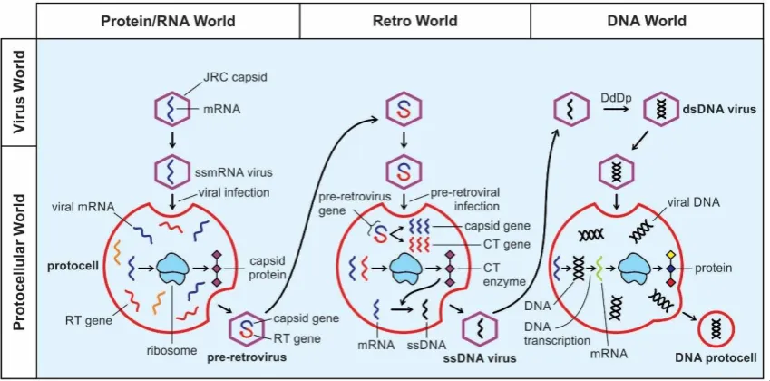
model of the origin of DNA [36,52] very attractive and plausible. In my view, retroid RNA such as pre-retrovirus might have invented DNA step-by-step by converting RNA with the help of the RT enzyme. I suggest three stages in the viral evolution, combining both Forterre’s and Koonin’s model that gave rise to DNA from RNA: the protein/RNA world, the Retro world, and the DNA world (Fig. 8). In my view, the existence of RNA-only cells (protocells) of Forterre [49] seem to meet formidable difficulties. A more parsimonious scenario includes the protein/RNA world [9].
.
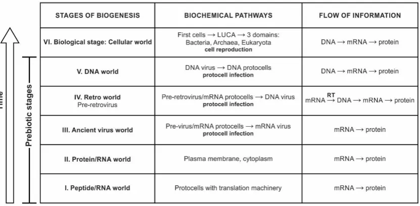
Figure 8. Postulated evolution of the biochemical pathwaysfrom the peptide/RNA world to the DNA world and the concurrent building of the information system to contemporary central dogma (DNA-makes-RNA-makes-proteins).
In the protein/RNA world, viral genes might have evolved de novo in the vent environment from pre-existing mRNA. These mRNA genes encoded JRC capsid protein for a protective shell for stability and durability to form the first virus inside protocells (Fig. 5). These ancient mRNA viruses began to infect protocells to increase their own populations in the gene pool (Fig. 6).
In the Retro world, pre-retroviruses developed the ability to stitch two genes into one. The first gene encoded the JRC capsid protein, but the second gene encoded the reverse transcriptase (RT) enzyme. The RT enzyme could generate complementary DNA from an mRNA template. The pre-retrovirus would play the crucial role for conversion of RNA to DNA during recurrent protocell infection, and subsequently incorporated into the host genome as a provirus. The reverse transcriptase enzyme, also called RNA-directed DNA polymerase, that catalyzed the conversion of RNA to DNA was not present in the host protocells, but was delivered by the pre-retroviruses that converted RNA of the host genome directly into DNA. The RT enzyme performed three sequential biochemical activities: RNA-dependent DNA polymerase activity, ribonuclease H (RNase H), and DNA-dependent DNA polymerase activity. Collectively these activities enabled the RT enzyme to convert single-stranded RNA (ssRNA) into single-stranded DNA (ssDNA). In our model, pre-retroviruses gradually modified its mRNA to DNA during recurrent retroviral infection. As soon as a pre-retrovirus invaded a protocell by endocytosis, its capsid coat was dissolved. The capsid gene segment would be translated into capsid protein and the RT gene segment into the RT enzyme, thus exploiting the ribosome of the host. The RT enzyme would then transcribe its own single-stranded RNA template to single-stranded DNA (ssDNA); the RT would make another strand of cDNA from another mRNA; two strands of cDNA then combined to make a double-stranded DNA (ssDNA). The conversion of ssDNA to double-stranded DNA was mediated by DNA polymerase (DdDp) (Fig. 9). The information contained in a retroviral gene is thus used to generate the corresponding protein via the sequence:
In the DNA world, once the dsDNA virus appeared, the role of the RNA pre-retrovirus was gradually replaced by DNA viruses. DNA viruses possessed a full set of independent DNA replication enzymes: a helicase to unwind the DNA helix, two DNA polymerases (DNA Pols) to replicate the two strands, and a primase to form RNA primers that DNA Pols extended. As dsDNA viruses infected an RNA protocell, its DNA genome was replicated into multiple copies. Each DNA genome then replaced the mRNA of the protocell, transcribed to the new generation of mRNA that translated into protein. This is the beginning of the DNA world, when DNA replaced mRNA as the major genome of the protocell. The DNA protocell followed a conventional pathway of flow information as in a cell: DNA––> mRNA ––> protein.
Figure 9. Coevolution of viruses and protocells. Viruses became important vectors for donating critical enzymes and modifying genomes of the protocells during recurrent infection. Later, some of those viruses evolved DNA as a way to defend their genomes from attack, and DNA-based viruses became incorporated into hosts. In between are the fundamental catalytic processes that allowed to stepwise generate viral deoxyribonucleotides from ribonucleotides by RNA polymerase (RdRp), reverse transcriptase (RT), and DNA polymerase (DdRp) enzymes.
The abundance of genetic parasites along with the presence of defense systems in all cellular life forms strongly suggest their coevolution might have started in the protocellular stage. The DNA viruses might have emerged as a novel survival strategy. When the RNA protocells were confronted with an invading pre-retrovirus, they might have protected themselves by a number of defense mechanisms and developed an ancestral immune system. Immunity of viral infection allowed the RNA protocells to proliferate. In response, pre-retroviruses might have invented DNA to ward off attacks from the hosts. For pre-retroviruses, DNA might have offered a very powerful, immediate benefit: That pre-retroviruses might have immediate fitness benefit for substituting DNA replacing its RNA genome [64].
It thus appears that the transition from RNA to DNA genomes occurred in the viral world, and that protocellular DNA and its replication machineries originated via transfers from DNA viruses to RNA protocells [62,64]. The DNA virus living in a carrier state in an RNA protocell probably lost the genes for capsid proteins and became established as DNA plasmids. These plasmids were later transferred to the RNA protocell and incrementally transformed into DNA by recurrent infection. In such a scenario, the RNA protocell was transformed, from within, into the DNA protocell cell [49]. With DNA genome and its transcribed mRNA and ribosome, protocells began to function as a DNA protocell that began to synthesize protein. The coded genetic information began to flow from DNA to RNA to protein, beginning the classical central dogma of molecular biology.
biological information. Once deoxyribonucleotide triphosphates (dNTPs) were available, it is likely that their assembly into DNA-like chains would have followed fairly rapidly. DNA has intrinsically higher replication fidelity, which allows genomes to increase in size and therefore complexity [16].
7. The Advent of DNA
DNA was derived from RNA [16]. In fact, these two molecules are so similar that transformation of RNA to DNA requires two constituents of RNA to be replaced by two close relatives: ribose by deoxyribose, and uracil (U) by thymine (T). As to the information, RNA could be transferred to DNA by reverse transcription, as happens in cells infected with retroviruses. We have already discussed the origin of the DNA virus from the RNA protocell by pre-retroviruses. Although the origin of DNA from RNA via retrovirus is widely accepted [32,49,52], there are some dissenters. The latest twist in the origin of DNA debate is that RNA and DNA might have appeared together in the prebiotic world from building blocks without the assistance of viruses.
Powner et al. [63] proposed a novel pathway for the prebiotic synthesis of several micro components in the assembly of DNA molecules from a mixture of the chemicals thought to have been present in the sulfur-rich prebiotic environment. In this environment both RNAs and DNAs emerged simultaneously, not one after another. The authors argued that the switching of RNA nucleotides into DNA nucleotides needed special enzymes that were costly to produce in terms of energy and material. On the other hand, if DNA molecules were present alongside RNA molecules, this problem of fundamental switching from conversion of RNA to DNA could be solved. The possibility of an abiotic route for the synthesis of deoxyribonucleic acid, according to these authors, provides a new perspective for the origin of DNA.
A similar view has been expressed by other authors. For example, Xu et al. [65] suggested de novo assembly of DNA from the building blocks of life in the prebiotic environment. They identified a compound presumably present in the prebiotic Earth called thiouridine that could have linked DNA nucleotides into chain-like DNA. Prebiotic phosphorylation of the 2-thiouridine molecule gave rise to nucleotides of DNA via photoreduction. In their view, both RNA and DNA may have arisen all at once in first life forms.
The possibility that DNA and RNA might have evolved concurrently appears to be the less parsimonious explanation than DNA following RNA. The DNA must have appeared later than the RNA molecule, because RNA degrades and mutates easily [6]. The backbone of a single-stranded RNA molecule is much less stable than the equivalent structure in a double-stranded DNA molecule. Its enhanced stability and longer molecular sequence give DNA greater fidelity and increased memory in its information storage system. RNA replication is intrinsically error-prone compared with DNA replication. DNA was selected over RNA based on its expanded capacity to store information and its dramatically improved error rate during replication [15].
Recent detection of ribose sugar in the Murchison meteorite and other primitive carbonaceous chondrites (NWA 801 and NWA 7020) by an international team of scientists clearly suggests that RNA evolved before DNA, not concurrently [66]. Ribose is a crucial component of RNA, which could have stored information and catalyzed reactions during prebiotic synthesis. The research provides the first direct evidence of ribose in space and the delivery of the sugar to Earth during impacts of carbonaceous meteorites about four billion years ago. It is remarkable that a molecule as fragile as ribose could be detected in such ancient meteorites. In contrast, the sugar in DNA (deoxyribose) was not detected in any of the meteorites analyzed in this study. This finding is important because there could have been a delivery bias of extraterrestrial ribose to the early Earth which is consistent with the hypothesis that RNA evolved first. According to the team, the next logical step would be to investigate the chirality of sugars in more carbonaceous chondrites to check whether the cosmic sugar is right-handed or not. Perhaps, homochirality evolved later in the hydrothermal vent environment [1]. The extraterrestrial ribose might have contributed to the formation of RNA on the prebiotic Earth, which possibly led to the origin of DNA by retroviral infection [36,49].