___________________________________________________________________________________________ 3(4): 2157-2172, 2013
SCIENCEDOMAINinternational
www.sciencedomain.org
A Possible Effect of Concentrated Oolong Tea
Causing Transient Ischemic Attack-Like
Symptoms
John W. Layher Jr.
1, Jon S. Poling
2, Mayumi Ishihara
3, Parastoo Azadi
3,
Gerardo Alvarez-Manilla
4and David Puett
5,6* 1Oconee Heart and Vascular Center, St. Mary`s Health Care System, Inc., Athens, GA30606, USA.
2Athens Neurological Associates, Athens, GA 30606, USA. 3Division of Analytical Services, Complex Carbohydrate Research Center, University of
Georgia, Athens, GA 30602, USA.
4Bioanalytical Lab, Pharmaceutical Product Development, Richmond, VA 23230, USA. 5Department of Biochemistry and Molecular Biology, University of Georgia, Athens, GA
30602, USA.
6Department of Biochemistry and Biophysics, University of North Carolina at Chapel Hill
School of Medicine, Chapel Hill, NC 27599, USA.
Authors’ contributions
This work was carried out in collaboration between all authors. Author JWL evaluated the cardiological performance of the patient and made recommendations for the interpretation of the clinical results and revisions to the manuscript. Author JSP was responsible for the neurological evaluation of the patient and made suggestions regarding the case and revisions to the manuscript. Author MI performed the HPLC and MS experimental work, made peak assignments, and prepared the figures and tables. Author PA was responsible for the analytical services, including the choice of instrumentation and laboratory in which the experimental studies were performed, as well as ensuring proper interpretation of the results. Author GAM made the initial purification of the tea before HPLC and MS, and supervised the early MS studies. Author DP coordinated the overall study, interacted with all co-authors, performed extensive literature searches, and wrote the manuscript. All authors had the opportunity to read and revise the manuscript.
ABSTRACT
Aims: Tea (green, oolong, and black) is the second most widely consumed beverage worldwide, second only to water. Aside from a few reported adverse effects, tea, particularly green tea, appears to be beneficial for human health. In the case described herein, a male experienced several transient ischemic attack-like symptoms immediately following the consumption of a cup of high quality oolong tea. A thorough medical evaluation uncovered no evidence of such an attack and leads to the suggestion of a heretofore unreported response to oolong tea.
Presentation of Case: A 72-year old male with hypertension and atrial fibrillation, who takes valsartan/hydrochlorothiazide to control hypertension and warfarin to reduce the risk of thrombosis and thromboembolism, presented at the emergency room of a local hospital describing several transient ischemic attack-like symptoms immediately after consuming a cup of oolong tea. His symptoms included presyncope, disequilibrium, bilateral hand parathesias, mild dysphasia, and visual problems (but apparently not presbyopia or amaurosis fugax), all of which had disappeared in approximately two hours after drinking the tea. (Mild presyncope was previously noted by the patient when ingesting a strong green tea.) No unusual features emerged from his physical examination, and his blood work was unremarkable except for elevation of his partial thromboplastin time (39 sec) and prothrombin time (22.5 sec), giving an international reference of 2.0, all consistent with the effects of warfarin. A battery of tests by the emergency room physician, a cardiologist, and a neurologist, e.g. electrocardiogram, brain computerized tomography, 2-dimensional transthoracic echocardiogram, brain magnetic resonance imaging, with and without 20 ml Gadolinium, and a magnetic resonance angiogram, confirmed the earlier diagnosis of atrial fibrillation but disclosed no additional malfunction in his heart. His brain showed no evidence of a prior hemorrhage, and his carotid arteries were clear.
Methodology and Results: Analysis of the oolong tea by high performance liquid chromatography and mass spectrometry identified the major catechins and two methylxanthines, caffeine and theophylline, as well as other constituents, but there was no evidence of any extraneous chemicals that could lead to the symptoms.
Conclusion:In view of the rapid onset of symptoms after the consumption of oolong tea, bilateral as opposed to unilateral parathesis, and the absence of any evidence of a hemorrhage or the presence of impurities in the tea, we suggest that the transient ischemic attack-like symptoms could possibly be attributable to one or more components of the oolong tea and was not an atypical magnetic resonance imaging-negative transient ischemic attack.
Keywords: Transient ischemic attack; oolong tea; mass spectrometry; high performance liquid chromatography.
ABBREVIATIONS
time-international normalized ratio, TFA-trifluoroacetic acid, TIA- transient ischemic attack, TTE-transthoracic echocardiogram, VKORC1- vitamin K epoxide reductase complex 1.
1. INTRODUCTION
Tea, one of the most widely consumed beverages worldwide, is derived from young buds and the leaves of several varieties of Camellia sinensis(Theaceae), first cultivated in China and Southeast Asia over 2,000 years ago [1]. The three major types of tea are green, oolong, and black. Oolong tea, upon which this paper is based, represents an intermediate in the preparation of green and black teas. Green tea is prepared by initial heating (pan firing or steaming) to inactivate the endogenous enzymes, while oolong and black teas are said to be `fermented,` that is, after moisture reduction (a process termed withering), the leaves are rolled and crushed, a procedure that begins the endogenous enzymatic reactions. Oolong tea is fired soon after crushing to inactivate these reactions, while black tea is prepared after increased `fermentation.` Oolong tea, like the green and black teas, is composed primarily of the four polyphenolic catechins, epigallocatechin gallate (EGCG), epigallate catechin, epicatechin gallate, and epicatechin, with EGCG being the most prevalent. While there are many studies on green tea, the literature on oolong tea is less detailed. There is an indication that oolong tea may contain several unique components resulting from the fermentation protocol, i.e. oolongtheanin, 8-ascorbyl EGCG, and oolonghomobisflavans [1], but no suggestions of physiological effects have been attributed to these products.
In addition to its popularity as a beverage, tea has been reported to have many beneficial effects to human health, including a reduction in the incidence of cancer and cardiovascular disease [2-7] as well as suggestions that tea may reduce the possibility of ischemic stroke [8-11]. Further, it has been shown that oolong tea is more effective than black tea, although less effective than green tea, in inhibiting an important enzyme in blood pressure regulation, angiotensin converting enzyme [12]. These reports notwithstanding, caution has been suggested in recommending tea to the population at large or to individual patients to lower cardiovascular risks [13]. Although there are some indications of adverse effects from concentrated green tea extracts, including liver toxicity and interactions with drugs and herbal supplements [14], there are no reported symptomatic responses to oolong, green, or black tea similar to those detailed in the present case study.
There are many neurologic symptoms accompanying a transient ischemic attack (TIA), and the etiology can be diverse, including hypertension, atrial fibrillation, hypercholesterolimia, diabetes, increasing age, and a family history of stroke. A TIA arises from ischemia, most often from an embolus originating in an atherosclerotic plaque in one of the carotid arteries or from a thrombus in someone with atrial fibrillation. Indicative of a possible future stroke, preventive action should be taken to minimize such an occurrence. Since atrial fibrillation is one of the factors responsible for a TIA, an anticoagulant such as warfarin or one of the newer anti-coagulants is often prescribed for such patients. Warfarin exerts its action on the vitamin K-dependent clotting factors, in particular by inhibiting the essential enzyme, vitamin K epoxide reductase complex 1 (VKORC1), that acts in the vitamin K cycle to catalyze the reduction of vitamin K epoxide to vitamin K quinone, thus diminishing the carboxylation of factors II, VII, IX, X, and the C and S anticoagulant proteins [15,16].
2. PRESENTATION OF CASE
2.1 Evaluation by an Emergency Care Physician
The patient, a 72 year-old Caucasian male in good physical condition, presented at the emergency department of a local hospital describing neurologic symptoms that had occurred about two hours earlier and were indicative of a TIA. He reported that while speaking with someone in his office as he finished drinking a cup of oolong tea he developed a number of worrisome symptoms. The patient had consumed a cup of green or oolong tea daily for many years prior to this incident, the usual amount being approximately 250 mg (dry weight of tea leaves) per 240 mL hot water. On this particular day the tea was prepared in a more concentrated blend, about 4-5 times the usual (1-1.2 g dry weight per 240 mL), seeped five minutes, the leaves remaining in the cup during the 30 min period of consumption. Immediately upon finishing the cup of tea, he experienced the following indicators of a TIA: mild visual problems (symptoms not descriptive, however, of presbyopia or amaurosis fugax), presyncope, disequilibrium, hand parathesias (albeit bilateral), and following that meeting noticed some mild dysphasia when speaking with someone else. He also noted that he had experienced some presyncope in the past when consuming either a greater quantity or a more concentrated Japanese green tea at a local restaurant. The symptoms had disappeared upon presentation at the hospital, but there was sufficient concern on the part of the patient, his wife, and attending physician that they felt an evaluation was needed in view of his past medical history. He had been diagnosed with hypertension and benign prostatic hypertrophy, and five years earlier with atrial flutter. The flutter was corrected by cardiac ablation in the right atrium, with atrial fibrillation one year earlier. His daily medications were warfarin sodium (coumadin, 5 mg six days a week and 7.5 mg once a week), taken in the evening about 9:00 PM, and valsartan/hydrochlorothiazide (diovan/ HCTZ, 160 mg/12.5 mg), alfuzosin HCl (uroxatral, 10 mg), rebeprazole sodium (aciphex, 20 mg for acid reflux), and bifidobacterium infantis (probiotic, 4 mg), all taken in the early morning between 5:00-6:00 AM. He was alert and had a temperature of 97.3ºF, a pulse rate of 57 beats per minute, a systolic/diastolic blood pressure of 160/90 that later decreased to 139-141/80-76 mm Hg during his stay in the emergency department, and an oxygen saturation of 97-99% throughout his stay as an outpatient. He mentioned that his blood pressure had been treated for several years and was generally lower than that measured in the hospital setting. Laboratory tests were mainly in the normal range, with a few being either marginally low or high. The prothrombin and partial thromboplastin times were elevated (22.5 and 39 sec, respectively), and the prothrombin time-international reference (PT-INR) was 2.0, attributable to warfarin. An x-ray of the chest demonstrated that the lungs were clear bilaterally and the cardiac silhouette and mediastinum were unremarkable; CT of the brain (axial imaging) showed no evidence of an acute process. Heart irregularity was noted, and an electrocardiogram (EKG) revealed atrial fibrillation with an average rate in the 50s, a normal axis, and no evidence of ST segment abnormalities. The patient was administered IV fluid and then released with a recommendation that he see a cardiologist for further evaluation.
2.2 Evaluation by a Cardiologist
using color and doppler flow imaging were as follows: there was mild right atrial enlargement with mild to moderate tricuspid insufficiency; the estimated right ventricular systolic pressure was slightly elevated; the left and right ventricles appeared normal in size, and the left ventricular systolic and diastolic function appeared normal; the aortic valve showed mild sclerosis without evidence of stenosis; and the mitral valve was structurally normal. An agitated saline contrast image (`bubble study`) showed no evidence of an intracardiac shunt. It was recommended that the patient begin 81 mg aspirin to be taken 3 times a week, along with warfarin. Overall there was no evidence of serious cardiac problems other than atrial fibrillation. In view of the TIA-like symptoms the patient originally presented with, it was suggested that he also be examined by a neurologist.
2.3 Evaluation by a Neurologist
A brain magnetic resonance imaging (MRI), with and without 20 ml Gadolinium, and a magnetic resonance angiogram (MRA) yielded the following information. The diffusion-weighted images were negative, and gradient echo studies showed no evidence for a prior hemorrhage. Overall, no acute intracranial abnormality was observed, and, importantly, there was no evidence for a prior cortically-based infarct; moreover, after Gadolinium administration a normal meningeal and vascular enhancement was observed. There was evidence of minimal small vessel ischemic cerebrovascular changes in the hemispheres, some slight fluid accumulation within the mastoid air cells, and mucosal lining thickening in the inferior maxillary sinuses and a few of the ethmoidal air cells. The MRA documented the absence of any hemodynamically significant stenosis involving the arteries of the neck.
2.4 Summary
A brain CT and MRI failed to detect any evidence of an infarct, and a MRA of the neck indicated normal flow. The 2D TTE of the heart, with and without agitated saline to ascertain bubble passage, detected no abnormality. Thus, atrial fibrillation appeared to be the only significant problem of the patient.
Based on these medical results, a study was undertaken to characterize the oolong tea that was consumed and, although there is no known precedent in the literature, could possibly have led to the acute TIA-like symptoms experienced by the patient. The goal was to determine the constituents in the tea, after extraction in hot water as was done for consumption. The techniques employed were liquid chromatography tandem mass spectrometry (LC-MS/MS) and high performance liquid chromatography (HPLC) using conditions similar to those described by others [17-20]. The methodologies used and the experimental data are reported in the Appendix.
3. RESULTS AND DISCUSSION
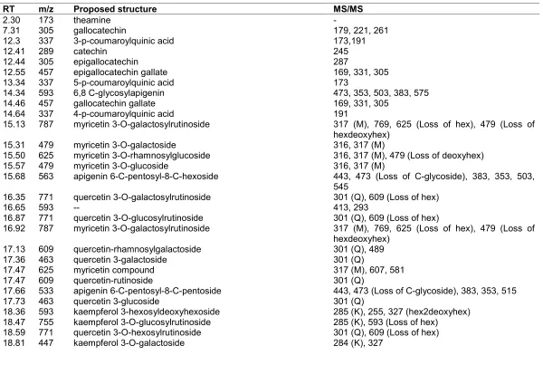
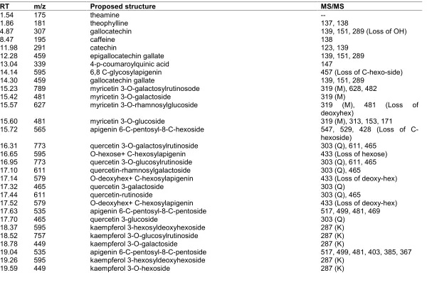
was found at m/z 349 and m/z 609. The m/z 349 component is attributed to a cross-ring cleavage of the hexose attached on quercetin at the 1 and 3 carbon bonds (1,3A/1,3X), whereas m/z 609 results from a neutral loss of the terminal hexose. This fragmentation pattern confirmed the structure of the carbohydrate moiety in the parent ion. A few flavonoids, including caffeine, were detected in just the positive ion mode because of their polarity. Also, in the positive ion mode acylated flavonol glycosides were observed in the H+, Na+, and K+ forms. For MS/MS analysis of the acylated flavonol glycosides in the positive ion mode, the H+form caused ion suppression during MS/MS fragmentation that resulted in poor signals; in these cases structural assignments were based on the Na+ adducts. The flavonoids identified in the tea sample are given in Appendix Tables 1 and 2 for results obtained in the negative and positive ion modes, respectively. For structural assignment the observed full mass and MS/MS were compared to those of known standards, those reported previously [18-20], and the theoretical full mass and MS/MS.
A sample of Yamamotoyama oolong tea was brewed according to the recommendations of the manufacturer, filtered, and then analyzed by MS. This control tea gave an almost identical profile to that of the oolong tea consumed by the patient, confirming that unusual or unexpected peaks were not present in the `experimental` tea.
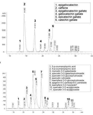
The HPLC chromatograms of the oolong tea extract are shown in Fig. 2 with detection at 270 nm (panel A) and 350 nm (Panel B), and assignments of the major peaks. Caffeine and the catchenins, EGCG and epicatechin gallate, are the major components identified at 270 nm, and the major phenolic components identified at 350 nm are myricetin 3-O-galactoside, quercetin glucosylrutinoside, quercetin rutinoside, and kaempferol 3-O-glucosylrutinoside; many other minor components are seen in each chromatogram.
The results obtained from MS and HPLC characterization of the oolong tea are in excellent agreement with findings reported by others [18-20], demonstrating the complexity and variety of components present in tea. A detailed study on 41 green teas and 25 fermented teas enabled Lin et al. to classify the teas into five categories based upon their compounds [18]. Oolong tea was in Group 3 that, compared to green tea, contained less EGCG, more theaflavins and other catechins, and comparable amounts of glycosylated flavonols. The majority of the health benefits attributed to tea are believed to arise from the flavonoids, particularly the catechins (polyphenols) that are the major constituent of tea. Present in much lower concentrations are the methylxanthines, including caffeine, theophylline, and methlyxanthine theobromine, the first two of which were detected in our sample. While caffeine, a well-known CNS and metabolic stimulant, may be suspect in leading to some of the TIA-like symptoms experienced by the patient, there is no documentation of such effects in the literature. Four caffeine-induced disorders are recognized by the American Psychiatric Association, none of which correspond to the symptomology of the patient discussed herein [21]. Interestingly, EGCG, the major catechin in tea and confirmed to be present in the tea in question in this study, has been suggested to counteract caffeine-mediated stimulant effects [22].
consumption of black tea, her INR increased to 5.0 but was restored to the 2-3 range when her warfarin dose was decreased [26]. While suggesting that tea can alter the INR of some individuals taking warfarin, it was not an issue in the present case since the patient was found to have an INR of 2.0 when he presented at the hospital.
It is unlikely that the response to the tea could arise from an acute metabolic disturbance of one or more medicines being taken by the patient, or vice versa, particularly since the onset of the symptoms appeared immediately after drinking the tea while the medications had been taken anywhere from 7-15 hours earlier. There are reports in experimental animals that quercetin enhances the bioavailability of valsartan [27]; however, the patient described herein had taken his daily valsartan some seven hours before drinking the concentrated tea. In another report based on studies in human hepatoma HepG2 cells and primary cultures of human hepatocytes, the authors concluded that EGCG is an antagonist to the aryl hydrocarbon receptor that regulates the expression of the cytochrome (CYP) P450 subfamily CYP1A, composed of two members, CYP1A1 and CYP1A2, that metabolize many chemical carcinogens [28]. At present, there is no apparent correlation of this effect with any of the symptomology experienced by the patient. Additionally, genotypes of CYP2C8 and CYP2C9, cytochrome P450s responsible for the metabolism of most angiotensin II receptor antagonists, impact on the pharmacokinetics of valsartan [29], but the relationship of these findings to the patient presented is not known. Of interest are the reports indicating that tea may reduce the onset of stroke [8-11], thus strengthening our suggestion that the symptoms observed in the patient were, in many respects, TIA-like, not an actual TIA.
Since (a) atrial fibrillation significantly increases a person`s risk of stroke or systemic embolization, with the risk steadily increasing with age, and (b) while warfarin reduces the risk some 62-68%, but does not eliminate it altogether, atrial fibrillation must be considered as a possible cause of a TIA in the patient. Moreover, brain and MR angiography imaging have known limitations, including (a) the ability to visualize some vessels but not others, and (b) the possibility that some microemboli could have been present but were too small to be imaged or the emboli may have broken up and resolved by the time the imaging was performed. On the other hand, the neurologic symptoms exhibited by the patient do not fit any cerebral vascular distribution involving, for example, a clot going to one area of the brain. In light of the fact that some of the symptoms were bilateral, e.g. hand parathesias, while others suggest a global cerebral process, e.g. presyncope, only a simultaneous diminished flow or pressure going to both sides of the brain could cause a similar picture, and this is ruled out by the normal or slightly elevated blood pressure of the patient in the Emergency Department. Thus, following a thorough evaluation of the patient and a critical review of the medical findings, no unequivocal evidence was found to indicate that a TIA had occurred, and it must be considered that the symptomology could be attributable to an unusual acute effect from the concentrated oolong tea.
4. CONCLUSIONS
tea-mediated hypotension could explain some of the symptoms experienced by the patient, there is no conclusive evidence that a drop in blood pressure could explain all the symptoms. Thus the effect may reflect both hemodynamic and CNS effects attributable to the oolong tea.
CONSENT
Written informed consent from the patient for publication of this case report copy was sent to the Chief Editor of this journal.
ETHICAL APPROVAL
Not applicable.
ACKNOWLEDGEMENTS
The authors thank Dr. Richard Panico for helpful discussions and suggestions. We also thank Dr. Gabriela Sanchez-Brambila for her assistance during the early phase of the experimental work. This research was supported in part by the National Institutes of Health (NIH/NCRR)-funded grant entitled `Integrated Technology Resource for Biomedical Glycomics` (grant no. 8P41 GM103490) to the Complex Carbohydrate Research Center of the University of Georgia, Athens, Georgia, USA.
COMPETING INTERESTS
Authors declare that they have no competing interests.
REFERENCES
1. Harbowy ME, Balentine DA. Tea chemistry. Crit Rev Plant Sci. 1997;16:415-480. 2. Balentine DA, Wiseman SA, Bouwens LCM. The chemistry of tea flavonoids. Crit Rev
Food Sci Nutr. 1997;37:693-704.
3. Higdon JV, Frei B. Tea catechins and polyphenols: Health effects, metabolism, and antioxidant functions. Crit Rev Food Sci Nutr. 2003;43:89-143.
4. Cabrera C, Artacho R, Gimenez R. Beneficial effects of green tea-A review. J Am Coll Nutr. 2006;25:79-99.
5. Hodgson JM, Croft KD. Tea flavonoids and cardiovascular health. Mol Aspects Med. 2010;31:SI 495-502.
6. Chow HHS, Hakim IA. Pharmacokinetic and chemoprevention studies on tea in humans. Pharmacol Res. 2011;64:105-112.
7. Zhang M, Li L, Liu P, Holman CD`AJ. Green tea for the prevention of cancer: Evidence of field epidemiology. Func Foods Health Dis. 2012;2:339-350.
8. Arab L, Liu W, Elashoff D. Green and black tea consumption and risk of stroke: A meta-analysis. Stroke. 2009;40:1786-1792.
9. Liang W, Lee AH, Binns CW. Tea drinking, diet and ischemic stroke prevention in China: A future perspective. Expert Rev Cardiovasc Ther. 2009;7:1447-1454.
11. Larsson SC, Mannisto S, Virtanen MJ, Kontto J, Albanes D, Virtamo J. Coffee and tea consumption and risk of stroke subtypes in male smokers. Stroke. 2008;39:1681-1687.
12. Dong J, Xu X, Liang Y, Head R, Bennett L. Inhibition of angiotensin converting enzyme (ACE) activity by polyphenols from tea (Camellia sinensis) and links to processing method. Food Funct. 2011;2:310-319.
13. Deka A, Vita JA. Tea and cardiovascular disease. Pharmacol Res. 2011; 64: 136-145. 14. Schonthal AH. Adverse effects of concentrated green tea extracts. Mol Nutr Food Res.
2011;55:874-885.
15. Wallin R, Wajih N, Hutson SM. VKORC1. A warfarin-sensitive enzyme in vitamin K metabolism and biosynthesis of vitamin K-dependent blood coagulation factors. In: Litwack G, editor. Vitamins and hormones. Book Series, Vol. 78. New York: Academic Press. 2008;227-246.
16. Lurie Y, Loebstein R, Kurnik D, Almog S, Halkin H. Warfarin and vitamin K intake in the era of pharmacogenetics. Brit J Clin Pharmaco. 2010;70:164-170.
17. Pellati F, Orlandini G, Pinetti D, Benvenuti S. HPLC-DAD and HPLC-ESI-MS/MS methods for metabolite profiling of propolis extracts. J Pharmaceut Biomed Anal. 2011;55:934-948.
18. Lin L-Z, Chen P, Harnly JM. New phenolic components and chromatographic profiles of green and fermented teas. J Agric Food Chem. 2008;56:8130-8140.
19. Del Rio D, Stewart AJ, Mullen W, Burns J, Michael EJ, Brighenti F, Crozier A. HPLC-MSn analysis of phenolic compounds and purine alkaloids in green and black tea. J Agric Food Chem. 2004;52:2807-2815.
20. Kite GC, Rowe ER, Lewis, GP, Veitch NC. Acylated flavanol tri- and tetraglycosides in the flavonoid metabolome of Cladrastis kentukea (Leguminosae). Phytochemistry. 2011;72:372-384.
21. First, MB. Editor. Diagnostic and statistical manual of mental disorders, 4th Edition, DSM-IV-TRTM. Washington, D.C.: American Psychiatric Publishing, See Caffeine-Induced Disorders, 292.85 (p. 655), 292.89 (p. 479), 292.90 (p. 234), 305.90 (p. 232); 2000.
22. Han JY, Kim CS, Lim KH, Kim JH, Kim S, Yun YP, Hong JT, Oh KW. Increases in blood pressure and heart rate induced by caffeine are inhibited by (-)-epigallocatechin-3-O-gallate: Involvement of catecholamines. J Cardiovasc Pharm. 2011;58:446-449. 23. Booth SL, Sadowski JA, Pennington JAT. Vitamin K1 (phylloquinone) content of foods:
A provisional table. J Food Comp Ana.l 1993;6:109-120.
24. Booth SL, Madabushi HT, Davidson KW. Tea and coffee brews are not significant dietary sources of vitamin K1(phylloquinone). J Am Diet Assoc. 1995; 95: 82-83. 25. Taylor JR, Wilt VM. Probable antagonism of warfarin by green tea. Ann Pharmacother.
1999;33:426-428.
26. Parker DL, Hoffmann TK, Tucker MA, Meier DJ. Interaction between warfarin and black tea. Ann Pharmacother. 2009;43:150-151.
27. Challa VR, Babu PR, Challa SR, Johnson B, Maheswari C. Pharmacokinetic interaction study between quercetin and valsartan in rats and in vitro models. Drug Devel Indus Phar. 2012 doi: 10.3109/03639045.2012.693502.
28. Williams SN, Shih H, Guenette DK, Brackney, W, Denison MS, Pickwell GV, Quattrochi LC. Chemico-Biol Interact. 2000;128:211-229.
APPENDIX
METHDOLOGY OF TEA ANALYSIS
Preparation of the tea for LC-MS/MS and HPLC:The oolong tea leaves (about 1.2 g of Tie Guan Yin Oolong Tea, Iron Goddess of Mercy, a high grade oolong tea) were seeped in 240 mL of hot water under conditions as close as possible to those used in the tea that was consumed (see Section 2.1). The leaves and any particulates were then separated from the liquid in the cup by filtration, and the filtrate was stored overnight at 5ºC.
A SepPak cartridge (1 g) was inserted into a vacuum manifold and fitted with a 50 mL plastic syringe and then activated with 10 mL methanol (MeOH) and equilibrated with 20 mL 0.1% (trifluoroacetic acid (TFA). The tea extract (50 mL) was added, and the unbound components were eluted with 200 mL 0.1% TFA. Following transfer of the cartridge to a collection manifold, the non-polar compounds were eluted with 3 mL MeOH and then dried in a Speed-Vac. Next the sample was dissolved in 1 mL MeOH, dried, and reconstituted in a mixture of 0.9 mL 0.1% formic acid and 0.1 mL acetonitrile (CH3CN). Following heating at 50ºC for about 5 min to ensure a homogeneous liquid phase, the sample was diluted 10-fold in 10% CH3CN in 0.1% formic acid.
LC-MS/MS: Several MS runs were made using MALDI-TOF-MS (Bruker Autoflex) and LC-MS (Perkin-Elmer SC/EX), after which it was decided to use LC-LC-MS/LC-MS on a LTQ mass spectrometer (ThermoFisher) equipped with an electrospray ion source. Using conditions similar to those described for separating polyphenols [17], the sample was diluted 5-fold with mobile phase A (0.2% acetic acid in water) and filtered with 0.2 µm filters (Nanosep, PALL). 5 µL of the sample was injected into a C18 column (2.1 x 100 mm, Grace Vydac) followed by separation using a 60 min gradient of increasing mobile phase B (80% CH3CN and 0.2% acetic acid in water) at a flow rate of 200 µL/min.
The instrument was tuned with EGCG and caffeine for negative ion mode and positive ion mode, respectively. The LTQ was run in the automatic mode collecting a MS scan (full mass spectrometry at 150-2,000 m/z) followed by data-dependent MS/MS scans of the six highest abundant precursor ions. The data were analyzed manually for structural assignment, and the observed full mass and MS/MS were compared to those of known standards, e.g. EGCG and caffeine, those reported by others [18-20], and the theoretical full mass and MS/MS.
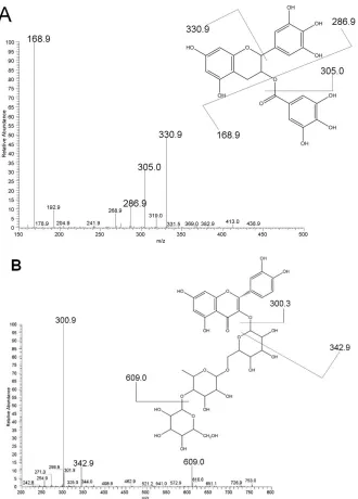
Fig. 1. MS/MS spectra of oolong tea in the negative ion mode documenting the presence of EGCG, m/z 457 (A), and quercetin 3-O-glucosylrutinoside, m/z 771 (B).
The main fragment ions identified in panel A are gallic acid (m/z 168.9) and epigallocatechin (m/z/ 305); the neutral loss of trihydroxybenzene is also indicated (m/z 331). In panel B the main fragment ion identified is quercetin (m/z 301). A neutral
loss of the carbohydrate portion is indicated at m/z 349, arising from a cross-ring cleavage of quercetin at the 1 and 3 carbon bonds, and at m/z 609, attributed to a
A
B
Table 1. Flavanoid analysis of oolong tea sample by LC-MS/MS in the negative ion mode
RT m/z Proposed structure MS/MS
2.30 173 theamine
-7.31 305 gallocatechin 179, 221, 261
12.3 337 3-p-coumaroylquinic acid 173,191
12.41 289 catechin 245
12.44 305 epigallocatechin 287
12.55 457 epigallocatechin gallate 169, 331, 305
13.34 337 5-p-coumaroylquinic acid 173
14.34 593 6,8 C-glycosylapigenin 473, 353, 503, 383, 575
14.46 457 gallocatechin gallate 169, 331, 305
14.64 337 4-p-coumaroylquinic acid 191
15.13 787 myricetin 3-O-galactosylrutinoside 317 (M), 769, 625 (Loss of hex), 479 (Loss of hexdeoxyhex)
15.31 479 myricetin 3-O-galactoside 316, 317 (M)
15.50 625 myricetin 3-O-rhamnosylglucoside 316, 317 (M), 479 (Loss of deoxyhex)
15.57 479 myricetin 3-O-glucoside 316, 317 (M)
15.68 563 apigenin 6-C-pentosyl-8-C-hexoside 443, 473 (Loss of C-glycoside), 383, 353, 503, 545
16.35 771 quercetin 3-O-galactosylrutinoside 301 (Q), 609 (Loss of hex)
16.65 593 -- 413, 293
16.87 771 quercetin 3-O-glucosylrutinoside 301 (Q), 609 (Loss of hex)
16.92 787 myricetin 3-O-galactosylrutinoside 317 (M), 769, 625 (Loss of hex), 479 (Loss of hexdeoxyhex)
17.13 609 quercetin-rhamnosylgalactoside 301 (Q), 489
17.36 463 quercetin 3-galactoside 301 (Q)
17.47 625 myricetin compound 317 (M), 607, 581
17.47 609 quercetin-rutinoside 301 (Q)
17.66 533 apigenin 6-C-pentosyl-8-C-pentoside 443, 473 (Loss of C-glycoside), 383, 353, 515
17.73 463 quercetin 3-glucoside 301 (Q)
18.36 593 kaempferol 3-hexosyldeoxyhexoside 285 (K), 255, 327 (hex2deoxyhex) 18.47 755 kaempferol 3-O-glucosylrutinoside 285 (K), 593 (Loss of hex)
18.59 771 quercetin 3-O-hexosylrutinoside 301 (Q), 609 (Loss of hex)
19.05 533 apigenin 6-C-pentosyl-8-C-pentoside 443, 473 (Loss of C-glycoside), 383, 353, 515 19.29 609 quercetin 3-O-deoxyhexosylhexoside 301 (Q)
19.33 593 kaempferol 3-hexosyldeoxyhexoside 285 (K)
19.33 625 myricetin compound 317 (M), 299, 607
19.63 447 --a 403, 249, 267
20.18 417 kaempferol 3-O-pentoside 284, 285 (K), 327
24.08 1063 quercetin 3-O-acylglycoside 917 (Loss of coumaric acid), 771, 615, 447, 301 (Q)
24.47 917 quercetin 3-O-p-coumaroylhexosyldeoxyhexosylhexoside 771 (Loss of coumaric acid), 301 (Q) 24.49 1063 myricetin 3-O-acylglycoside 917 (Loss of deoxy-hex), 777, 317 (M) 24.63 1049 quercetin 3-O-acylglycoside 903 (Loss of coumaric acid), 301 (Q) 24.71 917 quercetin 3-O-p-coumaroylhexosyldeoxyhexosylhexoside 771 (Loss of coumaric acid), 301 (Q)
24.78 1033 quercetin 3-O-acylglycoside 887 (Loss of coumaric acid), 741, 747, 447, 301 (Q)
24.86 901 quercetin 3-O-p-coumaroylhexosyldeoxyhexosylhexoside 755 (Loss of coumaric acid), 609, 447, 301 (Q) 25.00 1063 myricetin 3-O-acylglycoside 917 (Loss of deoxy-hex), 741, 451, 317 (M) 25.19 1049 quercetin 3-O-acylglycoside 903 (Loss of coumaric acid), 301 (Q) 25.41 1033 coumaric acid compound 887 (Loss of coumaric acid), 747, 597, 451 25.59 1063 quercetin 3-O-acylglycoside 917 (Loss of deoxy-hex), 771, 615, 447, 356, 301
(Q)
25.71 901 kaempferol 3-O-p-coumaroyldideoxyhexosylhexoside 755 (Loss of coumaric acid), 615, 433, 285 (K) 25.78 917 quercetin 3-O-p-coumaroylhexosyldeoxyhexosylhexoside 771, 753 (Loss of coumaric acid), 301 (Q)
25.86 1033 --a
--25.97 901 quercetin 3-O-p-coumaroylhexosyldeoxyhexosylhexoside 755 (Loss of coumaric acid), 615, 447, 301 (Q) 26.56 901 kaempferol 3-O-p-coumaroyldideoxyhexosylhexoside 755 (Loss of coumaric acid), 615, 285(K)
Table 2. Flavonoid analysis of oolong tea sample by LC-MS/MS in the positive ion mode
RT m/z Proposed structure MS/MS
1.54 175 theamine
--1.86 181 theophylline 137, 138
4.87 307 gallocatechin 139, 151, 289 (Loss of OH)
8.47 195 caffeine 138
11.98 291 catechin 123, 139
12.28 459 epigallocatechin gallate 139, 151, 289
13.04 339 4-p-coumaroylquinic acid 147
14.14 595 6,8 C-glycosylapigenin 457 (Loss of C-hexo-side)
14.30 459 gallocatechin gallate 139, 151, 289
15.23 789 myricetin 3-O-galactosylrutinosode 319 (M), 628, 482
15.42 481 myricetin 3-O-galactoside 319 (M)
15.57 627 myricetin 3-O-rhamnosylglucoside 319 (M), 481 (Loss of
deoxyhex)
15.60 481 myricetin 3-O-glucoside 319 (M), 313, 153, 171
15.72 565 apigenin 6-C-pentosyl-8-C-hexoside 547, 529, 428 (Loss of C-hexoside)
16.31 773 quercetin 3-O-galactosylrutinoside 303 (Q), 611, 465
16.65 595 O-hexose+ C-hexosylapigenin 433 (Loss of hexose)
16.95 773 quercetin 3-O-glucosylrutinoside 303 (Q), 611, 465
17.10 611 quercetin-rhamnosylgalactoside 303 (Q), 465
17.14 579 O-deoxyhex+ C-hexosylapigenin 433 (Loss of deoxy-hex)
17.32 465 quercetin 3-galactoside 303 (Q)
17.44 611 quercetin-rutinoside 303 (Q), 465
17.52 579 O-deoxyhex+ C-hexosylapigenin 433 (Loss of deoxy-hex)
17.63 535 apigenin 6-C-pentosyl-8-C-pentoside 517, 499, 481, 469
17.70 465 quercetin 3-glucoside 303 (Q)
18.37 595 kaempferol 3-hexosyldeoxyhexoside 287 (K)
18.52 757 kaempferol 3-O-glucosylrutinoside 287 (K)
18.78 449 kaempferol 3-O-galactoside 287 (K)
19.04 535 apigenin 6-C-pentosyl-8-C-pentoside 517, 499, 481, 403, 385, 367
19.26 595 kaempferol 3-hexosyldeoxyhexoside 287 (K)
20.04 419 kaempferol 3-O-pentoside 287 (K)
23.68 1065 (1086) quercetin 3-O-acylglycoside 941, 785 (Loss of Q), 639, 493, 331
24.10 1065 (1086) quercetin 3-O-acylglycoside 942, 787 (Loss of Q), 771, 640, 475, 330
24.25 1051 (1073) quercetin 3-O-acylglycoside 771(Loss of Q),639,607, 475
24.37 919 (941) quercetin 3-O-p-coumaroylhexosyldeoxyhexosylhexoside 639(Loss of Q),475
24.48 903 (925) kaempferol compound 779, 477
24.60 1065 (1086) kaempferol 3-O-acylglycoside 801 (Loss of K), 637, 621, 475, 391
24.79 1051 (1073) quercetin 3-O-acylglycoside 771(Loss of Q),621, 607, 475
25.06 1035 (1057) kaempferol 3-O-acylglycoside 771, 607, 475
25.25 1065 (1086) kaempferol 3-O-acylglycoside 941, 801 (Loss of K), 639, 475, 331
25.32 903 (925) kaempferol 3-O-p-coumaroylhexosyldeoxyhexosylhexoside 639(Loss of K),475
25.36 919 (941) quercetin 3-O-p-coumaroylhexosyldeoxyhexosylhexoside 639(Loss of Q),475
aUnknown. Abbreviations: Hex, hexose; K, kaempferol; M, myricetin; Q, quercetin; RT, retention time. MS/MS analysis of acyl glycosides (italicized)
were performed on sodium adducts, and the m/z values are italicized in parentheses.
© 2013 Layher Jr. et al.; This is an Open Access article distributed under the terms of the Creative Commons Attribution License (http://creativecommons.org/licenses/by/3.0), which permits unrestricted use, distribution, and reproduction in any medium, provided the original work is properly cited.
Peer-review history: