_____________________________________________________________________________________________________ *Corresponding author: E-mail: [email protected];
(Past name: British Journal of Medicine and Medical Research, Past ISSN: 2231-0614, NLM ID: 101570965)
Correlation of
GSTP1
Polymorphism with Severity of
Prostate Cancer in an Eastern Indian Population
Suparna Roy
1, Anindya Dasgupta
1*, Subarnarekha Chatterji
2and Dilip Karmakar
31
Department of Biochemistry, Calcutta National Medical College, Kolkata-14, India.
2
Department of Biotechnology, School of Life Sciences, Manipal University, Manipal, India.
3Department of Urology, Calcutta National Medical College, Kolkata-14, India.
Authors’ contributions
This work was carried out in collaboration among all authors. Authors SR, AD, SC and DK designed the study. Authors SR and SC performed the statistical analysis, wrote the protocol, and wrote the first draft of the manuscript. Authors AD and SR managed the analyses of the study. Author SC managed the literature searches. All authors read and approved the final manuscript.
Article Information
DOI: 10.9734/JAMMR/2019/v29i230062 Editor(s): (1) Dr. Mohammed Rachidi,Molecular Genetics of Human Diseases, French Polynesia, University Paris 7 Denis Diderot, Paris, France. (2) Dr. Sevgul Donmez, Faculty of Health Sciences, Gaziantep University, Turkey.
Reviewers: (1) Lawrence H. Lash, Wayne State University, USA. (2)Dr. Guven Aslan, Dokuz Eylül University, Turkey. (3)Dr. Alok Nahata, Ying Zhi Agricultural and Industries, Malaysia. Complete Peer review History:http://www.sdiarticle3.com/review-history/47755
Received 02 January 2019 Accepted 10 March 2019 Published 19 March 2019
ABSTRACT
Background: GSTP1 is one of the Glutathione-S-Transferases (GSTs) which suppress tumor genesis by detoxifying toxic carcinogens and reactive oxygen species (ROS). Prostate cancer is related to several mutations affecting the expression of GSTP1. A single nucleotide polymorphism (SNP: Ile105Val) in the GSTP1 gene results insignificant reduction in its anticancer activity. The current case control study was conducted to ascertain the risk of association of GSTP1polymorphism with risk of cancer prostate in an Eastern Indian population.
Materials and Methods: During a study period of 2 years, DNA was isolated using the phenol chloroform extraction method from the blood of 225 histopathologically diagnosed prostate cancer patients and 120 matched controls. The GSTP1 polymorphism was assessed by PCR amplification of the gene followed by restriction digestion with Alw261 (a restriction enzyme derived from
Roy et al.; JAMMR, 29(2): 1-10, 2019; Article no.JAMMR.47755
Acinetobactro lwoffi RFL26). Histopathological grading in the case group was performed using Gleason’s scores and International Society of Urological Pathology (ISUP) grading.
Results: Comparison of the distribution of different GSTP1 alleles between the case and control groups was performed by chi square test and odds ratio analysis. A χ2 value of 18.56 suggested significantly higher number of G alleles in the case group. An odds ratio of 2.25 with a confidence interval of 1.52 to 3.34 for 95% CI showed that the G allele in GSTP1 gene were linked with greater risk of prostate cancer. Post hoc ANOVA and logistic regression suggested that cases having G alleles had more progressive form of diseases as evident from ISUP grades.
Conclusion: From our study we can conclude that GSTP1 polymorphism is not only significantly associated with risk of prostate cancer but also with its severity in our Eastern Indian population. GSTP1 polymorphism should be considered as a prognostic indicator for prostate cancer patients along with planning for more aggressive management of the disease.
Keywords: Prostate cancer; GSTP1 polymorphism; ISUP grading; single nucleotide polymorphism; restriction digestion.
1. INTRODUCTION
Prostate cancer is the third leading cause of cancer death among men in the United States [1]. It has higher mortality rate among African-Americans compared to Caucasians [2]. However, in India, it has been reported to be the second killer cancer in large metro cities and among the top ten cancers in the rest of the country according to the population based cancer registries in India [3]. Recurrence rate and mortality rate of prostate cancer depend on Gleasonʹs grading and a higher serum prostate specific antigen (PSA) level [4] even after radical prostatectomy [5].
Initiation of cancer is attributable to several genetic disarrangements including chromosomal deletion, translocation, changes in DNA methylation and point mutations [6,7]. These genetic changes become particularly important when they affect the expression of tumor suppressor proteins. Glutathione-S-Transferases (GSTs) belong to one of such tumor suppressor proteins which restrict the initiation and progression of tumor genesis by detoxifying different toxic carcinogens and reactive oxygen
species (ROS). The GSTP1 gene is
approximately 4 kb in length, comprises 7 exons and 6 introns and codes for a 715 base mRNA. GSTs have several isozymes with almost similar functions in different tissues. They are responsible for metabolism and biosynthesis of various metabolites including detoxification of exogenous carcinogen chemicals like polycyclic aromatic hydrocarbon which are abundant in diesel fuels, cigarette smoke and grilled meats. Overall, they detoxify several carcinogenic xenobiotics by conjugation with glutathione during the phase II of detoxification process of
the electrophonic carcinogenic compounds [8,9]. Specific GST isoforms in the (M1), (T1) and (P1) classes are highly expressed in the prostate tissues [10]. Among the large family of their isoenzymes, the P1 class of enzyme GSTP1-1 is well studied in different types of cancers. GSTP1- 1expression has been found to be associated with resistance to cytotoxic drugs in breast cancer cells also [11]. Therefore any alteration in the genetic polymorphic profile of GSTP1may be associated with severity as well as the risk of recurrence of prostate cancer which has been strongly suggested by the present research works that found significant link between GSTP1 polymorphism and need for a repeat biopsy to evaluate a progression of prostate cancer [12].
GSTP1 is mainly expressed in the basal layer of normal prostate epithelium. Its expression has been found to be significantly down-regulated in the initial stages of majority of adenocarcinoma including the cancer prostate [13]. The potential
GSTP1 gene promoter site remains
unmethylated and an adenine (A) at the 303 position. Previous studies have shown that the CpG-rich promoter region of the p-class GSTP1is variably methylated producing multiple restriction sites in the majority of prostate cancers [7].
Another important single nucleotide
polymorphism (SNP) in the GSTP1 gene was found to be Ile105Val (rs1695 A > G) that replaced valine by isoleucine at the 105thpostion of the GSTP1 protein causing significant reduction in the detoxifying capability of this important GST isoenzyme [14].
varying indifferent regions of the world significantly. Some studies have reported strong association between it and prostate cancer whereas others reported their association to be negligible or nil. Two studies earlier reported more significant association of this SNP with prostate cancer in Caucasian people in comparison to the Asians and Americans [15,16], while recent meta-analyical studies have suggested a stronger association of prostate cancer with Ile105Val among the Asian population [17]. Keeping these factors in mind we hypothesized that prostate cancer is linked with this SNP of Ile105Val polymorphism (SNP rs1695 A > G) in our region and undertook the present study to ascertain its risk of association with the severity of prostate cancer in an Eastern Indian population.
2. MATERIALS AND METHODS
The present study was a hospital based cross-sectional observational study conducted in the Department of Biochemistry and Department of Urology of Calcutta National Medical College over a period of 2 years from November 2016 to October 2018.
2.1 Selection of Case Subjects
Patients suffering from adenocarcinoma of the prostate gland diagnosed on the basis of clinical investigations, histopathology and prostate specific antigen were selected. At first, cases were selected provisionally on the basis of clinical investigation at the Dept. of Urology by the method of convenience that was followed by their final inclusion by histopathology and PSA measurement. As PSA is specific for prostate tissue and not for prostate cancer only, there is a considerable overlapping of the PSA values between BHP and CA prostate. So, it is difficult to assign a PSA value with 100% sensitivity and specificity for CA prostate only. Values more than 4 µg/L increase the specificity but lowers the sensitivity. Hence, we selected the typically used value of 4 µg/L of PSA with for screening the CA prostate cases as recommended by National Comprehensive Cancer Network [18] followed by a definitive diagnosis by histopathological grading using the Gleason’s score and ISUP grading. During this period all patients suffering from prostate cancers were selected irrespective of the tumor stage and their localization status. Thus, patients with both localized and metastatic disease were considered which were further given appropriate Gleason’s score and ISUP
grading based on histopathology. Patients with any other malignancies, metabolic disorder, smoking and alcohol addiction or any other drug addiction were excluded.
2.2 Selection of Control Subjects
Control subjects were selected from those patients attending urology OPD for ailments other than prostate cancers. Before their final inclusion, prostate cancer were ruled out in them by clinical investigation and PSA estimation. Subjects, suffering from any chronic inflammatory disorders, malignant diseases, metabolic diseases and addiction to smoking, alcohol or any drug were excluded.
Both case and control population were selected from the same geographical area in age matched manner with more or less similar nutritional and socioeconomic status.
2.3 Ethical Considerations
The study was conducted following the guidelines and criteria for human studies as laid on by Helsinki declaration 1975 revised in 2000, and International Committee of Medical Journal Editors (ICMJE). Both informed and written consents were obtained in local language from all study participants in appropriately approved consent forms. The complete proposal was submitted to the Institutional Ethical Committee for the final approval and permission. The study was undertaken only after obtaining the written permission from the institutional ethical committee (vide CNMC/4, dated 26.10.16).
2.4 Study Technique
3 ml of venous blood was collected in aseptic way from the participants. 1.5 ml will be stored in EDTA vial for DNA separation and rest will be stored in clotted vial for serum separation. The EDTA blood was stored at minus 20 degree centigrade till DNA isolation from which DNA was isolated within a maximum period of seven days.
2.5 Isolation of DNA
Roy et al.; JAMMR, 29(2): 1-10, 2019; Article no.JAMMR.47755
polymorphism were assessed by amplification of the gene by PCR technique followed by restriction digestion.
2.6 PCR Technique for the GSTP1 Gene
For PCR we used the PCR mastermix from Thermofisher,USA. The forward and reverse
primers selected were
5ʹ-GTCTCTCATCCTTCCACGCA-3ʹ.and
5ʹ-CTGCACCCTGACCCAAGAA-3ʹ respectively.
We used 10 pmol of each primer in the final PCR mixture of 25µl. The PCR protocol was as follows. The initial preheating was at 95ºCfor 2 minutes, followed by 30 cycles of denaturation at 95ºC for 30 seconds ,annealing at 60ºC for 30 seconds and extension at 72ºC for 1minute. After completion of 30 cycles the final extension was programmed at 72ºC for 5min. the PCR process was performed using Veriflex PCR Thermocycler (ProflexTM) obtained from the Applied Biosystems, Thermofischer Scientific, USA.PCR products obtained were run in 1.2% agarose gel against 100 bp DNA ladder (Bangalore Genie, India) and were identified as 365 bp using the Gel Doc system obtained from Applied Biosystem, Thermofischer Scientific, USA.
2.7 Restriction Digestion
PCR products obtained were digested using the restriction enzyme Alw261 obtained from Thermofischer USA. Restriction digestion products were identified on 3% agarose gel against 100 bp DNA ladder using the gel doc system.
2.8 ISUP Grading
The grading system proposed by the
International Society of Urological Pathology (ISUP) have improved the overall Gleason grading system [20]. Accordingly, the prostate cancer patients in our study were divided into five distinct ISUP grades; Grade 1: Gleason’s score ≤6, Grade 2: Gleason’s score 3 + 4 = 7, Grade 3: Gleason’s score 4+3 = 7, Grade 4: Gleason’s score 4+4 = 8, and Grade 5: Gleason’s score 9 and 10.
2.9 Statistical Analysis
Comparison of the distribution of different GSTP1 alleles between the case and control groups was performed by chi square test and odds ratio analysis. Statistical comparison of ISUP grade distribution between the Ile/Ile, Ile/Val and
Val/Val was done by post hoc ANOVA with Bonferroni correction. Dependence of severity of prostate cancer as indicated by ISUP grading was done by logistic regression analysis. All statistical analyses were done using SPSS software version 20 for windows.
3. RESULTS
Following the inclusion and exclusion criteria, finally 225 cases and 120 control subjects were selected for data analysis for statistical interpretation.
Independent t test suggested that case (and control groups were age matched in our study (mean ± SD for case and control group were 69.3 ± 1.9 and 70.2 ± 5.2 respectively, P = 0.06, data not shown in Tables).
The pattern of digestion of the PCR products of different genotypes of GSTP1 gene and distribution of the digested fragments according to their base pair lengths through agarose gel electrophoresis is shown in the Fig. 1. When compared against 100 bp DNA ladder fragments, the PCR product of GSTP1 gene was reflected by the undigested wild AA genotype and was found to be of 365 bp as expected for the given set of primers (e.g. lane no. 1,6,8,9,11-15). The mutant GG genotype showed two digested products of 140 bp and 225 bp (e.g lane no. 3). On the other hand, the heterozygotes showed three bands of 365, 225 and 140 bp (e.g. lane nos. 2,4,5,10,16).
A significantly greater association of the GG genotype of the GSTP1 gene with the prostate cancer patients in comparison to the control group was reflected by the data in Table 1 with a chi square value of 18.56 (P < 0.001) against a degree of freedom (d.f) of 2. This observation was strengthened by an odds ratio of 16.9 (range of 1.5 to 3.3 at 95% CI) for G allele in the case group (Table 2).
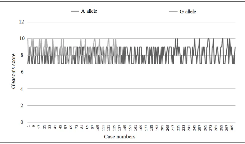
the risk of advanced stages of this cancer. The higher trend of Gleason score in the
was also evident from the boxplot shown in the Figs. 3 and 4, where the overall distribution of
Fig. 1. RFLP pattern of the polymorphic genotypes in 3 % aga
Fig. 2. Scatter plot showing the association between stratified grade
ISUP grading and stage of prostate cancer as indicated by Gleason’s score
the risk of advanced stages of this cancer. The higher trend of Gleason score in the G allele
he boxplot shown in the Figs. 3 and 4, where the overall distribution of
Gleason’s scores was shown according to the different genotypic variation among the case group.
Fig. 1. RFLP pattern of the polymorphic genotypes in 3 % agarose gele electrophoresis
Fig. 2. Scatter plot showing the association between stratified grade grouping as indicated by ISUP grading and stage of prostate cancer as indicated by Gleason’s score
P < 0.001
Pearson’s Correlation
coefficient = 0.923
Gleason’s scores was shown according to the different genotypic variation among the case
rose gele electrophoresis
Roy et al.; JAMMR, 29(2): 1-10, 2019; Article no.JAMMR.47755
Fig. 3. Boxplot showing the distribution of Gleason’s score between the II and VV allele in the prostate cancer patients
Table 1. Chi square test showing the distribution of wild and mutant variants of GST P1
genotypes among the Case (n = 225) and Control groups (n = 120)
Homozygote for AA (Ile/Ile)
Heterozygote AG (Ile/Val)
Homozygote GG (Val/Val)
Chi square (χ2) value
P value
Cases 105(46.6) 100(44.4) 20(9)
18.56 P<
0.001*
Controls 85(70.8) 30(25) 5(4.2)
*P valueis significant at P < 0.05; Percentage for the respective values is shown in parenthesis
Table 2. Odds ratio analysis for assessing the distribution of A and G alleles as risk factors between the case and control groups
A alleles G alleles
Cases 310(68.8) 140(31.2)
Controls 200(83.3) 40(16.7)
P < 0.001. OR = 2.25, Range = 1.52 to 3.34 at 95% confidence interval; Percentage for the respective values are shown in parenthesis
Table 3A. Simple one way ANOVA test to show the overall distribution of ISUP grading among all three genotypes of prostate cancer in the present study
All genotypes
Sum of squares d.f Mean square F value Sig (P value)
Between groups Within groups Total
10.44 286.48 296.93
2 222 224
5.22 1.29
4.04 .019
*P value significant at P < 0.05 for 95% confidence interval
Table 3B. Post hoc ANOVA with Bonferroni’s correction showing the distribution of ISUP grading between the heterozygote Ile/Val allele and the homozygote wild and mutant alleles
Bonferroni
Genotype Group means Mean difference (I-J) Standard error P value*
Ile/Ile and Ile/Val (AA and AG)
3.73; 4.06 -0.33333 0.15 0.11
Val/Val and Ile/Val (GG and AG)
4.40; 4.06 0.34000 0.27 0.66
Ile/Ile and Val/Val (AA and GG)
3.73; 4.40 -0.67333 0.27 0.04*
*P value significant at P < 0.05 for 95% confidence interval.
Table 4. Logistic regression analysis showing the effect of G allele on ISUP grading in prostate cancer patients
Step 1a Intercept Slope Exp(slope) Regression coefficient R2 P value
Allelic variation (A or G)
-2.11 0.31 1.37 0.50 0.0068*
a
: Dependent variable ISUP grading; *P value significant at P < 0.05
Both simple one way and Post hoc ANOVA results from the Table 3A and 3B respectively showed the individual comparison of different genotypes among the prostate cancer patients. A significantly high ISUP grade score in the mutant GG against the wild AA genotype (P = .04, Table
Roy et al.; JAMMR, 29(2): 1-10, 2019; Article no.JAMMR.47755
progressive cancer of prostate against the non mutant AA homozygotes. Furthermore, results of the logistic regression analysis in Table 4 suggested that the G allele has a significant contributory role in progression of the disease in contrast to the A allele both in its homozygous heterozygous form.
4. DISCUSSION
The current study was conducted to detect the association of GSTP1 genetic polymorphism with the risk of prostate cancer as well as its severity in an Eastern Indian population. A chi square value of 18.56 for a d.f of 2 in the Table 1 suggested that number of GG phenotypes were significantly higher in the case group. A significantly higher association of prostate cancer with the G allele in GSTP1 gene was further strengthened by an odds ratio of 2.25 with a confidence interval of 1.52 to 3.34 for 95% CI as shown in the Table 2. Our findings and outcomes are in well congruence with findings of some other studies where prostate cancers have been found to be associated with the G alleles or substitution of valine for isoleucine in the GSTP1 gene [21]. Expression of GSTP1 is regulated mainly at the transcriptional level. It has been suggested that replacement of isoleucine with the less bulkier but more hydrophobic valine in the protein results in the alteration in substrate binding capability of its catalytic site and hence reduction in its detoxifying capability of the pro-oxidant heterocyclic amine carcinogens [22]. Loss of detoxifying capability due to mutant GSTP1 gene has been reflected by a direct association between the lipid peroxidation product 4-hydroxynonenal and prostate cancer in some recent studies [23]. Results of our study not only support this view but in addition the one way (Table 3A) and post hoc ANOVA (Table 3B) suggest that the prostate cancer patients having the G alleles have more progressive form of the disease as evident from their higher ISUP grade. To find out the contribution of the mutant G allele on the ISUP grades in prostate cancer patients, we carried out logistic regression analysis considering the ISUP grade as a dependent variable on both A and G alleles of all three genotypes (Table 4). Results showed a significant predictive role of only G alleles on the ISUP score (regression coefficient 0.50, P = 0.0068).
Polymorphic changes from A to G not only alter the substrate binding property of this enzyme,
but also alter its signal transduction process related to the regulation of cell growth. GSTP1 enzyme protein is also closely linked to the signal transduction process involving Jun N terminal kinase (JNK pathway) [24]. Moreover, its specific inhibitor TER 199 has been found to stimulate the growth of granulocytes [25] suggesting an inhibitory effect of GSTP1 mediated intracellular signal transduction pathway on abnormal cell growth. These cellular mechanisms provide plausible biochemical explanations for the inducing effects of Ile105Val SNP (313 A to G) on prostatic carcinogenesis and its progression to more severe outcome. Hence, from our study we can conclude that GSTP1 polymorphism in the form of Ile105Val is not only significantly associated with risk of prostate cancer but also with its severity in our Eastern Indian population study group.
The major limitation of the present study is a relatively modest number of sample size of prostate cancer patients that could be included in the study according to the stipulated inclusion and exclusion criteria. Although, this limitation can be circumvented using larger study group, but keeping in mind several other studies performed worldwide, out study sample size was not at a lower level that much. Hence, based on our statistical calculations and result output we suggest that GSTP1 polymorphism at Ile105Val level should be explored in much wider areas involving different regions so that it can be considered as a prognostic indicator for prostate cancer in those study populations. Assessment of this polymorphism at the earlier stages of the disease may also help in planning more aggressive management of the disease for preventing further spread and its lethal outcome.
5. CONCLUSION
From our study we can conclude that GSTP1 polymorphism is not only significantly associated with risk of prostate cancer but also with its severity in our Eastern Indian population. GSTP1 polymorphism should be considered as a prognostic indicator for prostate cancer patients along with planning for more aggressive management of the disease.
CONSENT
ETHICAL APPROVAL
The study was conducted following the guidelines and criteria for human studies as laid on by Helsinki declaration 1975 revised in 2000, and International Committee of Medical Journal Editors (ICMJE). Both informed and written consents were obtained in local language from all study participants in appropriately approved consent forms. The complete proposal was submitted to the Institutional Ethical Committee for the final approval and permission. The study was undertaken only after obtaining the written permission from the institutional ethical committee (vide CNMC/4, dated 26.10.16).
COMPETING INTERESTS
Authors have declared that no competing interests exist.
REFERENCES
1. Jemal A, Siegel R, Ward E, Hao Y, Xu J, Murray T, et al. Cancer statistics, 2008. CA Cancer J Clin. 2008;58(2):71-96.
2. Thompson I, Tangen C, Tolcher A, Crawford E, Eisenberger M, Moinpour C. Association of African-American ethnic background with survival in men with metastatic prostate cancer. J Natl Cancer Inst. 2001;93(3):219-25.
3. Jain S, Saxena S, Kumar A. Epidemiology of prostate cancer in India. Meta Gene. 2014;2:596-605.
4. Grossfeld GD, Latini DM, Lubeck DP, Mehta SS, Carroll PR. Predicting recurrence after radical prostatectomy for patients with high risk prostate cancer. J Urol. 2003;169(1):157-63.
5. D'Amico AV, Cote K, Loffredo M, Renshaw AA, Chen MH. Pretreatment predictors of time to cancer specific death after prostate specific antigen failure. J Urol. 2003; 169(4):1320-4.
6. Knudson AG, Jr. Genetics of human cancer. Annu Rev Genet. 1986;20:231-51. 7. Millar DS, Ow KK, Paul CL, Russell PJ,
Molloy PL, Clark SJ. Detailed methylation analysis of the glutathione S-transferase pi (GSTP1) gene in prostate cancer. Oncogene. 1999;18(6):1313-24.
8. Rushmore TH, Pickett CB. Glutathione S-transferases, structure, regulation, and therapeutic implications. J Biol Chem. 1993;268(16):11475-8.
9. Buldak RJ, Buldak L, Kukla M, Gabriel A, Zwirska-Korczala K. Significance of selected antioxidant enzymes in cancer cell progression. Pol J Pathol. 2014;65(3): 167-75.
10. Elek J, Park KH, Narayanan R. Microarray-based expression profiling in prostate tumors. In Vivo. 2000;14(1):173-82. 11. Batist G, Reynaud A, Katki AG, Travis EL,
Shoemaker MC, Greene RF, et al. Enzymatic defense against radiation damage in mice. Effect of selenium and vitamin E depletion. Biochem Pharmacol. 1986;35(4):601-6.
12. Waterhouse RL, Jr., Van Neste L, Moses KA, Barnswell C, Silberstein JL, Jalkut M, et al. Evaluation of an epigenetic assay for predicting repeat prostate biopsy outcome in African American Men. Urology; 2018. 13. Cookson MS, Reuter VE, Linkov I, Fair
WR. Glutathione S-transferase PI (GST-pi) class expression by immunohistochemistry in benign and malignant prostate tissue. J Urol. 1997;157(2):673-6.
14. Sawers L, Ferguson MJ, Ihrig BR, Young HC, Chakravarty P, Wolf CR, et al. Glutathione S-transferase P1 (GSTP1) directly influences platinum drug chemosensitivity in ovarian tumour cell lines. Br J Cancer. 2014;111(6):1150-8. 15. Cai Q, Wu T, Zhang W, Guo X, Shang Z,
Jiang N, et al. Genetic polymorphisms in glutathione S-transferases P1 (GSTP1) Ile105Val and prostate cancer risk: a systematic review and meta-analysis. Tumour Biol. 2013;34(6):3913-22.
16. Yu Z, Li Z, Cai B, Wang Z, Gan W, Chen H, et al. Association between the GSTP1 Ile105Val polymorphism and prostate cancer risk: a systematic review and meta-analysis. Tumour Biol. 2013;34(3):1855- 63.
17. Zhang Y, Yuan Y, Chen Y, Wang Z, Li F, Zhao Q. Association between GSTP1 Ile105Val polymorphism and urinary system cancer risk: Evidence from 51 studies. Onco Targets Ther. 2016;9:3565-9.
18. National Comprehensive Cancer Network. Practice guidelines in oncology: Prostate cancer early detection, v1. 2010.
Available:http://www.nccn.org/network/busi ness_insights/flash_updates/
Roy et al.; JAMMR, 29(2): 1-10, 2019; Article no.JAMMR.47755
from eukaryotes. Nucleic Acids Res. 1976;3(9):2303-8.
20. Pierorazio PM, Walsh PC, Partin AW, Epstein JI. Prognostic Gleason grade grouping: data based on the modified
Gleason scoring system. BJU
international. 2013;111(5):753-60.
21. Wu K, Wang X, Xie Z, Liu Z, Lu Y. Glutathione S-transferase P1 gene
polymorphism and bladder cancer
susceptibility: an updated analysis. Mol Biol Rep. 2013;40(1):687-95.
22. Sundberg K et al. Differences in the catalytic efficiencies of allelic variants of glutathione transferase P1-1 towards carcinogenic diol epoxides of polycyclic
aromatic hydrocarbons. Carcinogenesis 1998;19:433-436.
23. Song X, Wang R, Xiao F, Zhang SX, Gong M, Wang XL, et al. [Expressions of glutathione S-transferase P1 and 4- hydroxynonenal and the progression of prostate cancer]. Zhonghua nan ke xue = National Journal of Andrology. 2017;23(5): 412-6.
24. Adler V, et al. Regulation of JNK signaling by GSTp. EMBO J. 1999;18:1321-1334. 25. Kauvar LM, Morgan AS, Sanderson PE,
Henner WD. Glutathione based
approaches to improving cancer treatment. Chem Biol Interact 1998;111/112:225– 238.
_________________________________________________________________________________
© 2019 Roy et al.; This is an Open Access article distributed under the terms of the Creative Commons Attribution License (http://creativecommons.org/licenses/by/4.0), which permits unrestricted use, distribution, and reproduction in any medium, provided the original work is properly cited.
Peer-review history: