____________________________________________________________________________________________________
*Corresponding author: Email: siegfriedhelage@yahoo.fr;
www.sciencedomain.org
A Simple Computed Tomography Scoring System to
Predict Histological Malignancy of Solitary Fibrous
Tumors of the Pleura
Siegfried Hélage
1*, Marie-Pierre Revel
1, Marco Alifano
2,
Audrey Mansuet-Lupo
3and Dominique Vadrot
11Department of Radiology, Hôpital Hôtel-Dieu de Paris (AP-HP), 1 Place du Parvis Notre-Dame,
75004 Paris, France. 2
Department of Thoracic Surgery, Hôpital Hôtel-Dieu de Paris (AP-HP), 1 Place du Parvis Notre-Dame, 75004 Paris, France. 3Department of Pathological Anatomy and Cytology, Hôpital Hôtel-Dieu de Paris (AP-HP), 1 Place du
Parvis Notre-Dame, 75004 Paris, France.
Authors’ contributions
This work was carried out in collaboration between all authors. All authors were involved in the management of the patients discussed. Author SH wrote the first draft of the manuscript and managed the literature searches. All authors read and approved the final manuscript.
Article Information
DOI: 10.9734/BJMMR/2015/13958 Editor(s): (1)Chulso Moon, The JS YOON Memorial Cancer Research Institute, USA and Department of Otolaryngology and Head and Neck Cancer, Research Institute, The Johns Hopkins University School of Medicine, Baltimore, USA.
Reviewers: (1) Anonymous, University of Sao Paulo Medical School, Sao Paulo, Brazil. (2)Anonymous, University of Medical Science, Japan. Complete Peer review History:http://www.sciencedomain.org/review-history.php?iid=717&id=12&aid=6795
Received 11th September 2014 Accepted 16th October 2014 Published 5th November 2014
ABSTRACT
Purpose: The aim of the present study was to define the very first score enabling discrimination between benign and malignant solitary fibrous tumors of the pleura (SFTPs), on the basis of reliable preoperative CT features.
Methods: Between December 2004 and November 2012, 56 patients underwent complete resection for SFTP at six institutes. CT scans were reviewed retrospectively, and a diagnostic scoring system for predicting malignant SFTP preoperatively was designed.
Results: Univariate analysis revealed seven significant predictors of malignant SFTP: tumor size ≥
10 cm (p=0.002), tumor heterogeneity spontaneously (p=0.019) or after contrast medium injection (p=0.029), existence of intratumoral fluid density areas (p=0.011), a pleural effusion (p=0.01),
measurable (diameter >1 mm) intratumoral vessels (p=0.019), a hypervascular character (visible intratumoral vessels and/or intense enhancement) (p=0.001). A scoring system based on these seven CT features, each assigned 1 point, and with a cut-off of 4 points, could predict malignant SFTP with a specificity of 85% and a sensitivity of 48%.
Conclusion: Our scoring system using seven CT features (tumor size ≥ 10 cm, tumor heterogeneity with or without contrast injection, intratumoral fluid density areas, pleural effusion, measurable intratumoral vessels, and a hypervascular character of the tumor) may be helpful for predicting histological malignancy of solitary fibrous tumors of the pleura.
Keywords: Solitary fibrous tumor; SFT; Pleura; pleural fibroma; CT scan; scoring system.
1. INTRODUCTION
Solitary fibrous tumors of the pleura (SFTPs) are very rare, if not exceptional, primary pleural neoplasms [1]. They originate from submesothelial connective tissue and are not associated with asbestos exposure [2]. Their prognosis is generally good, but up to a third behave in a malignant fashion [3]. Histological diagnosis of a focal malignant transformation, hidden by a prominent benign component intermixed with collagen fibers, depends greatly on the resected gross specimen sampling quality.
To date, no study was designed with a primary objective to assess CT features predictive of SFTP histological malignancy, even if mentioned in an allusive way [4]. Recognition of such CT features will yield several consequences. Concerning surgical treatment, such CT features will all the more compel the surgeon to strictly observe large safety margins, despite technical difficulties due to tumor location. Moreover, in patients with comorbidities that increase anesthetic risk, presence of such CT features will provide a valid argument supporting surgery during tumor boards, especially now that prosecution of medical professionals is becoming increasingly common. Concerning histological diagnosis, such CT features will strongly incite the pathologist to fulfill the widest possible sampling of the resected gross specimen, in order to find the small malignant focus lost in the major benign and/or fibrous component. This focus will be the source of a future tumor recurrence. This is for producing the most accurate diagnosis possible. Concerning imaging modalities, such CT features will provide a strong argument to plan a long-term CT scan follow-up to check for evidence of relapse, despite the fact that radiation protection of patients is coming into sharper focus. Indeed, late recurrences, sometimes more than fifteen years after complete resection, are a common feature of malignant SFTPs [5,6]. Additionally, CT features
suggestive of histological malignancy will strongly incite clinicians to prescribe a broad imaging assessment of cancer spread, especially in order to detect extrathoracic metastases.
Histopathology is not satisfactory in determining with certainty the malignant character of SFTPs [7]. This issue is due to the major heterogeneity of such tumors in terms of histological grade. Some pathologists circumvent this pitfall, talking about tumors of uncertain malignant potential. As a result of this histopathological diagnostic vagueness, percutaneous biopsies are not informative. Consequently, as a precaution, the standard treatment remains complete surgical excision with safety margins of 2 cm [8,9]. Hence, new preoperative tools are needed to justify such an aggressive treatment in fragile patients, and to support a long-term radiological irradiation for follow-up in relatively young patients.
The objective of the present study was to identify reliable CT features predictive of histological malignancy, and to build the very first preoperative CT scan diagnostic scoring system based on these features in SFTPs.
2. PATIENTS AND METHODS
2.1 Experimental Design
injection, 19 CT before and after contrast injection, 23 CT directly after contrast injection. Collected imaging data were analyzed for seven features: tumor size (the plane of section in which tumor diameter reached its maximum value was chosen, with mediastinal windowing), tumor heterogeneity (characterized as areas of low attenuation within the tumor) with or without contrast injection, intratumoral fluid density areas (-20 to +20 Hounsfield units), measurable intratumoral vessels (diameter >1 mm), a hypervascular character of the tumor (characterized as just visible intratumoral vessels and/or intense enhancement compared with paravertebral muscles), and presence of a pleural effusion.
All surgical specimens were classified and graded according to the World Health Organization nomenclature. Histopathology data were obtained from a review of original records. To predict postoperative histopathology results effectively, we designed a CT scan scoring system using the features found to be significant in our univariate analysis. Each CT feature was weighed for a point.
2.2 Statistical Analysis
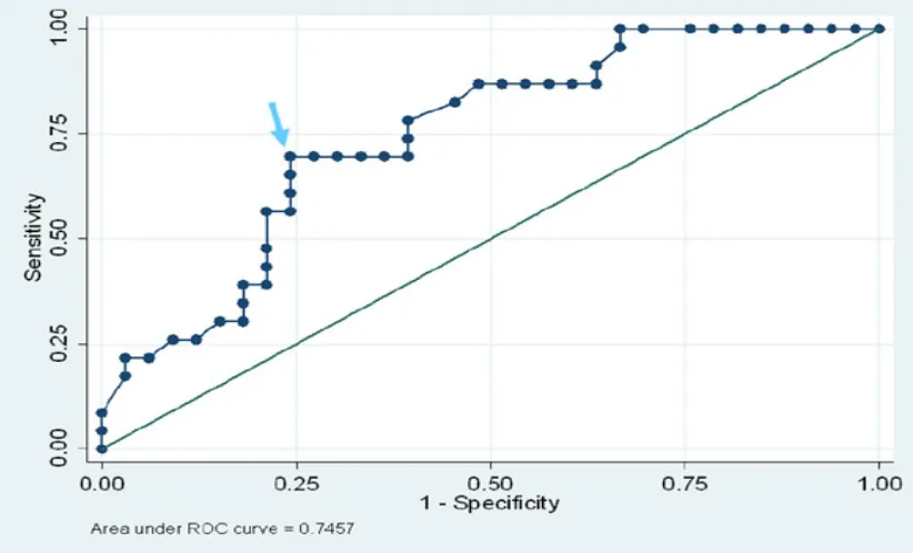
Given the relatively small number of patients, the following nonparametric tests were used to determine P values: Fisher's exact test for qualitative variables and Mann-Whitney test for quantitative variables. The receiver operating characteristics (ROC) curve and Youden index, integrating both sensitivity and specificity, were used to identify the best threshold for tumor size. A P value < 0.05 was considered statistically significant. XLSTAT software was used for all statistical analysis.
3. RESULTS
3.1 Demographic Features
The 56 patients included 33 women and 23 men, of mean age at diagnosis of 60 years (range: 27-82 years). Histological diagnosis included benign SFTPs in 33 patients and malignant SFTPs in 23.
3.2 Univariate Analysis of CT Features
Suggestive of Histological
Malignancy
Univariate analysis identified the seven chosen CT features to be statistically significant predictors of histological malignancy: tumor size
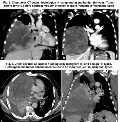
≥ 10 cm (p=0.002, maximum Youden index=0.453) (Fig. 1, Fig. 2), tumor heterogeneity without contrast injection (p=0.019) (Fig. 3), tumor heterogeneity with contrast injection (p=0.029) (Fig. 4), intratumoral fluid density areas (p=0.011), measurable intratumoral vessels (p=0.019) (Fig. 5), a hypervascular character of the tumor (p=0.001), presence of a pleural effusion (p=0.01) (Table 1).
3.3 Preoperative CT Scan Scoring System
for Predicting Histologically
Malignant SFTP
For simplicity in clinical application, we assigned 1 point to each of the seven variables found to be discriminatory between benign and malignant subtypes: tumor size ≥ 10 cm, tumor heterogeneity without contrast injection, tumor heterogeneity with contrast injection, intratumoral fluid density areas, measurable intratumoral vessels, a hypervascular character of the tumor, presence of a pleural effusion.
Assessment of our patients using this scoring system consisting of seven variables and a cut-off of 4 points resulted in a specificity of 85%, a sensitivity of 48%, and a positive predictive value (PPV) of 69%. Using a cut-off of 5, our scoring system could predict the possibility of histologically malignant SFTP with a specificity of 94%, a sensitivity of 30.5%, and a positive predictive value (PPV) of 78% (Table 2).
4. DISCUSSION
In the present study, a large tumor size, tumor heterogeneity either with or without contrast injection, a hypervascular character of the tumor, and a pleural effusion, were more frequent in histologically malignant subtypes.
Fig. 1. Chest sagittal CT scans: histologically malignant (a) and benign (b) types. A tumor whose maximum diameter is greater than or equal to 10 centimeters has a higher probability of
being malignant
Fig. 2. ROC (Receiver Operating Characteristic) curve of tumor size for the risk of being malignant. Youden index reaches its maximum value (YI=0.453) for a threshold size of 10
Fig. 3. Chest axial CT scans: histologically malignant (a) and benign (b) types. Tumor heterogeneity before contrast medium injection is more frequent in malignant types
Fig. 4. Chest coronal CT scans: histologically malignant (a) and benign (b) types. Heterogeneous tumor enhancement tends to be more frequent in malignant types
Fig. 5. Chest CT scan of a histologically malignant type. Pleural effusion is more frequent in malignant types, as well as measurable intratumoral vessels. a: axial section; b: coronal
1306
Table 1. Univariate analysis of the seven CT features predictive of histologically malignant solitary fibrous tumors of the pleura (SFTPs)
Histologically malignant Histologically benign P value Points
Number - n 23 33
Tumor size ≥ 10 cm - n (range in cm) 14 (4-24) 8 (0.9-20) 0.002 1
Tumor heterogeneity without contrast injection - % (n) 50 (7/14) 10.5 (2/19) 0.019 1
Pleural effusion - % (n) 52.2 (12/23) 18.2 (6/33) 0.01 1
Intratumoral fluid density areas - % (n) 56.5 (13/23) 21.2 (7/33) 0.011 1
Measurable intratumoral vessels (diameter >1 mm) - % (n) 52.9 (9/17) 16 (4/25) 0.019 1
Tumor heterogeneity with contrast injection - % (n) 76.5 (13/17) 40 (10/25) 0.029 1
Hypervascular character of the tumor - % (n) 64.7 (11/17) 12 (3/25) 0.001 1
Table 2. Sensitivity and specificity of the score (all patients included; an unavailable criterion was scored zero); positive (PPV) and negative (NPV) predictive values of different cut-offs for histological malignancy
Cut-off Histologically malignant Histologically benign Sensitivity Specificity PPV NPV
≥ 4 47.8% (11/23) 15% (5/33) 48% 85% 69% 70%
Because of the vagueness of histopathological diagnosis, the only recommended treatment is complete excision of the tumor burden for all patients, notwithstanding tumor grade [8,9]. Two stumbling blocks for this unequivocal therapeutic approach remain. Surgeons need valid arguments to justify an aggressive treatment in patients with comorbidities that increase anesthetic risk. Besides, considering radiation protection, a continued monitoring with CT after surgery should be supported by such arguments in relatively young patients. These arguments could be provided by CT features suggestive of histological malignancy on preoperative chest imaging.
Scarcity of SFTPs explains the small number of large patient series. Little has been written about CT appearance of malignant SFTPs [10]. There are only two reference studies, which included numerous patients. The first one, the histopathological study of England, included 223 SFTPs, 82 of them were malignant [3]. The gross examination criteria set for diagnosis of malignant subtypes match with some of the CT features we defined as suggestive of histological malignancy: tumor size ≥ 10 cm, as well as intratumoral necrosis and hemorrhage. The latter two characteristics correlate with intralesional areas of low attenuation on CT scans. The second reference study is radiological [4]. It included 82 SFTPs, 18 of them were malignant. As in our study, tumor heterogeneity either with or without contrast injection, intratumoral fluid density areas, and a pleural effusion were considered more common in malignant subtypes.
Our study is the first of its kind, because it was designed with a primary objective to assess CT features predictive of SFTP histological malignancy. These CT features allowed us to build the very first computed tomography scoring system to predict preoperatively the malignant character of SFTPs. Moreover, it’s a multicentric study, which included one of the largest number of patients in the literature, especially considering high scarcity of SFTPs. In addition, the number of meaningful variables was greater than in the cited studies.
In our CT scoring system, for predicting histological malignancy, specificity of a cut-off point of 4 is 85%, and sensitivity is 48%. This significant specificity means that there are few false positive results. In other words, it may be feasible to use our CT scoring system preoperatively, for selecting patients with very
likely malignant subtypes, with an acceptable level of confidence when the score is greater than or equal to 4. However, given its moderate sensitivity, our CT scoring system should only be used in "borderline" patients, whose comorbidities need a strong argument to optimize the benefit-risk balance of surgical treatment. Likewise, in relatively young patients, a high CT score on the initial chest imaging could provide a convincing argument supporting a long-term irradiating CT follow-up for more than 10 years. Indeed, late recurrences are one of the clinical characteristics of malignant SFTPs, their overall recurrence rate being at 63%, even with complete resection [10].
A cut-off point of 5 provides better specificity (94%), but its lower sensitivity (30.5%) leads to miss several malignant SFTPs. However, a score greater than or equal to 5 should all the more support surgical treatment in patients with comorbidities that may increase anesthetic risk.
Our study had a few limitations. Even if our study population was one of the largest in the literature for such a rare tumor, the relatively small number of patients might have caused loss of statistical power and prevented us to achieve multivariate analysis. A more accurate scoring system requires weighting of scores according to the odds ratios yielded by multivariate analysis. However, this is not realistic in a clinical situation, which requires a simpler scoring system. The retrospective aspect of the study explained variable scanning techniques. Thus, if a SFTP is suspected, it would be advisable to obtain a non-contrast CT, as well as post-non-contrast acquisitions in the early and late phases after starting injection. The criterion we used for malignancy assessment was initial histopathology report conclusions, notwithstanding follow-up data. However, our main purpose was to identify tumors that will have been deemed histologically malignant from the start.
Of course, our CT scoring system requires further corroboration, with larger series of patients, albeit SFTPs are extremely rare entities.
5. CONCLUSION
monitoring with CT. However, in patients with comorbidities that increase anesthetic risk, or if facing a relatively young age at diagnosis, the standard management could be questioned. A CT scoring system using our seven variables may be helpful in prediction of histologically malignant SFTPs, in order to strongly justify aggressiveness of both treatment and postoperative CT follow-up.
CONSENT
All authors declare that written informed consent was obtained for publication of this article.
ETHICAL APPROVAL
Not applicable.
ACKNOWLEDGMENTS
We would like to address special thanks to cases providers: Prof. Gilbert Ferretti (Grenoble, France), Prof. François Laurent (Bordeaux, France), Dr. Marie-Pierre Debray (Paris, France), Dr. Valeria Marini (Paris, France).
COMPETING INTERESTS
Authors have declared that no competing interests exist.
REFERENCES
1. Thorgeirsson T, Isaksson HJ, Hardardottir H, et al. Solitary fibrous tumors of the pleura: an estimation of population incidence. Chest. 2010;137:1005-6. 2. Travis WD, Brambilla E, Muller-Hermelin
HK, et al. World Health Organization classification of tumours. Pathology and
genetics of tumours of the lung, pleura, thymus and heart. Lyon: IARC Press. 2004;142.
3. England DM, Hochholzer L, McCarthy MJ. Localized benign and malignant fibrous tumors of the pleura: a clinicopathologic review of 223 cases. Am J Surg Pathol. 1989;13:640-58.
4. Rosado-de-Christenson ML, Abbott GF, McAdams HP, et al. From the archives of the AFIP: localized fibrous tumor of the pleura. Radiographics. 2003;23:759-83. 5. Baldi GG, Stacchiotti S, Mauro V, et al.
Solitary fibrous tumor of all sites: outcome of late recurrences in 14 patients. Clin Sarcoma Res. 2013;3:4.
6. Hoffmann H, Giger OT, Bubendorf L, et al. Contralateral recurrence of a malignant solitary fibrous tumor of the pleura. Interact Cardiovasc Thorac Surg. 2011;12:306-7. 7. Miyoshi K, Okumura N, Kokado Y, et al.
Solitary fibrous tumor of the pleura with a minute malignant component and diaphragmatic vascular supply. Surg Today. 2008;38:344-7.
8. Magdeleinat P, Alifano M, Petino A, et al. Solitary fibrous tumors of the pleura: clinical characteristics, surgical treatment and outcome. Eur J Cardiothorac Surg. 2002;21:1087-93.
9. Liu J, Cai C, Wang D, et al. Video-assisted thoracoscopic surgery (VATS) for patients with solitary fibrous tumors of the pleura. J Thorac Oncol. 2010;5:240-3.
10. Beecham Chick JF, Chauhan NR, Madan R. Solitary fibrous tumors of the thorax: nomenclature, epidemiology, radiologic and pathologic findings, differential diagnoses, and management. Am J Roentgenol. 2013;200:W238-48
© 2015 Hélage et al.; This is an Open Access article distributed under the terms of the Creative Commons Attribution License
(http://creativecommons.org/licenses/by/4.0), which permits unrestricted use, distribution, and reproduction in any medium,
provided the original work is properly cited.
Peer-review history: