_____________________________________________________________________________________________________
www.sciencedomain.org
Prognostic Implications of Expression of the Wilms
Tumor 1 (
WT1
) Gene in Acute Leukemia (Experience
from South Egypt)
Abeer Ibrahim
1, Hosny Badrawy
2*and Heba Sayed
31
Department of Medical Oncology and Hematological Malignancies, South Egypt Cancer Institute, Assiut University, Egypt.
2Department of Clinical Pathology, South Egypt Cancer Institute, Assiut University, Egypt.
3Department of Pediatrics medical oncology and hematological malignancies South Egypt Cancer
Institute, Assiut University, Egypt.
Authors’ contributions
This work was carried out in collaboration between all authors. Author AI wrote the manuscript and author HB designed the study, performed the practical analysis, review the manuscript and published
it. Author HS review the manuscript. All authors read and approved the final manuscript.
Article Information
DOI: 10.9734/BJMMR/2015/15834 Editor(s): (1)Salomone Di Saverio, Emergency Surgery Unit, Department of General and Transplant Surgery, S. Orsola Malpighi University Hospital, Bologna, Italy.
Reviewers: (1)Anonymous, Poland.
(2)Anonymous, Japan. Complete Peer review History:http://www.sciencedomain.org/review-history.php?iid=941&id=12&aid=7982
Received 21st December 2014 Accepted 20th January 2015 Published 2nd February 2015
ABSTRACT
Background: Wilms’ tumor (WT1) gene expression has been reported in the majority of acute leukemia patients at diagnosis and has been evaluated as a prognostic and minimal residual disease (MRD) marker but its role is still controversial.
Methods: Real-time quantitative polymerase chain reaction was used on bone marrow samples from 100 newly diagnosed adults and pediatrics acute leukemia patients (50 AML and 50 ALL patients). WT1 expression were examined at diagnosis and at the end of induction.
Results: WT1 was expressed in (14%) ALL and in (36%) AML patients. We found no statistically significant impact of WT1 expression at diagnosis on response p= 0.054, 0.057, DFS (P = 0.591, 0.858), or OS (p= 0.339, p= 0.331) in ALL and AML patients respectively. Persistence of WT1 expressions at the end of induction didn't show any effect on relapse rate in AML however, it
showed significant results in ALL p=0.045.
Conclusion: Our results suggest that WT1 expression in patients with acute leukemia doesn't have any implication on response or survival however, significant association was found in predicting ALL relapse but the small sample size should be considered.
Keywords: Wilms tumor gene 1; acute leukemia; real-time quantitative polymerase chain reaction.
1. INTRODUCTION
The prognosis of acute leukemia used to be dependent on its morphological immunophen-typical classification or on their chromosomal aberration [1]. In AML, it is typically divided into three different risk groups, based on the types of chromosomal aberrations. However, inconsistencies were found among this group of patients in their responses to chemotherapy and prognosis that sometimes makes it difficult to make the right decision for therapeutic treatment and/or assessment of the possible treatment outcome of the patients [1]. Adding examination of molecular aberrations is thought to be helpful in addressing these differences. However, they still controversial [2]. Thus, identification of novel markers for risk stratification and therapeutic targeting is still needed [2]. One potential marker is the mutational status of Wilms tumor 1 (WT1). which is a gene located at chromosomal band 11p 13 and encodes a transcription factor with an N-terminal transcriptional regulatory domain (exons 1 to 6) and a C-terminal 4-Cys2His2 zinc finger domain (exons 7 to 10) [3]. WT1 functions as a potent transcriptional regulator for genes involved in cellular growth and metabolism [4]. Although its role in normal hematopoiesis has not been clarified, disruption of WT1 function is currently considered to promote stem-cell proliferation and to hamper cell differentiation [3,5]. Acquired mutations of this gene have been reported in approximately 10% of cytogenetically normal AML (CNAML) patients [6,7] and have been associated with poor prognosis in both adult and pediatric CNAML patients [8-10]. However, another large study was done recently contradicted these results and found no prognostic impact of WT1 mutations in AML patients [11].
On the other hand, few studies have evaluated the prognostic relevance of WT1 expression in ALL and have suggested an association between high expression and an inferior outcome [12,13] while other studies showed non-significant affection on response or survival [14-16].
This study was conducted to evaluate the effect of expression of WT1 gene on the outcome of our patients with acute leukemia
2. METHODS
From May 2011 to May, 2014, we prospectively assessed the WT1 transcript expression of leukemic cells .Bone marrow samples were collected from 100 patients (50 ALL and 50 AML), from both Adults and Pediatrics Medical oncology and Hematological Malignancies Departments, South Egypt Cancer Institute. Assiut University. The age of the patients ranged from 2-60 years, all of them were de-novo acute leukemia.
The study was performed after obtaining approval from the local Institutional Review Board Committee and in accordance with the Declaration of Helsinki, the Good Clinical Practices, and local ethical and legal requirements. All patients provided written informed consents.
ALL patients were classified according to their immunophenotypying into B-lineage and T-lineage. The risk stratification was based mainly on each patient’s initial presenting features and immunophenotypical data. Patients with non-T cell immunophenotype, age between 1 year and <10 years in pediatric patients, and leukocyte counts <50×109 in B linage or< 100x109 in T lineage were assigned to the standard-risk group. Patients with t (9;22) or the presence of BCR–ABL fusion, were assigned as very high-risk group. The remaining patients were assigned to the high-risk group.
In AML patients the diagnosis was based on the presence of blast cells at ≥20% in bone marrow (BM) smears and French-American-British subtype was used for classification [17] and the diagnosis was confirmed by immunophenotyping.
2.1 Treatment Regimens
All patients were treated according to the protocols of the pediatrics and adults protocols of South Egypt Cancer Institute.
In ALL group adults patients received modified BFM regimen [18] while, patients with ALL were treated according to the institute treatment protocol modified from the study XIII-B of St. Jude Children Research Hospital [19]. Patients with very high risk who have t (9-22) received their standard protocol with addition to imatinib followed by bone marrow transplant after first remission.
Regarding AML patients, all patients received intensive induction therapy (cytarabine, and mitoxantron [20] , while pediatric received ADE protocol [21], if they achieved remission consolidation therapy was given in the form of (cytarabine, 3 g/ m2/12 h for 3 days repeated for 3-6 cycles).
CR was defined as the absence of clinical manifestations of acute leukemia accompanied with neutrophil count higher than 1.5x10⁹/L, platelet count higher than 150x109/L and hemoglobin levels higher than 100 g/L and morphological examination of bone marrow shows less than 5% of blast cells.
Patients with blast cells in BM greater than 5% at the end of the induction phase were considered induction failures [22].
Disease-free survival (DFS) was defined as survival without relapse or death from the date of first CR or censoring patients alive in continuous complete remission at last follow-up date. Overall survival was defined as the time from diagnosis until date of death or censoring patients' alive at last follow-up date.
2.2 RNA Extraction and cDNA Synthesis
Aspirated bone marrows were collected at South Egypt cancer Institute, Assiut Egypt. in EDTA tubes . Mononuclear cells (MNC) from bone marrow (BM) were isolated by density gradient centrifugation (LymphPrep), then the samples underwent total RNA extraction using the RNeasy mini kit, following the manufacturer’s instructions (Qiagen, GmbH, Hilden, Germany). cDNA was synthesized by reverse transcription using Transcriptor First Strand cDNA synthesis
kit (Roche Diagnostics GmbH, Mannheim Germany) and then stored at −80°C until use.
2.3 Real Time Quantitative PCR of WT1
RQ-PCR was performed using Light Cycler TaqManMaster [ready-to-use hot start reaction mix for PCR on the Light Cycler Carousel-based system with Hydrolysis TaqMan Probes (Roche Diagnostics GmbH)]. Using the primers and probe for WT1 (Roche, Genebank Accession: NM_000378) and Standard, primers and probes of ABL (Roche, Genebank Accession:M33197) and the Light Cycler TaqMan Master protocol
was used according to manufacture's
instructions.
2.4 Principles
Hydrolysis probes, also called the TaqMan assay, use a single probe containing two labels: a fluorescent reporter dye and a fluorescent quencher. While the probe is intact, the quencher is close to the reporter dye and suppresses reporter fluorescence by fluorescence resonance energy transfer. When the probe is hybridized to the target sequence, the 50 nuclease activity of the polymerase can cleave the hydrolysis probe, separating the reporter and quencher. With an increasing amount of target sequence during PCR, more probes are cleaved and the fluorescence signal of the unquenched reporter dye increases.
2.5 Standards and Normalisation of WT1 Expression
A standard curve for the housekeeping gene ABL was set up, normalized WT1 expression (WT1N) was determined as a ratio between WT1 and ABL levels assessed by RQ-real time PCR in each individual sample. Sensitivity of the assay reached 10-4 (= one cell among 104 normal pooled mononucleated cells) in all experiments, therefore all calculated WT1N values 10
-5
and lower were considered as zero ((Roche, Genebank Accession:M33197).
2.6 Statistics
clinical parameters. The probability of survival was estimated by the Kaplan–Meier method [23]. A p-value of <0.05 was considered to be statistically significant.
3. RESULTS
3.1 Patients' Characteristics
Bone marrow samples were collected from 100 consecutive adults and pediatric patients (65 males and 35 females). The age of patients ranged from 2-60 years, all of them are de-novo acute leukemia. We divided the patients into 2 groups according to the type of leukemia. ALL group: included 50 patients and AML group: included 50 patients. The expression of WT1 was analyzed in all patients according to their sex, age, total white blood cell, percentage of blast in bone marrow, and immunophentyping
classification in ALL patients, and FAB classification in AML patients (Tables 1 and 2).
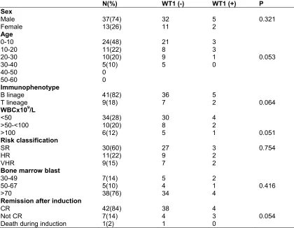
Regarding ALL group; included 34 males and16 females. 27 patients treated in pediatric department aged 2-16 years and 23 patients treated in adults department aged 17-40 years old. 82% of the patients were B lineage while, T cell lineage accounted for 18% .According to their risk we stratified the patients into 3 groups, "standard risk group" included 30 patient,' high risk group' group included 11 patients "very high risk group" included 9 patients (Table 1).
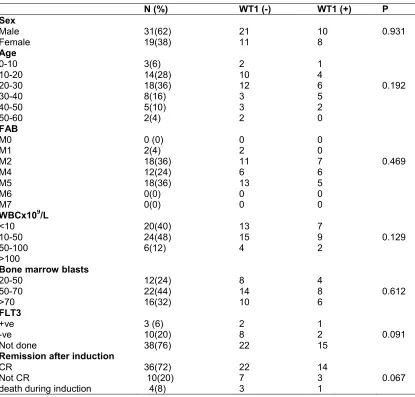
AML group, it included 31 males and 19 females, 11 patients treated in pediatric department aged 2-16 years old while, 39 patients aged 17-60 treated in adult department no one showed high risk feature of t (9-22), FLT3 was don in13 patients only 3 of them showed positive mutation (Table 2).
Table 1. ALL patients characteristics
N(%) WT1 (-) WT1 (+) P
Sex Male Female 37(74) 13(26) 32 11 5 2 0.321 Age 0-10 10-20 20-30 30-40 40-50 50-60 24(48) 11(22) 10(20) 5(10) 0 0 21 8 9 5 3 3 1 0 0.053 Immunophenotype B linage T lineage 41(82) 9(18) 36 7 5
2 0.064
WBCx109/L <50 >50-<100 >100 34(28) 10(20) 6(12) 30 8 5 4 2
1 0.051
Risk classification SR HR VHR 30(60) 11(22) 9(15) 27 9 7 3 2 2 0.754
Bone marrow blast 30-49 50-67 >70 7(14) 5(10) 38(76) 5 4 34 2 1 4 0.416
Remission after induction CR
Not CR
Death during induction
42(84) 7(14) 1(2) 38 4 1 4 3 0 0.054
Table 2. AML Patients characteristics
N (%) WT1 (-) WT1 (+) P
Sex Male Female 31(62) 19(38) 21 11 10 8 0.931 Age 0-10 10-20 20-30 30-40 40-50 50-60 3(6) 14(28) 18(36) 8(16) 5(10) 2(4) 2 10 12 3 3 2 1 4 6 5 2 0 0.192 FAB M0 M1 M2 M4 M5 M6 M7 0 (0) 2(4) 18(36) 12(24) 18(36) 0(0) 0(0) 0 2 11 6 13 0 0 0 0 7 6 5 0 0 0.469
WBCx109/L <10 10-50 50-100 >100 20(40) 24(48) 6(12) 13 15 4 7 9 2 0.129
Bone marrow blasts 20-50 50-70 >70 12(24) 22(44) 16(32) 8 14 10 4 8 6 0.612 FLT3 +ve -ve Not done 3 (6) 10(20) 38(76) 2 8 22 1 2 15 0.091
Remission after induction CR
Not CR
death during induction
36(72) 10(20) 4(8) 22 7 3 14 3 1 0.067
3.2 WT1 expression at diagnosis
WT1 expression at diagnosis was found in 14 % of ALL patients and in 36% of AML patients.
Our results showed no significant difference between WT1 expression among ALL patients regarding different prognostic variables (sex: p=0.321, age: p= 0.053 total white blood cell count: p= 0.051, immunophenotyping: p=0.064, percentage of BM blast: p=0.416 and different risk group: p=0.754).
Also in AML group we couldn't find any significant difference between WT1 expression and (sex: p= 0.931, age: p= 0.192, FAB subtypes: p=0.469, percentage of blast in BM: p=0.61, and FLT3 mutation status in examined patients: p=0.091.
Regarding remission after induction, no significant differences was found among patients with WT1 expression at diagnosis and the response to 1st induction in ALL group p= 0.054 and AML group p=0.067 p=0.067 (Table 1&2).
3.3 Status of WT1 Gene Expression (WT1) at the End of Induction
In AML After induction chemotherapy, of 18 patients who had positive baseline WT1 expression 15 of them achieved CR while 3 patients did not entered in CR and 1 patients died during treatment, WT1 expression persist in 12 (80%) out 14 patients who achieved CR Also it persist in 2(66%) out 3 of patients who didn't respond to first induction therapy p= 0.049.
After median follow up of 20 months, in ALL group, one patient out of 3 patients (33%) who had CR with persistence of WT1 expression relapsed after 6 months of finishing maintenance, while 5 out of 39 patients (12%) who had CR with negative WT1 expression at the end of induction relapsed after median 20 months of follow up p=0.042 Table 3, while in AML patients with same period of follow up relapse occurred in 4 patients out of 12 (33%) who had CR with persistence WT1 expression, while it occurred in
7 patients out of 21 (32%) who had CR with negative expression of WT1 p=0.988 (Table. 3).
3.4 Impact of WT1 Gene Expression on Survival
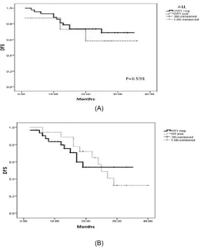
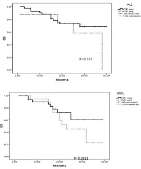
The median duration of follow-up was 20 months. We couldn't find any significant impact of positive WT1 expression at diagnosis on DFS in ALL group p=0.591 Figs. (1a and b) or AML patients p=0.858 Figs. (2a and 2b) and in OS p= 0.339 and 0.331 in ALL and AML groups respectively (Fig. 2).
4. DISCUSSION
In this work, WT1 expression was evaluated in ALL and AML patients at diagnosis and after induction chemotherapy to identify its impact on prognosis and relapse.
(A)
(B)
Fig. 2. OS in different WT1 expression at diagnosis a) in ALL, b) in AML
Table 3. WT1 expression at the end of induction remission and its affection on response and relapse
WT1 status at the end of induction
N (%) WT1 (-) WT1 (+) P
Response ALL group CR Not CR
Death during induction
42(84) 7(17) 1(2)
39 4 1
3 3 0
0.049
AML group CR Not CR
Death during induction
36(72) 10(20) 4(8)
24 8 3
12 2 1
0.097
Relapse
ALL group 7(16) 6 1 0.045
AML group 11(30) 7 4 0.965
Our study showed WT1 expression is less frequently expressed at diagnosis in BM samples in patients with ALL which is contradict many studies which reported more than 80% expression of WT1 [12,13,24], but our results agree to some extend with other studies [25-27]
T-ALL and B-ALL was found regarding WT1 expression both in adult and pediatric patients. Several studies showed controversial results with higher WT1 overexpression detected in B-ALL in some of them [29] and in T-ALL in others. [30] However, these studies investigated WT1 mostly in adult ALL patients or in heterogenous groups of children and adults, in PB samples, using potentially less sensitive PCR techniques for WT1 detection [31,32].
Our data shows the WT1 expression at diagnosis has no significant correlation to clinical complete remission rate or disease relapse rate in ALL, which is similar to the results of Gaiger et al. [31]. On the other hand, our data indicate that the WT1 level at the end of remission induction has significant relationship with disease relapse in ALL patients p= 0.045 and this correlated with Omaran et al. [25]. But contradict other study by Chen et al. [33] who didn't find any correlation between WT1 expression at the end of induction and relapse. However the small number of our study should be considered.
In the present work, patients were followed up for a median of 27 months, and the influence of WT1 expression levels at diagnosis on the DFS was determined. There was no statistically significant impact of WT1 gene’s expression on DFS in ALL patients. This comes consistent with the studies of Magyarosy et al. [14] Imashuku et al. [15] and Boublikova et al. [16] who reported that higher levels of WT1 gene expression at diagnosis were not associated with shorter DFS.
Regarding AML, the WT1 transcript was overexpressed in 34% of AML patients at diagnosis, which is much lower than other studies reported WT1 is overexpressed in approximately 70–90% of AML patients [34]. No
significant associations were encountered
between WT1 overexpression at diagnosis and other prognostic factors including age, total leukocyte count, and blast percentage. This is in accordance with the previous studies which could not find an association between the gene overexpression and prognostic factors. [35,36] Also FAB classification showed no statistical difference in WT1 expression which is in accordance with results reported by Grag et al. [37] and Noronha et al. [38] although this contradict the results of Weisser et al. [39] and others, [40,41] who found significant lower level in M5 subtype being more differentiated compared to more undifferentiated subtypes.
No observed significant difference in CR in patients expressing the gene compared to those without overexpression. This was similar to findings of Schmid et al. [42] Barragan et al. [43] and Cilloni et al. [35]. They reported no difference in WT1 transcript at diagnosis in patients resistance compared to responders to chemotherapy. Our study didn’t show any significant difference between WT1 expression and DFS and OS in AML patients which is in line with several studies which could not find a significant association between overexpression of the gene and DFS and OS, [44,45], in spite of other data reported worse outcome with WT1 overrexpression [30,38,46].
5. CONCLUSION
Our results suggest that WT1 expression in
patients with acute leukemia doesn't have any implication on response or survival however, significant association was found in predicting ALL relapse but the small sample size should be considered.
COMPETING INTERESTS
Authors have declared that no competing interests exist.
REFERENCES
1. Lyu X, Xin Y, RuihuaMi R, Ding J, Wang X, Hu J, et al. Overexpression of Wilms Tumor 1 Gene as a negative prognostic indicator in acute myeloid leukemia. PLOS ONE. 2014;9(3):92470-92472.
2. Schlenk RF, Do¨ hner K, Krauter J, et al. Mutations and treatment outcome in cytogenetically normal acute myeloid leukemia. N Engl J Med. 2008;358:1909-1918.
3. Hohenstein P, Hastie ND. The many facets of the Wilms’ tumour gene, WT1. Hum Mol Genet. 2006;15:196-201.
4. Yang L, Han Y, Suarez Saiz F, et al. A tumor suppressor and oncogene: The WT1 story. Leukemia. 2007;21:868-876.
5. Ariyaratana S, Loeb DM. The role of the Wilmstumour gene (WT1) in normal and malignant haematopoiesis. Expert Rev Mol Med. 2007;9:1-17.
7. Summers K, Stevens J, Kakkas I, et al. Wilms’ tumour 1 mutations are associated with FLT3-ITD and failure of standard induction chemotherapy in patients with normal karyotype AML. Leukemia. 2007; 20:2051-2054.
8. Paschka P, Marcucci G, Ruppert AS, et al.
Wilms’ tumor 1 gene mutations
independently predict poor outcome in adults with cytogenetically normal acute myeloid leukemia: A Cancer and Leukemia Group B study. J Clin Oncol. 2008; 26:4595-4602.
9. Hollink IH, van den Heuvel-Eibrink MM, Zimmermann M, et al. Clinical relevance of Wilms tumor 1 gene mutations in childhood acute myeloid leukemia. Blood. 2009;113:5951-5960.
10. Virappane P, Gale R, Hills R, et al. Mutation of the Wilms’ tumor 1 gene is a poor prognostic factor associated with chemotherapy resistance in normal karyotype acute myeloid leukemia: The United Kingdom Medical Research Council Adult Leukaemia Working Party. J Clin Oncol. 2008;26:5429-5435.
11. Gaidzik VI, Schlenk RF, Moschny S, et al. Prognostic impact of WT1 mutations in cytogenetically normal acute myeloid leukemia (AML): A study of the German-Austrian AML Study Group. Blood. 2009; 113:4505-4511.
12. Boublikova L, Kalinova M, Ryan J, Quinn F, O’Marcaigh A, Smith O, et al. Wilms’ tumor gene 1 (WT1) expression in childhood acute lymphoblastic leukemia: a wide range of WT1 expression levels, its impact on prognosis and minimal residual disease monitoring. Leukemia. 2006; 20:254-256.
13. Chiusa L, Francia di Celle P, Campisi P, Ceretto C, Marmont F, Pich A. Prognostic value of quantitative analysis of WT1 gene transcripts in adult acute lymphoblastic leukemia. Haematologica. 2006;91:270-271.
14. Magyarosy E, Varga N, Timar J, Raso E. Follow-up of minimal residual disease in acute childhood lymphoblastic leukemia by WT1 gene expression in the peripheral blood: the Hungarian experience. Pediatr Hematol Oncol. 2003;20:65-74.
15. Imashuku S, Terui K, Matsuyama T, Asami K, Tsuchiya S, Ishii E, et al. Lack of clinical utility of minimal residual disease detection in allogeneic stem cell recipients with childhood acute lymphoblastic leukemia:
multi-institutional collaborative study in Japan. Bone Marrow Transplant. 2003; 31:1127-1135.
16. Boublikova L, Kalinova M, Ryan J, Quinn F, O’Marcaigh A, Smith O, et al. Wilms’ tumor gene 1 (WT1) expression in childhood acute lymphoblastic leukemia: a wide range of WT1 expression levels, its impact on prognosis and minimal residual disease monitoring. Leukemia. 2006; 20:254–263.
17. Bennett JM, Catovsky D, Daniel MT, et al. Proposed revised criteria for the classification of acute myeloid leukemia. A report of the French-American-British Cooperative Group. Ann Intern Med. 1985;103:620-625.
18. Hoelzer D, Thiel E, Loffler H, Bodenstein H, Plaumann L, Buchner T, et al. Prognostic factors in a multicenter study for treatment of acute lymphoblastic leukemia in adults. Blood. 1988;71:123- 131.
19. Pui CH, Sandlund JT, Pei D, Campana D, Rivera GK, Ribeiro RC, Rubnitz JE, et al. improved outcome for children with acute lymphoblastic leukemia: results of Total Therapy Study XIIIB at St Jude Children's Research Hospital. Blood. 2004;104:2690-2696.
20. Löwenberg B, Suciu S, Archimbaud E, Haak H, Stryckmans P, de Cataldo R, Dekker AW, et al. Mitoxantrone versus daunorubicin in induction-consolidation chemotherapy--the value of low-dose cytarabine for maintenance of remission, and an assessment of prognostic factors in acute myeloid leukemia in the elderly: final report. European Organization for the Research and Treatment of Cancer and the Dutch-Belgian Hemato-Oncology Cooperative Hovon Group. J Clin Oncol. 1998;16:872-881.
21. Hann IM, Stevens RF, Goldstone AH, Rees JK, Wheatley K, Gray RG, et al. Randomised comparison of DAT versus ADE as induction chemotherapy in children and younger adults with acute myeloid leukaemia. Results of the Medical Research Council's 10th AML Trial (MRC AML10). Blood. 1997;89:2311-2318. 22. Creutzig U, Kaspers GJ. Revised
recommendations of the International
Working Group for diagnosis,
myeloid leukemia. J Clin Oncol. 2004; 22:3432-3433.
23. Kaplan EL, Meier P. Nonparametric estimation from incomplete observations. J Am Stat Assoc. 1958;53:457-481.
24. Miwa H, Beran M, Saunders GF.
Expression of the Wilms tumor gene (WT1) in human leukemias. Leukemia. 1992; 6:405-409.
25. AsgarianOmran H, Shabani M, Vossough P, Sharifian R, Tabrizi M, Khoshnoodi J, et al. Cross-sectional monitoring of Wilms' tumor gene 1 (WT1) expression in Iranian patients with acute lymphoblastic leukemia at diagnosis, relapse and remission. Leuk Lymphoma. 2008;49:281-290.
26. Chiusa L, Francia di Celle P, Campisi P, et al. Prognostic value of quantitative analysis of WT1 gene transcripts in adult acute lymphoblastic leukemia. Haematologica. 2006;91:270-271.
27. Menssen HD, Renk HJ, Rodeck U, et al. Presence of Wilms tumor gene (WT1) transcripts and the WT1 nuclear protein in the majority of human acute leukemias. Leukemia. 1995;9:1060-1067.
28. Spanaki A, Linardakis E., Perdikogianni C, et al. Quantitative assessment of WT1 expression in diagnosis of childhood acute leukemia. Leuk Res. 2007;31:570-572. 29. Neigemann E, Wehner S, Kornhuber B, et
al. Wilms tumor gene expression in childhood leukemias. Acta Hematol. 1999; 102:72-76.
30. Ozgen U, Amak S, Ozbek U, et al. Wilms tumor gene expression in childhood leukemias. Acta Hematol. 2000;103:229-230.
31. Gaiger A, Linnerth B, Mann G, Schmid D, Heinze G, Tisljar K, et al. Wilms’ tumour gene (wt1) expression at diagnosis has no prognostic relevance in childhood acute lymphoblastic leukaemia treated by an intensive chemotherapy protocol. Eur J Haematol. 1999;63:86–93.
32. Inoue K, Sugiyama H, Ogawa , Nakagawa M, Yamagami T, Miwa H, et al. WT1 as a new prognostic factor and a new marker for the detection of minimal residual disease in acute leukemia. Blood. 1994; 84:3071-3079.
33. Chen JS, Hsiao CC, Sheen JM, Cheng CN. Comparison of minimal residual disease (MRD) estimated by flow cytometry and by real-time quantitative PCR of Wilms tumor gene 1 (WT1) transcript expression in children with acute
lymphoblastic leukemia. Leuk Res. 2007;31(10):1351-1357.
34. Østergaard M, Olesen LH, Hasle H, Kjeldsen E, Hokland P. WT1 gene expression: an excellent tool for monitoring minimal residual disease in 70% of acute myeloid leukaemia patients - results from a single-centre study. Br J Haematol. 2004;125(5):590-600.
35. Cilloni D, Messa F, Arruga F, Defilippi I, Gottardi E, Fava M, et al. Early prediction of treatment outcome in acute myeloid leukaemia by measurement of WT1 transcript levels in peripheral blood samples collected after chemotherapy. Haematologica. 2008;93:921-924.
36. Trka J, Kalinova M, Hrusak O, Zuna J, Krejci O, Madzo J, et al. Real time quantitative PCR detection of WT1 gene expression in children with AML: prognostic significance, correlation with disease status and residual disease detection by flow cytometry. Leukemia. 2002;16:1381-1389.
37. Garg M, Moore H, Tobal K, Liu Yin JA. Prognostic significance of quantitative analysis of WT1 gene transcripts by competitive reverse transcription polymerase chain reaction in acute leukaemia. Br J Haematol. 2003;123:49-59.
38. Noronha SA, Farrar JE, Alonzo TA, Gerbing RB, Lacayo NJ, Dahl GV, et al. WT1 expression at diagnosis does not predict survival in pediatric AML: a report from the Children’s Oncology Group. Pediatr Blood Cancer. 2009;53:1136-9. 39. Weisser M, Kern W, Rauhut S, Schoch C,
Hiddemann W, Haferlach T, et al. Prognostic impact of RT-PCR-based quantification of WT1 gene expression during MRD monitoring of acute myeloid leukemia. Leukemia. 2005;19:1416-1423. 40. Cilloni D, Gottardi E, De Micheli D, Serra
A, Volpe G, Messa F, et al. Quantitative assessment of WT1 expression by real time quantitative PCR may be a useful tool for monitoring minimal residual disease in acute leukemia patients. Leukemia. 2002;16:2115–2121.
42. Schmid D, Heinze G, Linnerth B, Tisljar K, Kusec R, Geissler K, et al. Prognostic significance of WT1 gene expression at diagnosis in adult de novo acute myeloid leukemia. Leukemia. 1997;11:639-643. 43. Barragan E, Cervera J, Bolufer P, Ballester
S, Martín G, Fernández P, et al. Prognostic implications of Wilms’ tumor gene (WT1) expression in patients with de novo acute myeloid leukemia. Haematologica. 2004; 89:926-933.
44. Yanada M, Terakura S, Yokozawa T, Yamamoto K, Kiyoi H, Emi N, et al. Multiplex real-time RT-PCR for prospective evaluation of WT1 and fusion gene transcripts in newly diagnosed de novo
acute myeloid leukemia. Leuk Lymphoma. 2004;45:1803-1808.
45. Nowakowska-Kopera A, Sacha T, Florek I, Zawada M, Czekalska S, Skotnicki AB. Wilms’ tumor gene 1 expression analysis by real-time quantitative polymerase chain reaction for monitoring of minimal residual disease in acute leukemia. Leuk Lymphoma. 2009;50:1326-1332.
46. Gray JX, McMillen L, Mollee P, Paul S, Lane S, Bird R, et al. WT1 expression as a marker of minimal residual disease predicts outcome in acute myeloid
leukemia when measured
post-consolidation. Leuk Res. 2012;36:453-458.
© 2015 Ibrahim et al.; This is an Open Access article distributed under the terms of the Creative Commons Attribution License
(http://creativecommons.org/licenses/by/4.0), which permits unrestricted use, distribution, and reproduction in any medium,
provided the original work is properly cited.
The peer review history for this paper can be accessed here: Peer-review history: