_____________________________________________________________________________________________________
*Corresponding author: E-mail: [email protected], [email protected];
(Past name: British Journal of Medicine and Medical Research, Past ISSN: 2231-0614, NLM ID: 101570965)
Modification of the Postlethwait Method in
Improvement of Esophageal Bypass: An Essential
Treating Method
Masahiro Kimura
1*, Yasuyuki Shibata
1, Akira Mitsui
2and Yoshiyuki Kuwabara
21Department of Surgery, Nagoya City East Medical Center, 2-23 Wakamizu 1, Chikusa-ku, Nagoya,
Japan.
2
Department of Surgery, Nagoya City West Medical Center, Japan.
Authors’ contributions
This work was carried out in collaboration among all the authors. All authors read and approved the final manuscript.
Article Information
DOI: 10.9734/JAMMR/2018/38581
Editor(s):
(1) Georgios Tsoulfas, Assistant Professor of Surgery, Aristoteleion University of Thessaloniki, Thessaloniki, Greece. (2)James Anthony Giglio, Adjunct Clinical Professor, Oral and Maxillofacial Surgery, School of Dentistry, Virginia Commonwealth University, Virginia, USA. (3)Angelo P. Giardino, Professor, Texas Children’s Hospital, Houston, Texas, USA and Department of Pediatrics, Baylor College of Medicine, Houston, USA.
Reviewers:
(1) Yavuz Savas Koca, Suleyman Demirel University, Turkey. (2)Anastasios J. Karayiannakis, Democritus University of Thrace, Greece. (3)Jeffrey W. Clymer, USA. (4)Fernando Herbella, Federal University of Sao Paulo, Brazil. Complete Peer review History:http://www.sciencedomain.org/review-history/24463
Received 1st December 2017 Accepted 1st May 2018 Published 5th May 2018
ABSTRACT
The quality of life for patients with inoperative esophageal cancer is poor due to dysphagia caused by stenosis or development of a tracheoesophageal fistula. One option for these patients is bypass surgery. These patients are often medical compromised and have a poor prognosis. Consequently, any palliative surgical intervention should be definitiuve and minimally invasive. We have improved upon esophageal bypass surgery as originally described by Postlethwait by using a radial stapler to form the gastric tube.
1. INTRODUCTION
In patients with advanced esophageal cancer, severe dysphagia and recurrent pneumonia secondary to tracheoesophageal fistula are common complications. Placement of esophageal stents and bypass surgery can be performed on these patients as palliative treatment measures. Due to its minimally invasive nature and quick alleviation of symptoms, stent treatment is the preferred option at many hospitals [1,2]. However, complications from stent placement such as perforation or creation of a fistula are common, particularly when performed following esophageal radiation therapy. On the other hand, bypass surgery can be performed with minimal complications by experienced surgeons. Isoperistalitic gastric tube creation for esophageal bypass was first described by Postlethwait in 1979 [3]. The technique involves opening a small hole in the antrum and detaching this towards the cardia side of the stomach. In 2002, a method of hollowing out a circular portion of the stomach using a stapler was first described (Fig 1a). This method was a revolutionary development as it prevents gastric contamination and does not require sutures. We have performed bypass surgery for 40 patients with advanced esophageal cancer using this technique.
2. TECHNIQUE
The greater omentum is separated in the usual way for esophageal reconstruction using a gastric tube, preserving the right gastroepiploic artery and vein. The left gastroepiploic artery and vein are divided at their origin. The short gastric vessels are divided as close to the spleen as possible. A sealing device is used for this to easily and quickly divide the vessels to the left of the esophagogastric junction, allowing for creation of an adequate length of the stomach. This is not necessary along the lesser curvature side. A gauze is placed on the back of the spleen in order to avoid vascular damage. Mobilization of the stomach begins at the cardia as follows:
・ A 3 to 5 cm gastric tube along the greater curvature is developed.
・ The distal tube is created 3 cm from the pylorus.
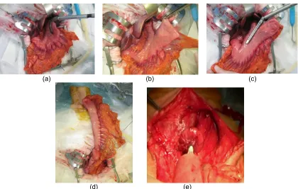
The GIATM Radial Reload (Covidien Japan, Tokyo) stapling system is used to place 3 rows of staples, 60 mm in length for the first stapler load (Fig. 2a). Regarding the stapler insertion
direction, two methods are used depending on the particular shape of the stomach. Careful attention is paid so that the tip of the gastric tube does not become too thin. The gastric tube is then easily mobilized out of the abdominal cavity (Fig. 2b). Sutures are placed using a linear type suturing device to complete creation of the gastric tube. (Fig. 2c,d).
The sections of the gastric tube with overlapping staple lines are reinforced with sutures. An approximately 5 cm left cervical incision is then made and the cervical esophagus is transected. We use the Radial Reload to secure even a long esophagus. With this stapler, longer esophagus can be dissected in the direction perpendicular to the long axis of the esophagus. If a Radial Reload is used for this dissection, it is easy to achieve esophageal separation at the level of the sternum horizontally. Subcutaneous tunneling of the gastric tube is used in our hospital. After tunneling from the abdomen to the neck in the subcutaneous tract, the gastric tube is brought cephalad. Finally, an end-to-side anastomosis between the cervical esophagus and the gastric tube is made using a circular stapler (Proximate Intraluminal Stapler CDH25; Ethicon Japan, Tokyo) (Fig 2e).
3. RESULTS
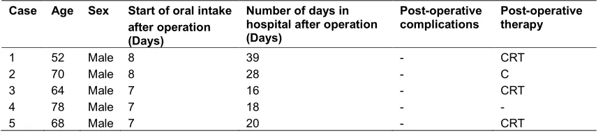
We performed this method on 5 patients and were able to reduce the number of staplers used as compared to the Postlethwait method. There were no operative complications. All patients could take oral intake after operation, and 4 out of 5 patients could receive post-operative therapy (Table 1).
4. DISCUSSION
Fig. 1a. Postlethwait method; After opening a small hole in the antrum with circular stapler 3 to 4 linear staplers were used towards the cardia side of the stomach. Fig. 1b. Type A; The Radial Reload was used for the first stapler load. Fig. 1c. Type B; the direction of Radial Reload can
be changed from Type A
(a) (b) (c)
(d) (e)
Fig. 2a. The GIATM Radial Reload is used for the first stapler. Fig. 2b. After the first stapler is placed. Fig. 2c. Attaching the second linear stapler. Fig. 2d. The completed gastric tube.
Table 1. Immediate post-operative course
Case Age Sex Start of oral intake after operation (Days)
Number of days in hospital after operation (Days)
Post-operative complications
Post-operative therapy
1 52 Male 8 39 - CRT
2 70 Male 8 28 - C
3 64 Male 7 16 - CRT
4 78 Male 7 18 - -
5 68 Male 7 20 - CRT
CRT: Chemo-radiation therapy C: Chemo therapy
In 2002, an improvement to the method of Postlethwait was first reported in Japan [6]. The method involves initiating the creation of the
gastric tube using a circular stapler as opposed to sutures, thus avoiding contamination
and the infections that result because of this. However, the hole created by the staple is 21
mm, which is problematic, as is the potentially compromised blood flow at the staple
line intersections. Another problem is the increased cost of staplers as opposed to conventional suturing.
These concerns were alleviated by the radial type suture separation device that appeared on the market in 2013 [7]. While it was developed for separation of the rectum in the pelvis in colorectal cases, it can also be used for separation of the gastric tube from the cardia. The key feature of this suture machine is that the stapler is at a right angle to the direction of the shaft of the instrument. Linear staplers can achieve at most 45 degrees of reticulation. If the left costal arch is elevated slightly, insertion of the stapler is easy using the radial stapling device. Because the intramural blood flow of the gastric tube is important as is the width of the apical portion of the gastric tube, it detaches in
the direction of type A (Fig 1b). If the separation direction with the 2nd stapler needs to
be different, it will detach in the direction of type
B (Fig 1c). Regardless of the direction in which the stapler is oriented, the insertion point
of the second stapler will easily be brought out of the abdominal cavity. This novel method is effective in creating ideal gastric tubes as it is possible to avoid the circular stapler currently used.
5. CONCLUSION
In this study, we have improved upon the esophageal bypass surgery as originally
described by Postlethwait. This novel
method is effective in creating ideal gastric tubes as it avoids the circular stapler currently
used.
CONSENT
The authors declared that informed consent was obtained from the participants of the study for publication of this paper.
ETHICAL APPROVAL
Authors confirmed that all necessary ethical approval from institutions were obtained.
COMPETING INTERESTS
Authors have declared that no competing interests exist.
REFERENCES
1. Cantero R, Torres AJ, Hernando F, others. Palliative treatment of esophageal cancer: Self-expanding metal stents versus Postlethwait technique. Hepato-Gastro-enterol. 1999;46:971-6.
2. Aoki T, Osaka Y, Takagi Y, others. Comparative study of self-expandable metallic stent and bypass surgery for inoperable esophageal cancer. Dis Esophagus. 2001;14:208-11.
3. Postlethwait RW. Technique for
isoperistaltic gastric tube for esophageal bypass. Ann Surg. 1979;
189:673-6.
4. Hanagiri T, Morita M, Shigematsu Y, others. Esophageal bypass using a gastric tube for a malignant tracheoesophageal/ bronchoesophageal fistula: A report of 4 cases. Int Surg. 2011;96:189-93.
oesophageal cancer: Early and late results in 124 cases. Br J Surg. 1988;75:283-6. 6. Takiyama W. Esophageal bypass surgery
formed using circular stapler and linear stapler. Shyujyutsu (Operation) 2002;56: 147-52.
7. Rivadeneira DE, Verdeja JC, Sonoda T. Improvement access and visibility during stapling of the ultra-low rectum: A comparative human cadaver study between two curved staplers. Ann Surg Innov Res. 2012;13:1164-6.
_________________________________________________________________________________ © 2018 Kimura et al.; This is an Open Access article distributed under the terms of the Creative Commons Attribution License (http://creativecommons.org/licenses/by/4.0), which permits unrestricted use, distribution, and reproduction in any medium, provided the original work is properly cited.
Peer-review history: