Impaired meiotic competence in putative primordial germ
cells produced from mouse embryonic stem cells
MARIANNA TEDESCO, DONATELLA FARINI and MASSIMO DE FELICI*
Department of Public Health and Cell Biology, Section of Histology and Embryology University of Rome “Tor Vergata”, Rome, Italy
ABSTRACT There are still several unanswered questions and problems about the recently claimed possibility of producing functional germ cells in vitro from pluripotent embryonic stem cells (ESCs). In the present paper, we compared by single-cell analysis the capability of putative primordial germ cells (PGCs), produced in vitro from ESCs, and that of endogenous PGCs isolated from embryos, to enter and progress through meiotic prophase I. Using a protocol previously reported to be suitable to produce female germ cells from mouse ESC monolayers, we first identified putative PGCs by analysing the expression pattern of several markers such as SSEA1, APase, OCT4, NANOG, MVH and SCP3 of pre-and post migratory PGCs. Next, after isolation of such cells from culture, we tested their meiotic capability. The evaluation at 2-5 days of culture of the number of cells showing meiotic nuclear SCP3 staining in cytospreads showed that it remained nearly constant in the putative PGCs, whereas it increased markedly in endogenous PGCs. Moreover, we observed that in putative PGCs, the nuclear distribution or expression of SCP3 and other meiotic markers such as DMC1, H2AX and SCP1 were always highly abnormal in comparison to that observed in endogenous cultured PGCs. We conclude that although the formation of cells showing characteristics of PGCs can occur efficiently from ESCs in vitro, these cells possess impaired capability to enter and progress through meiotic prophase I.
KEY WORDS:
embryonic stem cell, gametogenesis, meiosis, primordial germ cell
From the first report by Hübner et al. (2003) showing that XY mouse embryonic stem cells (mESCs) are able to produce follicle-enclosed growing oocytes, several studies have described the in vitro differentiation of female and male germ cells from mouse and human ESCs (hESC) in either the monolayer or the embryoid bodies (EBs) culture method (for reviews see Hua and Sidhu, 2008; Nagano, 2007; Aflatoonian and Moore, 2006). The experi-ments carried out to produce germ cells from ESCs are based on years of intense investigations on the in vivo and in vitro develop-ment of mouse primordial germ cells (PGCs), the oocyte and sperm precursors (for reviews see De Felici, 2009; De Felici et al., 2004; De Felici, 2001). In particular the recent progress in eluci-dating the process of germ line specification in mammals (Ohinata
et al., 2009 and references herein) and the possibility to mimic in vitro events of later PGC development including entering into meiosis (Farini et al., 2005), have given precious information for the derivation and characterization of germ cells produced from ESCs.
In the mouse embryo, PGC development occurs in about 7
BIOLOGY
www.intjdevbiol.com*Address correspondence to: Massimo De Felici. Dipartimento di Sanità Pubblica e Biologia Cellulare, Università di Roma "Tor Vergata", Via Montpellier, 1, 00173 Roma, Italy. Fax: +39-06-7259-6172. e-mail: defelici@uniroma2.it
Accepted: 26 October 2010. Final author corrected PDF published online: 8 June 2011.
ISSN: Online 1696-3547, Print 0214-6282
© 2011 UBC Press Printed in Spain
Abbreviations used in this paper: APase, alkaline phosphatase; bFGF, basic fibroblast growth factor; BLIMP, B-lymphocyte induced maturation; BMP, bone morphogenetic protein; Daz, deleted in azoospermia; Dazl, deleted in azoospermial-like; DMC, disrupted meiotic cDNA; dpc, days post coitum; EB, embryoid body; EG, embryonic germ; eGFP, enhanced green fluorescent protein; epiSC, epiblast stem cvell; FACS, fluorescence-activated cell sorting; GFP, green fluorescent protein; ePGC, endogenous primordial germ cell; hEG, human embryonic germ; hESC, human embryonic stem cell; KL, kit ligand; IF, immunofluorescence; LIF, leukemia inhibitory factor; MEF, mouse embryonic fibroblast; mESC, mouse embryonic stem cell; MLH, mutL homolog; MVH, mouse VASA homolog; PRDM, PR domain containing; PGC, primordial germ cell; pPGC, putative primordial germ cell; RA, retinoic acid; SMC, structural maintenance of chromosome; STAG, stomalin antigen; SCP, synaptonemal complex protein; SSEA, stage-specific embryonic antigen; SDF, stromal cell-derived factor.
occur (for a review, see Western, 2009). Elegant recent studies have shown that mouse PGCs are derived from a few precursors located at the proximal posterior epiblast, which around 6.25 dpc express the transcription factor B-lymphocyte induced matura-tion protein1 (BLIMP1 also known as PR domain containing 1 (PRDM1)) (Ohinata et al., 2009 and references herein). PGCs are finally specified at the base of the allantois around 7.5 dpc and move into the gonadal ridges between 8.5 and 11.5 dpc (for reviews, see Hayashi et al., 2007; De Felici, 2009). By 12.5 dpc, the ovary and testis become morphologically distinguishable and PGCs initiate sex-specific development. During migration and for a couple of days after gonad colonization, the somatic status of imprinted genes in PGCs is progressively erased (Szabo and Mann, 1995; Szabo et al., 2002; Hajkova et al., 2002; Lee et al., 2002; Kato et al., 1999). Besides the expression of genes and other markers shared from PGCs and ESCs (see below), this epigenetic modification is a valid criterion to distinguish PGCs from ESCs. At 13.5 dpc female PGCs enter meiosis becoming primary oocytes which pass through the meiotic prophase I stages (leptotene/zygotene/pachytene) until around and early after birth become arrested at the diplotene stage. Differently, male PGCs enter mitotic arrest and developed as spermatogonial will be reactivated to initiate spermatogenesis only after birth. Recent studies reported that the decision of meiotic entry or mitotic arrest of post migratory PGCs is regulated by retinoic acid (RA). Male PGCs do not enter meiosis because an enzyme (CYP26b1) expressed in somatic cells in the male gonadal ridge degrades RA. In contrast, the lack of CYP26b1 expression in the embryonic ovary allows PGCs to enter meiosis (for a review, see Bowles and Koopman, 2007). Under various in vivo and in vitro
conditions, male PGCs until 12.5 dpc can be, however, induced to enter meiosis likely if exposed to RA at the right time (for a review, see Kocer al., 2009). This indicates that germ cell sex depends on sex-determining signalling from the environment rather than the sex chromosomes. RA is considered the meiotic inducer also in spermatogonia at later postnatal stages (Pellegrini
PGCs can be actually produced both from mouse and human ESCs and that their formation recapitulates many of the crucial developmentally-timed events that occur in the embryo (Wei et al., 2008; West et al., 2010; Kee et al., 2009). The assessment of properly differentiated PGC status requires, however, the demon-stration that these cells are able to enter and progress into meiotic prophase I after reaching the premeiotic stage. While the expres-sion of premeioitic markers such as mouse Vasa homolog (MVH) and synaptonemal complex protein 3 (SCP3) or other proteins involved in meiosis (i.e. disrupted meiotic cDNA 1, DMC1, and mutL homolog 1, MLH1) is readily detected in putative PGCs produced form ESCs both at mRNA and protein level, their meiotic capability has been accurately analysed in a few papers only likely for the lack of a suitable method to quantify such process in single PGCs in vitro. Novak et al. (2006) using XY mouse ESC (mESC) monolayers and a panel of meiosis-specific markers and cytospreads to identify stages of meiotic prophase I found that up to 40% of the cells within colonies grown onto the culture dish or in suspension considered putative PGCs at 14-16 days of culture expressed the synaptonemal complex protein3 (SCP3). However, they also found that these cells did not express other two synaptonemal complex proteins SCP1, SCP2 and some meiotic proteins, such as stromalin antigen3 (STAG3), Rec8 (REC8) and structural maintenance of chromosome1 (SMC1). Moreover, in cytospreads such cells showed highly abnormal distribution of SCP3. Nicholas et al. (2009) reported that 1-3% of putative PGCs formed within mouse XX EBs were able to enter into meiosis in culture as evidenced by SCP expression, but showed limited chromosome alignment and mei-otic progression. Similar results have been obtained in hESCs. Tilgner et al. (2008) found that SCP3 protein detected in stage-specific embryonic antigen 1 (SSEA1) positive putative PGCs formed within hESC monolayers was concentrated in cytoplasmic foci outside the nuclear envelope. Likewise, in cells positive for SCP3 observed throughout differentiating hEBs, the protein was localized predominantly in the cytoplasm (Clark et al., 2004). But
Fig. 1. Expression patterns of various ESC and PGC markers in ESC-D3 cultured for 10 days. The expression was analyzed using FACS (SSEA1), IF (OCT4, MVH, NANOG, SCP3) or cytochemistry (APase) directly onto the monolayers or on cell samples detached from the monolayer. Similar results were obtained from ESCs cultured for 12 days.
et al., 2008; Anderson et al., 2008). Intriguing, RA can stimulate mitotic proliferation of sex undifferentiated PGCs (Koshimizu et al., 1995; De Felici, 2000) and prevent mitotic ar-rest of male prospermatogonia in the fetal testis (Trautmann et al., 2008). Moreover, in combination with leukemia inhibitory factor (LIF) and kit ligand (KL), RA favours the for-mation of embryonic germ (EG) cells from sex undifferentiated PGCs (Koshimizu et al., 1997). Thus indi-cating that germ cell response to RA is strictly developmental stage de-pendent.
it has also been reported (West et al., 2008) that when hESCs are cultured for 16 days onto mouse embryonic fibroblast (MEF) monolayers in medium supplemented with basic fibroblast growth factor (bFGF), 90% became positive for MLH1 and SCP3 protein and staining was localized to the nucleus. Finally, in monolayers of XX or XY hESCs cultured for 7 days, Kee et al. (2009) reported the presence of putative PGCs with punctuate (resembling pro-leptotene/leptotene stages) or elongated (considered zygotene/ pachytene/diplotene stages) SCP3 nuclear staining in the range from 3 to 27% and 0-10%, respectively, but only after inducing
deleted in azoospermia (Daz), deleted in azoospermia-like (Dazl)
and/or Boule over expression.
The lack of crucial information about the meiotic capability of the ESC-derived putative PGCs and the recent development of an
in vitro culture method in which the majority of the mouse PGCs isolated from the gonadal ridges of 11.5-12.5 dpc embryos enter and progress through meiotic prophase I stages in the absence of
sorting (FACS) and the enzyme alkaline phosphatase (APase) by cytochemistry (Donovan and de Miguel, 2003). Such analyses were performed from the be-ginning of culture up to 12 days under spontaneous differentiation of ESCs as monolayers. In addition, during the same period, we studied by immunofluores-cence (IF) the expression of the tran-scription factors OCT4 and NANOG, also expressed both in ESCs and PGCs (Elliot
et al., 2007) and of MVH and SCP3, two proteins specific for pre-and meiotic PGCs (Tanaka et al., 2000; Di Carlo et al., 2000). In line with several other works (Hübner et al., 2003; Wei et al., 2008; Nicholas et al., 2009; West et al., 2009; Haston et al., 2009), we observed that the number of cells positive for SSEA1, APase, OCT4 and NANOG undergo a progressive decrease during the culture time although with different kinetics (Figs. 1-2). While at the beginning of culture 90-100% of cells were positive for all markers, at the end of the culture period examined (12 days), only about 20% of the cells expressed these markers. On the contrary, the number of cells expressing MVH increased from less than 5% to approximately 20%. Cells showing SCP3 appeared around 3 days of culture and from then onward repre-sented about 7% of the cultured cells. These analyses together with the results reported in previous works reported above al-lowed considering the SSEA1+ cells after 7-12 days of culture or a subpopulation within them as putative PGCs.
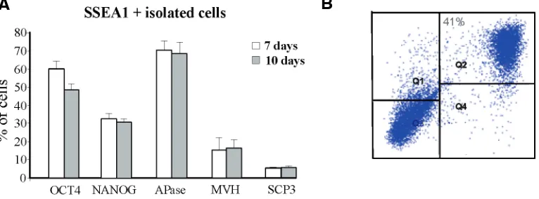
The phenotype of such putative PGCs was further character-ized by immunoisolation of SSEA1+ cells from D3-ESC monolay-ers at 7 and 12 days of culture and carrying out immunofluores-cence on single cells for OCT4, APase, NANOG, MVH and SCP3. The results showed that at 7 days of culture the majority of the SSEA1+ cells were also APase+ (70%7) and OCT4+ (60%5), while only a subset of these cells were NANOG+ (33%5) and Fig. 2. Representative immunofluorescence for MVH (A) and SCP3 (B,C) performed on ESC
monolayers after 10 days of culture. Nuclear staining with Hoechst is also shown. Similar results were obtained from ESCs cultured for 12 days.
Fig. 3. Characterization of putative PGCs (pPGCs) after 7-10 days of culture. (A) Expression of OCT4, NANOG, APase, MVH and SCP3 protein in SSEA1+ cells immunoisolated from the ESC monolayer by TG-1 after 7 and TG-10 days of culture. The evaluation was performed with IF in cell samples attached to poly-l-lysine-coated slides. (B) A representative FACS analysis of Oct4-GFP ES-OG2+ cells after 10 days of culture for SSEA1 positivity. Note that the most part of gcOct4-GFP+ cells are also SSEA1+ (Q2). Similar results were obtained from putative PGCs isolated from ESCs cultured for 12 days.
somatic cells (Farini et al., 2005), prompted us to precisely evalu-ate the capability of the putative PGCs produced from mouse ESC monolayers to enter and progress through the stages of meiotic prophase I.
Results
In a first series of experiments, we sought to reproduce the pro-cess of PGC formation from mESCs following the protocol used by Hübner and coll. (2003). To this aim, we used the D3-ESC and analyzed the expression pat-tern of two ESC and PGC mark-ers, the surface antigen SSEA1 by fluorescence-activated cell
B
C
A
MVH+ (15%10); less than 10% of the SSEA1+ cells were stained for SCP3 (Fig. 3A). Similar results were obtained at 12 days of culture (not shown).
Parallel experiments carried out under the same culture con-ditions using the Oct4-GFP ES-OG2 cells used by Hübner and coll. (2003), showed expression pattern of the gcOct4 transgene similar to that of endogenous OCT4 reported above (not shown) and almost complete coexpression of the transgene and SSEA1 marker after 10-12 days of culture (Fig. 3B).
All these results are consistent with the notion that at 7-12 days of culture around 20% of SSEA1+ putative PGCs are present among cultured ESCs and a significant fraction of them, approxi-mately 10%, express premeiotic MVH+ SCP3+ proteins. We reasoned that by isolating the SSEA1+ or OCT4+ putative PGCs from the culture at 7-12 days and maintaining them under suitable culture conditions previously used with endogenous PGCs immunoselected with antibodies against SSEA1 from gonads (Farini et al., 2005), we should be able to induce those at earlier stages of differentiation (MVH- and SCP3-) to progress at premei-otic stages and those about to begin meiosis (MVH+ and SCP3+) to enter and progress through the meiotic prophase I stages. In fact, the capability to enter into meiosis in response to RA is normally acquired by PGCs around 12.5-13-5 dpc about 7 days after their specification and is associated to MVH and SCP3 expression (Toyooka et al., 2000; Di Carlo et al., 2000). In vivo, SCP3 expression is first detected as punctuate nuclear staining at meiotic leptotene. Subsequently, SCP3 localizes along the entire length of the sister chromatids as part of the lateral element of the synaptonemal complex (Di Carlo et al., 2000). PGCs at early stages of differentiation can acquire the capability to enter meiosis following a similar timeline outside the gonadal ridges in
mouse and human ESCs the precise sequence of the events leading to the germ cell development remains elusive. In addition the efficiency of such process is not clear and the functionality of the produced germ cells is often not proven. In fact so far only two papers reported germ cells derived from mESCs able to produce male germ cells at advanced stages of differentiation and to induce apparently normal fertilization and em-bryo development after injection into oocytes (Nayernia et al., 2006; Geijsen
et al., 2003). Since in the embryo game-togenesis begins with the formation of PGCs it is logical to assume that this process should first occur when ESCs differentiate through the germ line. Sev-eral recent papers have actually reported convincing evidence that mouse and human ESCs can spontaneously or af-ter stimulation with specific growth fac-tors, namely bone morphogenetic pro-tein 4 (BMP4), give rise to cells showing phenotypic and molecular characteris-tics of pre-and post-migratory PGCs (see Introduction).
The results obtained in the present culture (Chuma and Nakatsuji, 2001; McLaren and Southee, 1997).
Evaluation of the percentage of cells showing meiotic nuclear SCP3 staining in cytospreads of putative PGCs and endogenous PGCs immunoselected with antibodies against SSEA1 from the ESC monolayers at 7-12 days or from 12.5 dpc gonads, respec-tively, at the beginning and after 2-5 days of culture in the Farini’s medium (Farini et al., 2005) is reported in Fig. 4A. It appeared that while during this culture period the number of SCP3+ cells in endogenous PGCs increased markedly, it remained nearly con-stant in putative PGCs. Moreover, the nuclear distribution of SCP3 in putative PGCs was always highly abnormal and not clearly associated with chromosomes as instead observed in control PGCs in the most part of which various apparently mor-phologically normal prophase I stages were recognizable (Fig. 4B). Other markers used to determine entering (DMC1 and
H2AX) and progression (SCP1) into meiosis also showed abnor-mal or no expression. In particular, putative PGCs in culture showed only a few foci of DMC1 and small spots of cH2AX staining instead of the numerous DMC1 foci and the large, cloud-like regions of H2AX labelling observed in the nucleus of meiotic PGCs; no SCP1 staining, indicative of the zygotene stage, was detectable in the putative PGGs (Fig. 5).
Putative OCT4-GFP+ PGCs isolated from Oct4-GFP ES-OG2 by FACS also showed the same features of impaired meiotic competence (data not shown).
Discussion
While it is unquestionable that it is possible to produce in vitro
cells with some characteristics of sperm and oocytes both from
B
A
Fig. 4. Capability of putative PGCs (pPGC) and endogenous 12.5 dpc PGCs (ePGCs) to enter and progress through meiotic prophase I. (A) Entering into meiosis of TG-1 immunoselected SSEA1 positive cells (pPGCs) obtained from 7 day ESC monolayers and PGCs isolated from 12.5 dpc gonads (ePGCs) during 5 days of culture evaluated as percentage of SCP3 positive cells in cytospreads. (B) Cytospreads stained for SCP3. Right panel:
paper are in accord with the reports that putative PGCs can arise from ESC monolayers after relatively short time of culture reflect-ing timreflect-ing of PGC formation from the epiblast (Wei et al., 2008; West et al., 2010; Kee et al., 2009). Since the formation of putative PGCs in culture is likely asynchronous it is difficult to properly follow the differentiated status reached by single cells. For this reason, we decided to isolate cells expressing markers of early and pre-meiotic PGCs at 7-12 days and to evaluate their capabil-ity to enter and progress through meiotic prophase I stages using cytospreads immunolabeling for meiotic markers including the synaptonemal complex proteins SCP1 and SCP3 and the recom-bination proteins DMC1 and H2AX. The results showed that, at this time, putative PGCs originated from cultured ESCs are basically meiotic incompetent or showed severe meiotic anoma-lies. So far the meiotic competence of ESC-derived putative PGCs has been little investigated likely for the lack of a suitable assay to analyse and quantify such event in comparison to endogenous PGCs isolated from the embryo. Here we report that when exposed to a cocktail of growth factors including RA, considered to be the physiological inducer of meiosis in PGCs (for a review, see Bowles and Koopman, 2007), putative PGCs spontaneously produced from ESC monolayers have a much lower capability to enter and progress through meiotic prophase I in vitro in comparison to endogenous PGCs. As reported in the Introduction, human putative PGCs formed in ESC monolayers appear to possess a quite variable meiotic capability ranging from about 3 to 20% but only after over expression of specific genes (Kee et al., 2009). Moreover, we observed in line with previous works carried out in mouse (Novak et al., 2006; Nicholas et al., 2009) and in human ESCs (Clark et al., 2004; Tilgner et al., 2008), a severe impairment in the capability of such putative PGCs to organize a normal meiotic synaptonemal complex. From these
cannot exclude that some cells able to enter meiosis were undetected in our assay or will enter meiosis after longer period in culture or if transferred into a favourable environment. In this regard, strong evidence exists that both mouse and human ESCs can give rise to haploid germ cells resembling spermatocyte/ spermatides or even tailed sperm after relatively long culture time (mouse:Toyooka et al., 2003; Geijsen et al., 2003; Najernia et al., 2004 and 2006; Kirkis et al., 2007; Yu et al., 2009; human: Tilgner
et al., 2008; Aflatoonian et al., 2009) Thus suggesting that PGCs produced in ESC culture more frequently differentiate towards male rather female germ line characterized by entering into meiosis only at later postnatal developmental stages. Consider-ing only the more convincConsider-ing results obtained in the mouse, it appears that efficient differentiation of the ESC-derived germ cells through advanced spermatogenic stages requires trans-plantation into a favorable testicular environment and does not necessitate stage-matched testicular cells. Interestingly, these characteristics also belong to endogenous PGCs which from as early 8.5 dpc have been reported to colonize the testis of infertile newborn mice and enter spermatogenesis (Chuma et al., 2005). How our observations may conciliate with the studies reporting the formation of oocyte-like cells in the ESC culture? None of these studies clearly documented the presence of meiotic PGCs preceding the appearance of the oocyte-like cells, nor the func-tionality of these latter. Moreover, the frequency of formation of the oocyte-like cells was very low or not reported. In fact, although oocyte-like cells enclosed within follicle-like structures were ob-served by Hübner and coll. (2003) after about 1 month of culture of D3-ESCs, the frequency, the meiotic stage and functional data concerning these cells were not provided. Lacham-Kaplan et al. (2006) reported that EBs cultured in the conditioned medium derived from newborn mouse testis germ cells developed ovarian and the present results we can speculate that the ability of PGCs to respond to the meiotic induction by RA requires the acquisition of “mei-otic competences” not properly or rarely achieved after their formation in the ESC culture systems em-ployed so far. It is known, that en-dogenous PGCs can acquire such capability outside the gonads fol-lowing a precise timeline in culture covered by the culture periods used in the present work (Chuma and Nakatsji, 2001).This suggests de-fective or incomplete PGC differen-tiation from ESCs under a variety of different culture conditions. In line with this notion, micro-array analy-ses have shown that many “PGC signature” genes (i.e. Dazl, Mvh
and developmental pluripotency-as-sociated protein 2, Dppa2) were not fully up-regulated in putative PGCs obtained from Mvh-LacZ or Mvh-GFP knock-in mouse ESCs (Mise
et al., 2008).
Despite the present results, we Fig. 5. Cytospread staining for DMC1, cH2AX and SCP1 in putative PGCs (pPGCs) isolated from
structures containing unspecified number of putative oocytes; the oocyte-like cells were surrounded by one to two layers of flattened cells and did not have a visible zona pellucida; oocyte-specific markers, such as Fig and Zp3, were expressed by the ovarian structures. The same two papers cited above showing the forma-tion of tailed sperm reported the presence of oocytes-like cells in their mouse ESC culture systems (Kirkis et al., 2007 and Yu et al., 2009). Outgrowth of 4 day EB cells containing putative PGCs onto newborn ovarian granulosa cells formed a few oocyte-like cells 25 in size after 10 days of culture (Qing et al., 2007). These cells expressed some oocyte specific genes such as Fig, Gdf9 and
Zp1-3, but showed abnormal SCP3 staining in the cytoplasm. In ESCs stably transfected with the promoter region of the mouse
Gdf9 coupled to enhanced green fluorescent protein (eGFP) and cultured on feeder cell layers, large eGFP-positive cells, sur-rounded by a structure resembling a zona pellucida appeared transiently and very rapidly on the first days of culture. How-ever, in EB cultures, eGFP-positive cells appeared transiently and then reappeared in regional clusters after a relatively long culture of 30–45 days (Salvador et al., 2008). Finally, putative PGCs obtained from 21 day EBs reaggregated with newborn ovarian tissues and transplanted under the kidney capsule of recipient mice give rise to oocytes enclosed in primary follicle at very low frequency (0.023%) (Nicholas et al., 2009). In humans, as far as we know, only two papers reported the occasional formation of oocytes-like cells from ESCs (Chen et al., 2006; Aflatoonian et al., 2009).
The lack of evidence of proper beginning and progression through meiotic prophase I by putative PGCs and the available information about the characteristics of the oocyte-like cells produced in vitro raise doubts that continuous female gameto-genesis can be correctly reproduced in the present ESC culture systems. In fact, even PGCs isolated from the embryo as early as 10.5 dpc although able to enter and progress through meiotic prophase I outside the gonad are unable to reach the growing oocyte stage (Chuma and Nakatsuji, 2001; Farini et al., 2005). This can be achieved by isolated fetal oocytes providing avail-ability of the growth factor kit ligand (KL) and subsequently of follicular cell support (Klinger and De Felici, 2002). Moreover, several studies indicate that synchronized interactions be-tween oocytes and ovarian somatic cells are important to ensure normal ovogenesis (Qing et al., 2008 and references therein).
In conclusion, we postulate that the formation of oocyte-like cells reported in ES culture may bypass the normal entering and progression of the putative PGCs through meiotic prophase I and is occasionally induced only when a contemporary differen-tiation of follicular-like cells from the surrounding ESCs occurs. Finally, two recent papers reported that putative PGCs can be formed in vitro with high efficiency from epiblast and epiblast stem cells (EpiSCs), the cell type from which endogenous PGCs originate (see Introduction) (Ohinata et al., 2009; Hayashi and Surani, 2009). Such PGCs appear to posses gene expres-sion and epigenetic signature very close to endogenous migra-tory PGCs. While the meiotic competence of the epiblast-derived cells were not investigated, EpiSC-epiblast-derived PGCs after 7 days of culture form undetermined number of SCP3+ cells when co-cultured with 12.5 dpc ovarian cells and oocyte-like after about 40 days of culture at low frequency (around 0.02%).
All together these results demonstrate a robust spontaneous or inducible formation of bona fide PGCs from ESCs but a relatively low or defective formation of germ cells in the culture dishes at later stages of differentiation mainly in the female germ line. Here we show that such defect may originate from abnormal or incomplete in vitro PGC specification relatively to the acquisition of the meiotic competence at the time proper of the female germ line. Recent papers indicate that epigenetic status (Wang and Tilly, 2010) and elevated expression of certain genes (i.e. Dazl) (Kee et al., 2009; Lin et al., 2008; Linher et al., 2009) is crucial for the meiotic commitment of PGCs. Such processes could be altered in putative PGCs. One of the major challenges for future studies will be the identifica-tion of the molecular basis of the PGC meiotic competence and the compounds and/or conditions able to induce such compe-tence in PGCs produced from ESCs or epiblast cells in vitro.
Materials and Methods
ESC culture
The XY ESC-D3 (ATCC) and the Oct4-GFP ES-OG2 (a kind gift of Prof. H. Shöler, Max Plank Institute, Monster) lines were maintained on mitomycin C-treated MEFs in 0.1% gelatin-coated tissue culture plates in Dulbecco’s modified Eagle’s medium (D-MEM) containing 4.5 g/l glu-cose, 100 mM non-essential amino acids, 0.1 mM 2-mercaptoethanol, 2 mM L-glutamine, 0.25 mM pyruvate, N-acetyl-l-cysteine, 75 mg/L penicil-lin-G, 50 mg/L streptomycin (Sigma) supplemented with 15% fetal bovine serum (Invitrogen) and 1,000 U/ml LIF (ESGRO). Cultures are trypsinized and expanded at a ratio of 1:7 on fresh feeder cells every 48 h, when the colony size reaches approximately 80 to 100 m in diameter.
ESC differentiation
In order to induce germ cells differentiation of ESCs the protocol described in Hübner et al. (2003) was employed. Briefly the ESCs were grown in medium without LIF and MEFs. The ability of MEFs to reattach faster to a tissue culture plate than ESCs was used for MEF removal. After two round of preplating, the cell suspension contains about 98% ES cells and only about 2% MEFs. The ES cells were plated at a density of 1 to 2.5x10⁴ cells/cm2 in gelatinized 6 cm tissue culture plates in ESC medium
without LIF. The medium was replaced on day 3, if the cultures were confluent. Over the next few days the cell layer overgrows, and substan-tial cell death took place. The medium was then changed every day and care was be taken to remove as many dead cells and debris as possible.
Isolation and culture of putative PGCs
(0.2 ml and 0.7 ml above and below the membrane, respectively) of high glucose D-MEM (Gibco), containing non-essential amino acids, 0.1 mM 2-mercaptoethanol, 2 mM glutamine, 0.25 mM pyruvate, 75 mg/L penicil-lin-G, 50 mg/L streptomycin (Sigma), 15% fetal calf serum (Gibco), recombinant mouse KL or stem cell factor (SCF), human bFGF, mouse stromal cell-derived factor 1 (SDF1) and human BMP4 (R&D System) and N-acetyl-l-cysteine, forskolin and RA (Sigma) (Farini’s medium). Growth factors and compounds were added at the time of seeding at the concentration reported in Farini et al. (2005) and changed every day of culture (2 or 5 days). Cultures were carried out in a humidified incubator at 37C and 5% CO2 in air.
Flow cytometry
Culture plates of differentiating ES-D3 and Oct4-GFP ES-OG2 cells were EDTA-trypsinized 5 min at 37C, washed and resuspended in PBS with 0.4% bovine serum albumin (PBS-BSA). After filtration through a 40
cell strainer to remove aggregates, single cell suspensions were incu-bated for 30 min on ice in PBS-BSA containing monoclonal antibody against the surface protein SSEA1 conjugated with allophycocyanin (APC) or with an APC-isotype control and then rinsed twice in PBS-BSA. FACS analyses or GFP+ cell sorting were performed using Becton Dickinson FACScalibur and FACScan flow cytometers (Becton Dickinson).
Immuno- and APase staining
IF was performed on fixed adherent cells or trypsinized single cells detached from the cell monolayers and left to adhere to poly-l-lysine-coated slides before fixation. All samples were fixed with 4% paraform-aldehyde in PBS for 10 minutes at room temperature and then permeabilized for 10 minutes in PBS/0.1% Triton X-100. After 1 hour in PBS/5% BSA, the cells were incubated overnight at 4C with the primary antibody to OCT4 (1:300, mouse monoclonal IgGs, SantaCruz), NANOG (1:200, rabbit polyclonal IgGs, SantaCruz) MVH (1:300, rabbit polyclonal IgGs, AbCam), DMC1 (1:200, rabbit polyclonal IgGs, Santa Cruz), H2AX (1:104, mouse monoclonal IgG1s,Upstate), SCP1 (1:100, goat polyclonal
IgGs, Santa Cruz) or SCP3 (1:100, rabbit polyclonal IgGs, SantaCruz). The next day cells were rinsed three times in PBS and incubated with the appropriate TRITC or FITC conjugated secondary antibodies (1:500 in PBS, AlexaFluor, Molecular Probes) for 45 min in the dark. For nuclear staining, fixed cells were incubated in PBS for 10 min with Hoechst 33349 dye (1 g/ml). Cells with only secondary antibodies staining were nega-tive controls. APase staining was performed by cytochemistry as re-ported in Farini et al. (2005).
The meiotic prophase I stages were determined by characteristics patterns of SCP3 immunolabeling in cytospreads as described in Farini
et al. (2005).
Statistical analysis
All experiments were replicates at least three times. The means were tested for homogeneity of variance, and analyzed by ANOVA. The level of significance was set at P < 0.05 and P < 0.01.
Acknowledgements
We are in debt with Prof. H. R. Schöler and Dr D. Sabour of the Max-Planck-Institute for Molecular Biomedicine, Münster, for the gift of the Oct4-GFP ES-OG2 cell line and their kind advice and assistance in carrying out part of the experiments. We thank Dr M. Stehling for FACS analyses; Dr M. Barchi and C. Antinozzi for generously providing the SCP1 and DMC1 antibodies; Dr F.G. Klinger for help in cytospread preparations.This work was supported by grants from MIUR PRIN 2008 and ASI I/065/08/0.
References
AFLATOONIAN, B., and MOORE, H. (2006). Germ cells from mouse and human
embryonic stem cells. Reproduction 132: 699-707.
AFLATOONIAN B., RUBAN L., JONES M., AFLATOONIAN R., FAZELI A. and MOORE H.D. (2009). In vitro post-meiotic germ cell development from human embryonic stem cells. Hum Reprod 24: 3150-3159.
ANDERSON, E.L., BALTUS, A.E., ROEPERS-GAJADIEN, H.L., HASSOLD, T.J., DE ROOIJ, D.G., VAN PELT, A.M. and PAGE, D.C. (2008). Stra8 and its inducer, retinoic acid, regulate meiotic initiation in both spermatogenesis and oogenesis in mice. Proc Natl Acad Sci USA 105: 14976-14980.
BOWLES, J. and KOOPMAN, P. (2007). Retinoic acid, meiosis and germ cell fate in mammals. Development 134: 3401-3411.
CHEN, H.F., KUO, H.C., C.-L.CHIEN, C.L., SHUIN, C.T., YAO, Y.L., IP, P.L., CHUANG, C.Y., WANG, C.C., YANG, Y.S. and HO, H.N. (2007). Derivation, characterization and differentiation of human embryonic stem cells: comparing serum-containing versus serum-free media and evidence of germ cell differen-tiation. Hum Repr 22: 567–577.
CHUMA, S. and NAKATSUJI, N. (2001). Autonomous transition into meiosis of mouse fetal germ cells in vitro and its inhibition by gp130-mediated signaling.
Dev Biol 229: 468-479.
CHUMA, S., KANATSU-SHINOARA, M., INOUE, K., OGONUKI, N., MIKI, I., TOYOKUNI. M.K., HOSOKAWA, M., NAKATSUJI, N., OGURA, A. And SHINOARA, T. (2005). Spermatogenesis from epiblast and primordial germ cells following transplantation into postnatal mouse testis. Development 132: 117-122.
CLARK, A. T., BODNAR, M. S, FOX, M., RODRIGUEZ, M.T., ABEYETA, M.J., FIRPO, M. T. and PERA, R. A. R. (2004). Spontaneous differentiation of germ cells from human embryonic stem cells in vitro. Hum Mol Genet 13: 727-739. DE FELICI, M. (2000). Regulation of primordial germ cell development in the
mouse. Int J Dev Biol 44: 575-580.
DE FELICI, M. (2001). Twenty years of research on primordial germ cells. Int J Dev Biol 45: 519-22.
DE FELICI, M. (2009). Primordial germ cell biology at the beginning of the XXI Century. Int J Dev Biol 53: 891-894.
DE FELICI, M., SCALDAFERRI, M.L., LOBASCIO, M., IONA, S., NAZZICONE, V., KLINGER, F.G., and FARINI, D. (2004). Experimental approaches to the study of primordial germ cell linear and proliferation. Hum Reprod 10: 197–206. DE FELICI, M. and PESCE, M. (1995). Purification of muose primordial germ cells
by MiniMACS magnetic separation system. Dev Biol 170: 722-727.
DI CARLO, A., TRAVIA, G. and DE FELICI, M. (2000). The meiotic specific synaptonemal complex protein SCP3 is expressed by female and male primor-dial germ cells of the mouse embryo. Int J Dev Biol 44: 241-244.
DONOVAN, P.J. and DE MIGUEL, P. (2003). Turning germ cells into stem cells.
Curr Opin Genet Dev 13: 463–471.
ELLIOTT, A., DE MIGUEL, P, REBEL, V. and DONOVAN PJ. (2007). Identifying genes differentially expressed between PGCs and ES cells reveals a role for CREB-binding protein in germ cell survival. Dev Biol 311: 347-358.
FARINI, D., SCALDAFERRI, M.L., IONA, S., LA SALA, G. and DE FELICI M. (2005). Growth factors sustain primordial germ cell survival, proliferation and entering into meiosis in the absence of somatic cells. Dev Biol 285: 49-56.
GEIJSEN, N., HOROSCHAK, M., KIM, K., GRIBNAU, J., EGGAN, K. and DALEY, G.Q. (2004). Derivation of embryonic germ cells and male gametes from embryonic stem cells. Nature 427: 148–154.
HAJKOVA, P., ERHARDT, S., LANE, N., HAAF, T., EL MAARRI, O., REIK, W., WALTER, J. and SURANI, M.A. (2002). Epigenetic reprogramming in mouse primordial germ cells. Mech Dev 117: 15–23.
HASTON, K.M., TUNG, J.Y. and PERA, R.A.R. (2009). Dazl functions in mainte-nance of pluripotency and genetic and epigenetic progress of differentiation in mouse primordial germ cells in vivo and in vitro. PLoS One 4: 1-15. HAYASHI, K. and SURANI, M.A. (2009). Self-renewing epiblast stem cells exhibit
continual delineation of germ cells with epigenetic reprogramming in vitro.
Development 136: 3549-56.
HAYASHI, K., DE SOUSA LOPES, S.M. and SURANI, M.A. (2007). Germ cell specification in mice. Science 316: 394-396.
HUBNER, K., FUHRMANN, G., CHRISTENSON, L.K., KEHLER, J., REINBOLD, R., DE LA FUENTE, R., WOOD, J., STRAUSS, J.F. III, BOIANI, M. and SCHOLER, H.R. (2003). Derivation of oocytes from mouse embryonic stem cells. Science 300: 1251–1256.
KATO, Y., RIDEOUT, W.M., III, HILTON, K., BARTON, S.C., TSUNODA, Y. and SURANI, M.A. (1999). Developmental potential of mouse primordial germ cells.
Development 126: 1823–1832.
KERKIS, A., FONSECA, S.A.S., SERAFIM, R.C, LAVAGNOLLI, T.M.C., ABDELMASSIH, S., ABDELMASSIH R. and KERKIS, I. (2007). In vitro Differ-entiation of Male Mouse Embryonic Stem Cells into Both Presumptive Sperm Cells and Oocytes. Cloning Stem Cells 9: 535-548.
KEE, K., ANGELES, V.T., FLORES, M., NGUYEN, H.N. and PERA, R.A. (2009). Human DAZL, DAZ and BOULE genes modulate primordial germ-cell and haploid gamete formation. Nature 462: 222-225.
KLINGER, F.G. and DE FELICI, M. (2002). In vitro development of growing oocytes from fetal oocytes: stage-specific regulation by stem cell factor and granulose cells. Dev Biol 244: 85-95.
KOCER, A., REICHMANN, J., BEST, D. and ADAMS, I.R. (2009). Germ cell sex determination in mammals. Mol Hum Repr 15:205-213.
KOSHIMIZU, U., WATANABE, M. and NAKATSUJI, N. (1995). Retinoic acid is a potent growth activator of mouse primordial germ cells in vitro. Dev Biol
168:683-685.
KOSHIMIZU, U., TAGA, T., WATANABE, M., SAITO, M., SHIRAYOSHI, Y., KISHIMITO, T. and NAKATSUJI N. (1997). Functional requirement of gp-130 mediated signaling for growth and survival of mouse primordial germ cells in vitro and derivation of embryonic germ (EG) cells. Development 122: 1235-1242.
LACHAM-KAPLAN, O., CHY, H. and TROUNSON, A. (2006). Testicular cell conditioned medium supports differentiation of embryonic stem cells into ovarian structures containing oocytes. Stem Cells 24: 266–273.
LEE, J., INOUE, K., ONO, R., OGONUKI, N., KOHDA, T., KANEKO-ISHINO, T., OGURA, A. and ISHINO, F. (2002). Erasing genomic imprinting memory in mouse clone embryos produced from day 11.5 primordial germ cells. Develop-ment 129: 1807–1817.
LINN, Y., GILL, M.E., KOUBOVA, J. and PAGE, D.C. (2008). Germ cell-intrinsic and extrinsic factors govern meiotic initiation in mouse embryos. Science 322: 1684-1687.
LINHER, K., DYCE, P. and LI, J. (2009). Primordial germ cell-like cells differentiated
in vitro from skin-derived stem cells. PLoS ONE 4: 1-10.
Mc LAREN A. and SOUTHEE D. (1997). Entry of mouse embryonic germ cells into meiosis. Dev Biol 187: 107-113.
MISE, N., FUCHIKAMI, T., SUGIMOTO, M., KOBAYAKAWA, S., IKE, F., OGAWA, T., TADA, T., KANAYA, S., NOCE, T. and ABE, K. (2008). Differences and similarities in the developmental status of embryo-derived stem cells and primordial germ cells revealed by global expression profiling. Genes Cells 13: 863-877.
NAGANO, M.C. (2007). In vitro gamete derivation from pluripotent stem cells: Progress and perspective. Biol Repr 76: 546–551.
NAYERNIA, K., LI, M., JAROSZYNSKI, L., KHUSAINOV, R., WULF, G., SCHWANDT, I., KORABIOWSKA, M., MICHELMANN, H.W., MEINHARDT, A. and ENGEL, W. (2004). Stem cell based therapeutical approach of male infertility by teratocarcinoma derived germ cells. Hum Mol Gen 14: 1451-1460. NAYERNIA, K., NOLTE, J., MICHELMANN, H.W., LEE, J.H., RATHSACK, K., DRUSENHEIMERN, D. A., WULF, G., EHRMANN, I.E., ELLIOTT, D.J., OKPANYI, V., ZECHNER, U., HAAF, T., MEINHARDT, A. and ENGEL, W. (2006). In vitro-differentiated embryonic stem cells give rise to male gametes that can generate offspring mice. Dev Cell 11: 125–132.
NICHOLAS, C.R., CHAVEZ, S.L., BAKER, V.L. and REIJO PERA, R.A. (2009). Instructing an embryonic stem cell-derived oocyte fate: lessons from endog-enous oogenesis. Endocr Rev 30:264-283.
NOVAK, I., WANG, L., ERIKSSON, A., MAHDY, E. and HÖÖG, C. (2006). Mouse embryonic stem cells form follicle-like ovarian structures but do not progress through meiosis. Stem Cells 24: 1931–1936.
OHINATA, Y., OHTA, H., SHIGETA, M., YAMANAKA, K., WAKAYAMA, T. and SAITOU, M. (2009). A signaling principle for the specification of the germ cell lineage in mice. Cell 137: 571-584.
PELLEGRINI, M., FILIPPONI, D., GORI, M., BARRIOS, F., LOLICATO, F., GRIMALDI, P., ROSSI, P., JANNINI, E.A., GEREMIA, R. and DOLCI, S. (2008). ATRA and KL promote differentiation toward the meiotic program of male germ cells. Cell Cycle 7: 3878-3888.
QING, T., SHI, Y., QING, H., YE, X., WEI, W., LIU, H., DING, M. and DENG, H. (2007). Induction of oocyte-like cells from mouse embryonic stem cells by co-cultured with ovarian granulose cells. Differentiation 75: 902-911.
QING, T., LIU, H., WEI, W., YE, X., SHEN, W., ZHANG, D., SONG, Z., YANG, W., DING, M., and DENG, H. (2007). Mature oocytes derived from purifiued mouse fetal germ cells. Human Repor 23: 54-61.
SALVADOR, L. M., SILVA, C.P., KOSTETSKII, I., RADICE, G.L. and STRAUSS, J.F. (2008). The Promoter of the Oocyte-Specific Gene, Gdf9, is Active in a Population of Cultured Mouse Embryonic Stem Cells with an Oocyte-Like Phenotype. Methods 45: 172–181.
SZABO, P.E. and MANN, J.R. (1995). Biallelic expression of imprinted genes in the mouse germ line: implications for erasure, establishment, and mechanisms of genomic imprinting. Genes Dev 9: 1857–1868.
SZABO, P.E., HUBNER, K., SCHOLER, H. and MANN, J.R. (2002). Allele-specific expression of imprinted genes in mouse migratory primordial germ cells. Mech Dev 115: 157–160.
TANAKA, S.S., TOYOOKA, Y., AKASU, R., KATOH-FUKUI, Y., NAKAHARA, Y., SUZUKI, R., YOKOYAMA, M. and NOCE, T. (2000) The mouse homolog of
Drosophila vasa is required for the development of male germ cells. Genes Dev
14: 841-853.
TILGNER K., ATKINSON S.P., GOLEBIEWSKA A., STOJKOVIC, M., LAKO, M., ARMSTRONG, L. (2008). Isolation of Primordial Germ Cells from Differentiating Human Embryonic Stem Cells. Stem Cells 26: 3075–3085.
TOYOOKA, Y, TSUNEKAWA, N., AKASU, R. and NOCE, T. (2003). Embryonic stem cells can form germ cells in vitro. Proc Natl Acad Sci USA 100: 11457–11462. TRAUTMANN, E., GUERQUIN, M.J., DUQUENNE, C., LAHAYE, J.B., HABERT, R.
and LIVERA, G. (2008). Retinoic acid prevents germ cell mitotic arrest in mouse fetal testes. Cell Cycle 7:656-664.
WANG, N. And TILLY, J.L. (2010). Epigenetic status determines germ cell meiotic commitment in embryonic and postnatal mammalian gonads. Cell Cycle 9: 1-11. WEI, W., QUING, T., YE, X., LIU, H., ZHANG, D. YANG, W. and DENG, H. (2008). Primordial Germ Cell Specification from Embryonic Stem Cells. PLoS One 3: 4013
WEST, F.D., MACHAACEK, D.W., BOYD, N.L., PANDYIAN, K., ROBBINS, K.R., STICE S.L. (2008). Enrichment and differentiation of human germ-like cells mediated by feeder cells and basic fibroblast growth factor signaling. Stem Cells
26: 2768–2776.
WEST, J.H., VISWANATHAN, S.R., YABUCHI, A., CUNNIFF, K., TAKEUCHI, A., PARK, I., SERO, J.E., ZHU, H., PEREZ-ATAYDE, A., FRAZIER, A.L., SURANI, M.A. and DALEY, G. Q. (2009). A role for Lin28 in primordial germ-cell development and germ-cell malignancy. Nature 460: 909-914.
WEST, F.D., ROCHE-RIOS, M.I., ABRAHAM, S., RAO, R.R., NATRAJAN, M.S., BACANAMWO, M. and STICE, S.L. (2010). KIT ligand and bone morphogenetic protein signaling enhances human embryonic stem cell to germ-like cell differ-entiation. Hum Repr 25: 168-178.
WESTERN P. (2009). Foetal germ cells: striking the balance between pluripotency and differentiation. Int J Dev Biol 53: 393-409.
Further Related Reading, published previously in the
Int. J. Dev. Biol.
Interleukin-2 induces the proliferation of mouse primordial germ cells in vitro
Cristina Eguizabal, Maria D. Boyano, Alejandro Díez-Torre, Ricardo Andrade, Noelia Andollo, Massimo De Felici and Juan Aréchaga Int. J. Dev. Biol. (2007) 51: 731-738
Primordial germ cell migration
Kathleen Molyneaux and Christopher Wylie Int. J. Dev. Biol. (2004) 48: 537-543
Myogenic potential of mouse primordial germ cells
Francesca-Gioia Klinger, Maria-Lucia Scaldaferri, Anna Di Carlo, Marta Baiocchi, Marcello Coletta, Giulio Cossu and Massimo De Felici Int. J. Dev. Biol. (2003) 47: 303-305
Differentiation of mouse primordial germ cells into female or male germ cells N Nakatsuji and S Chuma
Int. J. Dev. Biol. (2001) 45: 541-548
Primordial germ cell biology at the beginning of the new century (A tribute to Anne McLaren) (2009). De Felici M.
Int. J. Dev. Biol. (2009) 53: 891-894