On the transition from the meiotic to mitotic cell cycle during
early mouse development
JACEK Z. KUBIAK
1,*, MARIA A. CIEMERYCH
2,3, ANNA HUPALOWSKA
2,5, MARTA SIKORA-POLACZEK
2,4and ZBIGNIEW POLANSKI
41Institute of Genetics & Development, UMR 6061 CNRS, Mitosis & Meiosis Group, IFR 140 GFAS, University of Rennes 1, France, 2Department of Embryology and 3Department of Cytology, Institute of Zoology, University of Warsaw, Poland,
4Department of Genetics & Evolution, Institute of Zoology, Jagiellonian University, Cracow, Poland and 5Laboratory of Cell Biology, International Institute of Molecular and Cell Biology, Warsaw, Poland
ABSTRACT Here, we outline the mechanisms involved in the regulation of cell divisions during oocyte maturation and early cleavages of the mouse embryo. Our interest is focused on the regulation of meiotic M-phases and the first embryonic mitoses that are differently tuned and are characterized by specifically modified mechanisms, some of which have been recently identified. The transitions between the M-phases during this period of development, as well as associated changes in their regulation, are of key importance for both the meiotic maturation of oocytes and the further development of the mammalian embryo. The mouse is an excellent model for studies of the cell cycle during oogenesis and early development. Nevertheless, a number of molecular mechanisms described here were discovered or confirmed during the study of other species and apply also to other mammals including humans.
KEY WORDS:
meiosis, mitosis, oocyte, embryo, MPF, CSF, spindle assembly checkpoint, Mad2, Rec8
Cell cycle is often modified in different cell types during develop-ment and aging of the individual. These adaptations seem to be of a particular importance for the success of the early develop-mental program. The cell cycle must be modified during meiosis, when DNA replication is suppressed, during spermatogenesis in males and in oocytes undergoing the process of meiotic matura-tion in females. The oocyte enters the first meiotic prophase that can take several years. Then, in sexually mature females, the oocyte resumes meiosis and progress through two M-phases of the meiotic maturation, each of them differently regulated, to assure correct separation of maternal chromatin. Fertilized egg transits from meiotic to the mitotic cell cycle and resume DNA replication.
In the current review we will focus on M-phase modifications that are the subject of the research in our laboratories. We choose three examples of specific M-phase adjustments i.e. metaphase I, and metaphase II of oocyte meiotic divisions and the first mitosis of mouse embryo. We center our attention on the temporal regulation of metaphase I progression and the singularities of the spindle assembly checkpoint during this period, mechanisms of sustaining and exiting metaphase II-arrest, and finally the unique regulation of the first embryonic mitosis. The correct timing of
BIOLOGY
www.intjdevbiol.com*Address correspondence to: Jacek Z. Kubiak. Institute of Genetics & Development, UMR 6061 CNRS, Mitosis & Meiosis Group, IFR 140 GFAS, University of Rennes 1, Faculty of Medicine 2 Ave. Prof. Léon Bernard, 35043 Rennes cedex, France. Fax. 33-(0)2-2323-4478. e-mail: jacek.kubiak@univ-rennes1.fr
Published online: 14 February 2008
0214-6282/2008/$35.00 © UBC Press
Printed in Spain
Abbreviations used in this paper: APC/C, anaphase promoting complex/ cyclosome; CDK1, Cyclin-Dependent Kinase 1; CSF, cytostatic factor; GVBD, germinal vesicle breakdown; MPF, M-phase Promoting Factor; MI, metaphase I; MII, metaphase II; NEBD, nuclear envelopm breakdown; SAC, spindle assembly checkpoint.
events during these periods is of the key importance for the success of development. Therefore, it is carefully controlled and precisely measured by specific molecular mechanisms described in this paper.
The main M-phase regulatory mechanisms
Each meiotic as well as mitotic M-phase is initialized through activation of the M-phase Promoting Factor (MPF) (Masui, 2001). MPF is a complex of the enzymatic subunit CDK1 (Cyclin-Dependent Kinase 1) and the regulatory one - cyclin B (Fig. 1). Activation of MPF requires stoichiometric association of cyclin B with CDK1, and a series of phosphorylations (Thr-161 or equiva-lent) and Cdc25-mediated dephosphorylations (Thr 14/Tyr 15; Gautier et al., 1990, Nurse, 1990). Inactivation of MPF requires
degradation of the latter. Dissociation of CDK1 and cyclin B is mediated by the proteasome (Nishiyama et al., 2000). The
com-plex is targeted to the proteasome via ubiquitin pathway due to the polyubiquitination of the cyclin B (tagging the protein with the ubiquitin chain) still within the MPF complex (Fig.1). Dissociation of cyclin B and CDK1 is mediated by the lid of the proteasome. CDK1, once dissociated from cyclin, becomes inactivated by dephosphorylation of Thr-161 and phosphorylation of Thr 14/ Tyr15. Simultaneously Cyclin B is deubiquitinated and degraded within the proetasome. Its neosyntesis and accumulation is nec-essary for the subsequent MPF activation before the next G2/M transition (Murray et al., 1989). In general, MPF activity is
tran-sient and detectable only during M-phase, which in majority of somatic cells takes on average 40-50 min (Howell et al., 2004;
Jones et al., 2004; Meraldi et al., 2004). However, under certain
conditions MPF activity can be stabilized for several hours and the way in which this stabilization can be achieved varies.
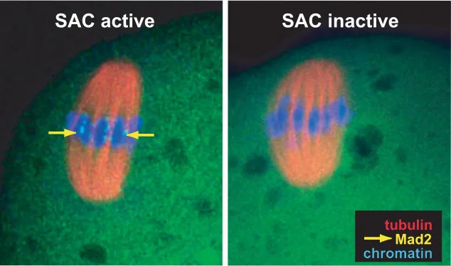
The potent MPF stabilizing factor operating in many cell types is spindle assembly checkpoint (SAC; Fig. 2) (Lew and Burke, 2003; Homer et al., 2005a; Malmanche et al., 2006). SAC
be-comes activated during prometaphase when chromosomes are not aligned in the spindle and emanate signals to the cytoplasm informing the checkpoint machinery that the kinetochores are not under proper tension. When all chromosomes become stably attached with spindle microtubules via kinetochores (thus fulfilling the key requirement for correct segregation), this signal is switched off, and the cell enters metaphase i.e. becomes competent to start the anaphase and complete the division. Thus, SAC activity prevents precocious inactivation of MPF which could result in abnormal separation of sister chromosomes (or chromatids). The main components of SAC are Mad2, Mad1, BUB1, and BUBR1 kinetochore proteins (Fig. 2) (Brady and Hardwick, 2000; Shah and Cleveland, 2000; Nasmyth, 2005). Mad2 is the factor respon-sible for binding Cdc20 that is an activator of anaphase promoting complex/cyclosome (APC/C) (Fang et al., 1998; Yu, 2002). APC/
C, a multiprotein complex, is a specific E3 ligase mediating
poluybiqitination and targeting different mitotic proteins including cyclin B to degradation in the proteasome (Peters, 2002; Passmore, 2004; Fry and Yamano, 2006). The association of Cdc20 with Mad2 prevents APC/C activity and the SAC remains active. The dissociation of the complex triggers SAC inactivation. Recently it was documented that the complex of Mad2 with Cdc20 is regu-lated by the equilibrium between polyubiquitination and deubiquitination of Cdc20 (Reddy et al., 2007; Stegmeier et al.,
2007). Thus, SAC inactivation is caused by the polyubiquitination of Cdc20 and does not require Cdc20 degradation, what explains its very rapid inactivation.
Another mechanism of MPF stabilization, described as cyto-static activity (CSF), functions specifically in oocytes that reached metaphase II of the second meiotic division (Fig. 3) (Masui, 2001; Tunquist and Maller, 2003; Schmidt et al., 2006,). In these
oocytes stabilization of MPF does not require SAC activity (Tsurumi
et al., 2004) and CSF alone is responsible for sustaining MPF
activity and preventing anaphase onset. The molecular nature of CSF is complex. One of the first factors discovered to be involved in this activity are Mos/MEK/MAP (ERK1/ERK2) kinase i.e. MAP kinase pathway (Fig. 3) (Colledge et al., 1994; Hashimoto et al.,
1994). Action of this pathway is vital for both proper functioning of meiotic spindle (via Doc1R and MISS; Lefebvre et al., 2002;
Terret et al., 2003a) and for MPF stabilization (Brunet and Maro,
2005). Recently, the crucial role in the CSF activity has been attributed to a member of Emi protein family – Emi2 (Endogenous meiotic inhibitor 2), that has been shown to bind Cdc20, and in consequence, prevent APC/C activation and anaphase onset (Liu and Maller, 2005; Rauh et al., 2005; Schmidt et al.; 2006, Shoji et al., 2006). Details concerning CSF and its components are
described in the next chapters.
As already mentioned, cyclin B degradation is crucial for the long term MPF inactivation. However, except cyclin B, numerous other proteins are degraded sequentially during the whole mitotic process (e.g. early mitotic proteins Nek2A, cyclin A), and their sequential degradation regulates the M-phase progression. Among
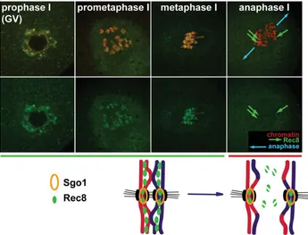
these proteins are also cohesins which act as a ‘glue’ holding together homologous chromosomes during the first meiotic divi-sion or two chromatids during second meiotic dividivi-sion or mitosis (Haering and Nasmyth, 2003; Uhlmann, 2004). Rec8 is a crucial cohesin involved in the binding of homologous chromosomes in meiotically dividing oocytes and spermatocytes (Fig. 4) (Lee et al., 2003; Prieto et al., 2004; Lee et al., 2006b). Its degradation
catalyzed by separase is a prerequisite step for the proper chromosome segregation (Siomos et al., 2001; Terret et al.,
2003b; Uhlmann, 2003). However, during the M-phase, until the moment of APC/C activation, separase remains inactive due to its binding to specific inhibitor – securin (Herbert et al., 2003; Pines,
2006). Securin degradation occurs via Cdc20 - APC/C mediated manner (Hagting et al., 2002). Thus, during oocyte meiotic
matu-ration the release of Cdc20 from its inhibitors allows activation of APC/C leading to the degradation of securin, activation of separase and in consequence Rec8 proteolysis (Herbert et al., 2003; Terret et al., 2003b; Homer et al., 2005b). Importantly upon the first
meiotic division Rec8 localized at centromeres is protected from degradation by shugoshin what secures cohesion of sister chro-matids (Watanabe and Kitajima, 2005). Finally, during the second meiotic division, this portion of Rec8 is also degraded (Fig. 4). MPF inactivation and cohesin degradation are separate events mediated, simultaneously by APC/C. Therefore, proper chromo-somes separation, which is the main goal of each M-phase, is achieved only when well ordered sequence of events is re-spected. Modifications of meiotic and mitotic events, described below, enable such a fine control to be achieved with high precision during each specific M-phase.
The first meiotic M-phase in oocytes: cyclin
B-associ-ated machinery governs meiotic timing
Like the M-phases in other cell types, the first meiotic M-phase is promoted by high activity of MPF. However, unlike in other cells the first meiotic M-phase in mammalian oocytes is particularly long lasting 6-10 hours (Polanski, 1997a; Polanski, 1997b). It is obvious that such prolonged M-phase may be associated with unique and complex events which have to occur to ensure proper progression through meiosis. It seems that special control of metabolism of cyclin B, the regulatory subunit of MPF, may help to extend the duration of the first meiotic M-phase (MI). In mitotic cells the bulk of cyclin B is accumulated and complexed with CDK1 at late G2 phase which results in the fast establishment of M-phase upon complex activation (Pines and Hunter, 1991; Jackman et al., 2003). Recently, cyclin A2, probably associated
with both CDK1 and CDK2, was found to participate in this process in HeLa cells (Gong et al., 2007). In the mouse oocyte the
situation is different since only a limited pool of cyclin B is present at the onset of meiotic maturation and no information about a potential role of cyclin A2 despite its presence in oocytes is available (Winston et al., 2000).
The pool of cyclin B complexed with CDK1 constitutes so called pre-MPF (Fig. 1), and is maintained stable in prophase oocyte through Emi1-dependent inhibition of APC/C (Reis et al., 2006;
Marangos et al., 2007). The first stimulus to resume meiotic
maturation comes through Cdc25-dependent dephosphorylation of pre-MPF (Hoffmann et al., 1993), leading to its activation and
the increase in the MPF activity to the level sufficient to induce
Emi2
Cdc20
Bub3
Mad1 Mad2 BubR1
Cdc20
APC
Cdc20
APC
cohesin degradation cyclin B
degradation
MPF inactivation
securin degradation
separase activation Cdc20
Emi2
SAC active
SAC inactive
accumulation and in the arrest of MPF activity at the intermediate level. This shows a biphasic control of MPF activity with the initial phase independent of cyclin B synthesis relying on activation of pre-MPF, and the second one requiring cyclin B synthesis to induce full MPF activity necessary to achieve complete M-phase characteristics. Additionally, protein synthesis inhibition results in anomalies of spindle formation and chromosome condensation. Such anomalies are also observed when oocytes mature in the absence of the nuclear factor(s) and cannot be repaired by restoration of normal level of MPF activity through exogenous expression of cyclin B (Polanski et al., 2005). Clearly, such
spindle and chromatin aberrations are not linked directly to poor cyclin B synthesis but rather reflect the inhibition of the synthesis of other cell cycle regulating proteins. Since nuclear factor(s) appear to stimulate cyclin B translation through its mRNA 3’UTR dependent control mechanism (Hoffmann et al., 2006) it is
pos-sible that a similar mechanism could initiate translation of other proteins crucial for spindle assembly, for example Cdc6 (Anger et al., 2005), and chromosome condensation. Such mechanism
might couple the intensive translation of cyclin B (and possibly other proteins) with the initiation of meiotic maturation, thus, securing that appropriate synthesis of necessary proteins occurs at the right time (Fig. 5).
Regardless of other proteins translated during meiotic matura-tion cyclin B synthesis seems to play the key role in determining the duration of the first meiotic M-phase. Striking differences in the level of cyclin B synthesis and in corresponding MPF activity were reported in mouse oocytes differing in the length of the first
APC
Emi2
Cdc20mos
MEK1
MAPK
(ERK1/ERK2)
Doc1R
MISS
meiotic spindle
stabilization
active
MPF
cyclin B
degradation
?
Fig. 3. Cytostatic factor (CSF) activity in metaphase II-arrested mouse oocytes. MAP kinase pathway is involved in meiotic spindle stabilization via phosphorylation of the spindle proteins Doc1R and MISS and independently, via an unknown mechanism, influences MPF activity. Emi2 binds Cdc20 and prevents APC/C activation, thus blocking cyclin B degradation and MPF inactivation.
nuclear envelope breakdown (also described as Germinal Vesicle Breakdown, GVBD) and chromatin condensation (Fig. 5). Thus, the oocyte enters M-phase already at this initial phase of meiotic maturation; however it is still a long way to establish conditions of a full M-phase. Resumption of meiotic maturation through pre-MPF activation results in crucial events ensuring further progres-sion of the M-phase. The induction of a massive synthesis of cyclin B (Hampl and Eppig, 1995, Winston, 1997) is controlled by nuclear factor(s) released into the cytoplasm upon nuclear enve-lope disassembly play important role in this process (Hoffmann et al., 2006). Either inhibition of the protein synthesis (Hampl and
Eppig, 1995; Polanski et al., 1998) or removal of such nuclear
factor (Hoffmann et al., 2006) results in the lack of cyclin B
meiotic M-phase. The oocytes with long M-phase showed clear biphasic profile of the increase of MPF activity correlated with lower level of cyclin B synthesis, whereas the oocytes with short M-phase reached the full MPF activity quickly and in association with high level of cyclin B synthesis (Polanski et al., 1998).
Moreover, a mild overexpression of cyclin B in ‘slow’ oocytes resulted in M-phase shortening (Polanski et al., 1998). This is
consistent with the well documented dependence between APC/ C and MPF (Cohen-Fix and Koshland, 1997) suggesting that in oocytes in which MPF activity increases slowly (as a conse-quence of slow accumulation of cyclin B) APC/C is activated later. Accordingly, the retarded activation of APC/C would delay the destruction of securins and cyclin B, the events marking the end of the first meiotic M-phase. On the other hand, fast increase of MPF activity seems to result in fast activation of APC/C and thus earlier destruction of its targets. Our recent results show that in the oocytes in which both CSF and SAC are disrupted the periodic changes in cyclin B level still occur, thus, suggesting the possibil-ity of regulation via an oscillator driven by simple feedback between MPF and APC/C activity (Hoffmann, Kubiak and Polanski, in preparation). Therefore, the high level of cyclin B synthesis correlates with shortening of the first meiotic M-phase likely due to earlier activation of APC/C (Polanski et al., 1998; Marangos
and Carroll, 2004). However, this dependence may be easily reversed since the strong overexpression of cyclin B saturates APC/C what leads to the hampering of efficient degradation of its excess. This in turn results in a metaphase arrest (Ledan et al.,
2001; Marangos and Carroll, 2004). All these data put cyclin B translation in the centre of the control of meiotic timing. This notion becomes especially clear in the light of observations that cyclin B synthesis during meiotic maturation leads to immediate formation of active MPF, probably with no need of any posttranslational modifications (Hampl and Eppig, 1995).
Another main mechanism involved in the control of the timing of the first meiotic anaphase is the SAC. Requirement for stable connection between kinetochores and the spindle for the anaphase signaling or disappearance of SAC proteins from the kineto-chores of the first meiotic spindle before anaphase suggested SAC function in oocyte meiosis (Brunet et al., 1999; Brunet et al.,
2003). These data were recently confirmed by interfering with Mad2 and Bub1 proteins, the key elements of the SAC pathway (Wassmann et al., 2003; Tsurumi et al., 2004; Homer et al., 2005c;
Yin et al., 2006; Wang et al., 2007). Knocking down Mad2
(Tsurumi et al., 2004; Homer et al., 2005c; Wang et al., 2007) or
Bub1 (Tsurumi et al., 2004; Yin et al., 2006) resulted in premature
induction of the first meiotic anaphase and elevated aneuploidy providing direct proof for SAC function during the first meiotic division of the mouse oocyte. Interestingly, upon SAC inactivation the first meiotic division is accelerated for only 2-3 hours still leaving 5-7 hours period of the M-phase (Tsurumi et al., 2004;
Homer et al., 2005c). It seems, however, that even upon SAC
disruption a period necessary to the extrusion of first polar body is not shorter than 5 hours, since the degradation of APC/C inhibitor Emi1 initiated at the M-phase entry may require about 3 hours (Marangos et al., 2006) and the proteolysis of APC targets
takes at least two more hours as revealed by the kinetics of cyclin B-GFP and securin-GFP degradation in mouse oocytes (Ledan et al., 2001; Hyslop et al., 2004; Homer et al., 2005b; Homer et al.,
2005c; Hoffmann, Kubiak and Polanski, in preparation). Surprisingly, SAC does not detect some chromosomal anoma-lies in female mouse meiosis (LeMaire-Adkins et al., 1997).
Moreover, the trisomies in humans resulting from meiotic errors are 8 times more frequent for the female meiosis than for the male one (Hassold et al., 1993). These data suggest that SAC in
mammalian oocytes may not be as efficient as in other cell types. On the other hand, even a single unattached bivalent is capable
CSF MAP kinase
MPF
?
GV GVBD MI telophase I MII fertilization interphase
cyclin B synthesis
cyclin B degradation
Emi1 Emi2 APC/Cdc20 SAC
tubulin
chromatin
sperm heads
time after GVBD 0h 5h 9h 12h
Fig. 5. Control of the cell cycle during meiotic maturation of mouse oocyte. (Upper lane) Microtubules (green) and chromatin (red) in maturing oocytes.
to establish SAC-mediated inhibition of cyclin B proteolysis in mouse oocytes showing that at least some elements of SAC control work effectively (Hoffmann, Kubiak and Polanski; in prepa-ration).
It is not clear whether CSF, which could theoretically modulate the timing of the first meiotic anaphase, is already active at meiosis I (Ciemerych and Kubiak, 1998). However, in mice deprived of mos gene and protein, whose oocytes clearly lack
CSF activity, the duration of the first meiotic M-phase remains unaffected (Verlhac et al., 1996). This observation questions CSF
as a possible candidate regulating the timing of anaphase I of meiosis.
MII-arrest and exit: Emi2, calcium and APC/C do the job,
but what about ERK1/2 MAP kinases?
The completion of the first meiotic division results in the metaphase II-arrest of the oocyte (MII), in which chromosomes and the second meiotic spindle remain in a stable state for hours. However, the nuclear maturation of oocytes can be decoupled from the cytoplasmic one. In other words, MII oocyte needs some time to become fully “fertilizable” despite that it remains still in the same stage of meiosis. Different reactions of mouse oocytes, depending on their age after the first polar body extrusion, were described following fertilization or parthenogenetic activation (Kubiak, 1989). Right after the extrusion of the first polar body MII oocytes are not capable to inactivate MPF (Fulton and Whittingham, 1978; Polanski, 1995). Then, they develop this capacity. How-ever, paradoxically, MPF inactivation is transitory. It is rapidly followed by the subsequent reactivation of MPF in maturing oocytes, and these oocytes enter the next stable M state, so called MIII (Kubiak, 1989). Such reaction clearly depends on the type of the activation stimulus. In vitro fertilization rarely results in
MIII, but parthenogenetic stimuli induce an abortive meiotic MIII
stage more frequently (Kubiak, 1989). Shortly after the second polar body extrusion, the MII oocytes are therefore programmed to reactivate the M-phase state after MPF inactivation. Such meiotic “phenotype” perpetuating M-phases with-out interphases is, therefore, not directly linked to the unique characteristic of the MII state, but rather to the age of the oocyte and the stage of its cytoplasmic maturation. The capacity to mobilize calcium stores seems to be the key event in the response of the oocyte to activation stimulus (Vincent et al., 1992; Vitullo and Ozil, 1992; Dupont,
1998). In fertilized oocytes Ca2+ increase is caused by the sperm-brought PLCζ that catalyzes hy-drolysis of PIP2 to inositole triphosphate (IP3) and diacylogrycerol (DAG) (Cox et al., 2002; Saunders et al., 2002; Halet, 2004; Swann et al., 2004).
Next, IP3 binds to its endocytoplasmic reticulum receptors (IP3R) and induces the release of Ca2+ oscillations (Markoulaki et al., 2003; Markoulaki et al., 2004). Moreover, in MII oocytes during early
periods after the second polar body extrusion, Mad2 localizes on their kinetochores, as clear-cut fluorescent dots visible on immunofluorescence
SAC active
SAC inactive
tubulin
Mad2
Fig. 6. Mad2 localization on kinetochores suggests that the spindle assembly checkpoint (SAC) is active in early metaphase II mouse oocytes. SAC seems active one hour after the completion of the first meiotic division, as Mad2 localizes clearly on kinetochores. It becomes inactivated within the next 6-10 h, when Mad2 disappears from the kinetochores, despite the oocyte remaining arrested in metaphase II due to CSF activity.
images. However, this location of Mad2 disappears gradually from the kinetochores during MII-arrest progression, showing that SAC is inactivated (Fig. 6, Sikora-Polaczek et al., 2006). This
behavior of Mad2 protein may correlate with the changes of the potential of oocytes to undergo normal activation. There are, however, no evidences that the changes in Mad2 localization (it seems that Mad2 disappearing from the kinetochores is translo-cated to the cytoplasm, where it could participate in the general inhibition of APC/C; Meraldi et al., 2004) could play any role in
cytoplasmic maturation.
Abortive activation of MII oocytes leading to MIII was described for the first time in the mouse. Since then it was reported in other mammals, including rat (Zernicka-Goetz, 1991), hamster (Tateno and Kamiguchi, 1997), rabbit (Collas et al., 1995), cow (Li et al.,
2005, Liu et al., 1998), pig (Ito et al., 2003), and human (Balakier et al., 2004). The phenomenon of reactivation of MPF following
abortive activation of a MII oocyte suggests also that its triggering mechanism remains in a delicate equilibrium. In some cases even M III-arrested oocyte can react to an activating stimulus again by the decrease and the subsequent rise in MPF activity entering long-lasting MIV (Kubiak, 1989). This suggests the efficiency of the mechanism stabilizing MPF activity even under repeated abortive activations of oocytes.
The M-phase arrest is due to the action of CSF identified in MII oocytes of Rana pipiens (Masui and Markert, 1971) by
experi-mental transfer of cytoplasm between mature oocytes and two-cell stage blastomeres (parallel to the discovery of MPF involving the cytoplasmic transfer between mature and non-mature oo-cytes; Masui and Markert, 1971) (Fig. 7). The formal proof for the CSF presence in mammalian oocyte came later from studies of mouse oocytes (Kubiak et al., 1993), while the presence of MPF
strain (Ciemerych and Kubiak, 1998). LT/Sv oocytes have pro-longed MI and therefore it was possible to demonstrate the CSF presence by cell fusion experiments, that revealed the capacity of metaphase I oocyte to arrest the mitotic cycle (Fig. 8). Similar experiments with wild type MI oocytes as fusion partners did not prove the presence of CSF during first meiotic M-phase simply because such oocytes spontaneously completed first meiotic division and reached the metaphase II stage during their matura-tion. However, these results do not contradict the possibility that CSF develops during MI. Thus, we believe that in maturing wild-type oocytes the CSF activity may develop during the first meiotic M-phase similarly as in the LT/Sv strain (how such oocytes could undergo MPF inactivation and complete the first meiotic division will be discussed below, after the description of the molecular identity of CSF). Alternatively, CSF development in LT/Sv matur-ing oocytes, as the cytoplasmic maturation itself, could be un-coupled from the nuclear one. If the CSF development were not affected in these oocytes, this activity would appear at the right time, i.e. when wild type oocytes reach the MII-arrest. In this case, the above comparison between LT/Sv MI oocytes and wild-type MI oocytes in terms of CSF activity could be erroneous. Future studies of molecular components of CSF should resolve this issue. However, as we mentioned previously, normal progression of the first meiotic division in mos-null oocytes questions the role
2005). Additional problem with Mos/MEK/ERK pathway lies in its permanent activity during all transitional periods in meiosis, when MPF is inactivated i.e. upon anaphase of the first and the second meiotic division. Apparently, the Mos/MEK/ERK pathway does not fulfill the postulate by Markert & Masui (1971) that CSF should stabilize MPF, and thus, should be inactivated before cyclin B degradation and MPF inactivation. Some review articles seemed to ignore this point (e.g. Dupont, 1998).
The real breakthrough in the “CSF science”, and at least a partial explanation for the paradox described above, came from studies devoted to APC/C inhibition. Reiman et al. described a
new protein acting as a potent APC/C inhibitor (Reimann et al.,
2001a; Reimann et al., 2001b). They called it Emi1 - Early Mitotic
Inhibitor. Soon, further studies have shown that Emi1 is a member of family of Early Mitotic Inhibitors when Emi2/Erp1 was discov-ered (Tung et al., 2005). Emi2 was shown to have the CSF activity
both in Xenopus (Liu and Maller, 2005; Rauh et al.; 2005; Liu et al., 2006) and in the mouse (Shoji et al., 2006).
Identification of Emi2 as a principal molecule of the CSF pathway was possible when the mechanism of its inactivation was understood (Liu and Maller, 2005; Rauh et al., 2005). It was
revealed that Emi2 must be degraded to enable the full activation of APC/C which in turn allows cyclin B degradation and therefore MPF inactivation (Madgwick et al., 2006). The mechanism of
Fig. 7. Discovery of M-phase promoting factor (MPF) and cytostatic factor (CSF) in oocytes of Rana pipiens. (MPF) In 1971, Masui and Markert induced frog prophase I oocytes to mature with progesterone (left), removed a portion of its cytoplasm (red arrow out) and transferred it to the cytoplasm of an uninduced prophase I oocyte (red arrow in; Masui and Markert, 1971). The injected oocyte resumed meiosis despite the lack of progesterone induction and reached the metaphase stage. These changes were attributed to the activity of the factor named as Maturation Promoting Factor. (CSF) In the same paper, they reported that the cytoplasm of metaphase II arrested frog oocytes injected into one of the two blastomeres of a dividing 2-cell embryo caused a permanent M-phase arrest. The control, uninjected blastomere followed cleavages and formed a multicellular morula.
of CSF in MI (Verlhac, et al., 1996)
CSF induces the M-phase arrest by MPF stabilization. The molecular nature of CSF and its mode of action on MPF are not fully resolved. However, a great progress in understanding of CSF nature was marked recently when few key molecules and crucial events were discovered. The first molecule clearly involved in the CSF pathway was identified as Mos protooncogene in Xenopus
oo-cytes (Watanabe et al., 1989). Mos is MAP kinase kinase
kinase activating MAP kinase kinase i.e. MEK1/2 and further ERK1 and ERK2 MAP kinases by series of phos-phorylations (Kosako et al., 1994) This pathway was
shown to be of the key importance for the CSF activity both in Xenopus (Sagata et al., 1988; Watanabe et al.,
1989) as well as in the mouse (Colledge et al., 1994;
Hashimoto et al., 1994; Araki et al., 1996; Verlhac et al.,
1996; Phillips et al., 2002). However, the final interaction
of this pathway with MPF resulting in stabilization of the latter was not so far clearly identified. In Xenopus, it
seems that Mad2 and Bub1 checkpoint proteins down stream in the Mos/MAP kinase pathway) could play a direct role of APC/C inhibitors (Schwab et al., 2001).
However, in the mouse these proteins are clearly not involved (Tsurumi et al., 2004). Similarly, the role of
p90rsk1, the direct substrate of ERK1 and ERK2 MAP kinases was shown to have the CSF activity in Xenopus
(Gross et al., 1999) but not in the mouse (Dumont et al.,
2005). These differences might be likely linked to spe-cies-dependent variants in the identity of CSF. It was also postulated by Dumont et al. (2005) that differences in
functioning of MAPK mediated signal transduction in
Xenopus and mouse oocytes might result from the
Emi2 degradation is rather complicated. It requires sequential phosphorylation of Emi2 by CaMKinase II and Plk1 (Schmidt et al., 2005). Ca+2/calmodulin dependent kinase II (CaMKII) is
activated as a result of IP3 to IP3R binding and the burst of ER-derived Ca2+ (Markoulaki et al., 2003; Markoulaki et al., 2004).
Plk1 is active in MII oocyte, but cannot phosphorylate Emi2 before the CaMKII phosphorylation (Liu and Maller, 2005; Rauh et al.,
2005, Schmidt et al., 2005).
Such a serial phosphorylation triggers polyubiquitination of Emi2 and its targeting for the proteasome-mediated degradation.
MPF
1977 - Balakier and Czolowska - maturing mouse oocytes
Fig. 8. Discovery of M-phase promoting factor (MPF) and cytostatic factor (CSF) in mouse oocytes.(MPF) In 1977, Balakier and Czolowska bisected a GV stage oocyte of the mouse into nucleate and anucleate halves (Balakier and Czolowska, 1977). When nucleate halves resumed meiosis and reached metaphase, their anucleate counter-parts (supposedly also entering into maturation) were fused with one of the blas-tomeres of a 2-cell mouse embryo. In the resulting hybrids, blastomere nuclei underwent nuclear envelope breakdown (NEBD) and chromatin condensation. (CSF)
In 1994, Kubiak and collaborators (1994) induced fusion between a metaphase II-arrested mouse oocyte and a 1-cell parthenogenetic mouse embryo. In the resulting hybrids, the pronucleus underwent NEBD, condensed chromatin and remained perma-nently arrested in the M-phase. Luckily, Kubiak et al. used 1-cell embryos that resulted from parthenogenetical activation. In 1995 Zernicka-Goetz et al. and Kono et al.
documented that hybrids of metaphase II-arrested oocytes with 1-cell parthenoge-netic, but not with 1-cell zygotes, permanently arrest in M-phase. (Kono et al., 1995, Zernicka-Goetz et al., 1995). In hybrids of MII-arrest oocytes with zygotes, Ca2+ spikes occurring within zygote cytoplasm inactivate CSF present within the cytoplasm of the metaphase II-arrested component of the hybrid. In 1998, Ciemerych and Kubiak analyzing oocytes of the LT/Sv mouse strain, which are characterized by a prolonged first meiotic division, documented that CSF is also present at this stage of maturation by fusion of an MI oocyte with a partenogenetic one-cell embryo (Ciemerych and Kubiak, 1998).
In Xenopus, its proteolysis is necessary to trigger
polyubiquitination of MPF complex on its cyclin B component (Nishiyama et al., 2000), separation of
both cyclin B and CDK1 resulting in MPF inactivation and further degradation of cyclin B by the proteasome (Nishiyama et al., 2000; Chesnel et al., 2006).
More-over, cyclin B dissociation from CDK1 appears to be a primary cause of MPF inactivation followed by CDK1 dephosphorylation on Thr-161 in Xenopus laevis cell-free extracts (Chesnel et al., 2007). The
latter was shown in cell-free extract during the first embryonic M-phase, but judging by similarities be-tween the molecular mechanisms of MPF inactiva-tion between meiosis (Nishiyama et al., 2000; Chesnel et al., 2005a) and mitosis (Chesnel et al., 2006;
Chesnel et al., 2007) we believe that it is also the
case for the activated MII oocytes both in Xenopus
and in the mouse.
If Emi2 serves in MII oocytes as a major APC/C inhibitor what role could be played by Mos/MEK/MAP kinase pathway as the CSF components? Studies showing that CSF inactivation is a two-step event in mouse oocytes could suggest a connection between these two pathways. Indeed, cell fusion experiments between freshly activated mouse oocytes and mitotic one-cell embryos demonstrated that after rapid inac-tivation of the CSF its activity is restored and main-tained for the additional 60 min, even in effectively activated oocytes (Ciemerych and Kubiak, 1999). This interval corresponds to the period of ERK1 and ERK2 inactivation in mouse oocytes (Verlhac et al.,
1994). The recent data concerning the mode of Emi2 degradation facilitate understanding of MPF inacti-vation in oocytes without Mos/MEK/MAP kinase path-way inactivation. The results concerning Emi2 sug-gest that the degradation of this protein corresponds to the first wave of CSF inactivation. Once Emi2 becomes degraded MPF inactivation occurs, how-ever, still active Mos/MAK/MAP kinase pathway tran-siently restores the CSF activity, i.e. it remains active until Mos degradation and ERK1 and ERK2 inactiva-tion (Weber et al., 1991; Verlhac et al., 1994). How
this pathway could restore the CSF activity remains unclear and needs further studies (see below the hypothesis that Mos/MEK/MAP kinase pathway could interact with Emi2). Alternatively, Mos/MEK/MAP kinase pathway may stabilize Emi2.
The activities of ERK1 and ERK2 are necessary to slow down both the remodeling of the paternal and maternal chromatin via nuclear lamins phosphoryla-tion (Peter et al., 1990; Kubiak et al., 1991; Prather et al., 1991) and the reorganization of the interphase
microtubule cytoskeleton (Szollosi et al., 1993;
II-arrested oocyte. The restoration of CSF activity seems there-fore to be a kind of a side effect of ERK1 and ERK2 activity. During physiological oocyte activation i.e. upon fertilization, the reap-pearance of the CSF activity has no role since MPF is definitely inactivated and becomes reactivated only at the end of the first embryonic cell cycle i.e. approximately 20 hours later. The em-bryo seems therefore to have a sufficient time to eliminate the meiotic activities present within the oocyte from which it origi-nated. However, in the next chapter we will document that it is not necessary the whole truth of CSF inactivation.
Let’s come back to the hypothesis that the first meiotic division (anaphase I) proceeds in the presence of CSF activity already developed in the oocyte to further elucidate the enigma of this factor. This apparent paradox is not more paradoxical than the onset of anaphase of the second meiotic division (anaphase II). Mos/ERK1/ERK2 MAP kinases pathway is not inactivated imme-diately upon fertilization, but some 60-90 min later (Weber et al.,
1991; Verlhac et al.; 1994). Thus, MPF inactivation and the
anaphase II proceed in the presence of the activity of this key pathway for the CSF activity. The resolution of the mystery comes again from understanding Emi2 degradation which immediately, but transitionally removes the CSF activity. So, Emi2 is most probably also degraded upon anaphase I of meiosis in mouse oocytes (its degradation was recently shown to be essential for MI/MII transition in Xenopus laevis; Ohe et al., 2007), and the
presence of active MAP kinase pathway only helps to restore the CSF activity in MII oocyte following the first polar body extrusion. It seems therefore, that the CSF activity is a complex network of different pathways including direct APC/C inhibitors (like Emi2) and creating the appropriate meiotic environment (like Mos/MAP kinase one). It would be of great importance to understand the molecular relationship between the two major CSF pathways and specially to verify a speculation that Mos/MEK/MAP kinase path-way could activate or stabilize Emi2 (see below).
Another important characteristic of the CSF action is its capa-bility to stabilize cyclin B and MPF activity in a dynamic manner. In MII oocytes of the mouse, a clear turnover of cyclin B was found (Kubiak et al., 1993). Such a turnover was later confirmed in Xenopus oocytes as well (Thibier et al., 1997). Cyclin B
degrada-tion is equilibrated by the continuous cyclin B synthesis. Upon oocyte activation this equilibrium is destroyed in favor to cyclin proteolysis, and the level of this protein dramatically falls down
(Kubiak et al., 1993). Moreover, during both the MII-arrest and
upon oocyte activation cyclin B degradation requires a functional meiotic spindle (Kubiak et al., 1993; Winston et al., 1995). Spindle
microtubules could be likely necessary to bring cyclin B and ubiquitine molecules together or the polyubiquitinated complex CDK1/cyclin B to the proteasome since both cyclin B and proteosomes are highly concentrated within the spindle. How-ever, data concerning the role of SAC and Mad2 protein in mouse oocytes suggest another possibility (Tsurumi et al., 2004;
Sikora-Polaczek et al., 2006). It seems that in MII oocytes two separate
pathways stabilize cyclin B and MPF activity: namely SAC and CSF. We propose that the CSF (Emi2, Mos/MEK/MAP kinases pathway) efficiently slows down cyclin B degradation assuring the APC/C only partially active and this action is independent on microtubules. Disassembly of microtubules, as well as probably other spindle anomalies, switches the SAC pathway on, without affection the CSF activity, and results in a double inhibitory system with SAC as an emergency brake (Tsurumi et al., 2004).
SAC activation causes supplementary inhibition of APC/C via Mad2 (on kinetochores or in the cytoplasm) in a microtubule-dependent manner. Elucidation of the details of the interrelation-ship between CSF and SAC will certainly enable to verify this hypothesis.
It was shown recently in Xenopus laevis CSF extracts that
CDK1 phosphorylates and inhibits Emi2 (Wu et al., 2007).
CDK1-phosphorylated sites are deCDK1-phosphorylated by PP2A phosphatase, which antagonizes the effect of CDK1. According to Wu and colleagues, such equilibrium would be responsible for maintain-ing stable cyclin B levels and moderate APC/C activity (i.e. controlled cyclin B turnover) during the CSF arrest (Wu et al.,
2007). However, this mechanism might also be completed or reinforced by the Mos/MEK/MAP kinase pathway. This could explain a role of this pathway in the CSF arrest of oocytes. Mos/ MEK/MAP kinase pathway could potentially be involved in phos-phorylation of Emi2 and could affect it specifically in MII-arrested oocytes. Recently, such a hypothesis was successfully tested in
Xenopus laevis extracts. Emi2 was shown to be a direct substrate
of p90rsk kinase (Inoue et al., 2007; Nishiyama et al., 2007). Moreover, phosphorylation of Emi2 is necessary for the Emi2-dependent CSF activity. Therefore, this protein, at least in Xeno-pus oocytes, is the most downstream identified substrate in the
Mos/MEK/MAP kinase ERK1 and ERK2 pathway. It is, however,
still unclear which kinase phosphorylates Emi2 in MII mouse oocytes, since, as explained above, p90rsk kinases were excluded as CSF component (Dumont et al., 2005). Thus, identification of
such kinase remains an open issue. On the other hand, it was also not investigated whether CDK1/PP2A balance operates also in mouse oocytes.
Finally, to complete our vision of CSF we have to mention a potential role od CDK2-cyclin E complex. It was shown to play a part of CSF activity in Xenopus oocytes via checkpoint protein
Mps1 (Grimison, et al., 2006) and is supposed to be a third,
parallel component next to Mos/MEK/MAP kinase and Emi2 (Liu,
et al., 2007). However, there is no data suggesting such a role in
the mouse oocyte. Moreover, there is no evidence that oocytes of mice lacking cyclin E1 and cyclin E2 (Geng, et al., 2003; Parisi et al., 2003) or CDK2 (Berthet, et al., 2003, Ortega, et al., 2003)
exhibit any abnormalities in the regulation of MII arrest. Thus, we assume that the CDK2-cyclin E is not involved in the CSF activity in mouse oocytes.
First embryonic mitosis: meiosis-like mitosis, or how
and why to prolong cell division of the zygote?
Several lines of evidence document that the first embryonic cell cycle differs from the subsequent ones. Progression of the first cell cycle relies on maternal factors i.e. mRNAs and proteins accumulated during growth phase of GV oocyte i.e. during the long arrest in the prophase of the first meiotic division (Bachvarova and De Leon, 1980; Pratt et al., 1983; Bachvarova, 1985; Nothias et al., 1995). The intense transcription ceases once oocyte
reaches its maximal volume and becomes competent to resume meiotic division (Zuccotti et al., 1995; Bouniol-Baly et al., 1999;
De La Fuente, 2006). Fertilization that triggers oocyte activation does not terminate transcriptional silencing. Despite that the first mRNAs transcribed from embryonic genome is detectable in G2 phase of the first cell cycle (Ram and Schultz, 1993; Bouniol et al.,
1995; Aoki et al., 1997; Thompson et al., 1998; Hamatani et al.,
2004) little is known about genes activated at that period of development. Currently, only single pieces of evidence document that proteins encoded by embryonic genome are both synthe-sized and active in one-cell embryo. Studies involving mice lacking functional RGS14 gene (a regulator of G protein signaling) showed that this mitotic spindle protein is necessary for the first mitotic division of the mouse embryo (Martin-McCaffrey et al.,
2004). Nevertheless, the burst of the major transcription, also described as zygotic genome activation (ZGA), occurs in the mouse during the second cell cycle when proteins originated from the embryonic genome become detectable (Flach et al., 1982;
Bensaude et al., 1983; Bolton et al., 1984; Schultz, 1993). Thus,
the regulation of the first embryonic cell cycle depends largely on the mRNA and proteins accumulated already during meiosis. Consequently, during the early mitotic cycles of the embryo some meiotic pathways could also be present and eventually active.
Transcriptional silence is not the only characteristic that distin-guishes the first cell cycle from the subsequent cycles. Among special features of the one-cell embryo one can list rapid deforma-tion of its surface i.e. cortical activity that precedes the first, but not the next mitotic divisions (Waksmundzka et al., 1984; Ciemerych,
1995; Ciemerych et al., 1998; Liu et al., 2002; Alikani et al., 2005).
Moreover, some phenomena such as cyclic activity of K+ ion
channels (Day et al., 1998) or cytoplasmic MPF activation are
induced autonomously, i.e. independently from the nucleus, ex-clusively during the first cell cycle (Ciemerych et al., 1998; Kubiak
and Ciemerych, 2001). All these phenomena point to a high autonomy of the one cell embryo cytoplasm, comparable to the autonomy of the oocyte cytoplasm (Czolowska and Balakier, 1977; Hoffmann et al., 2006).
Detailed analyses of cleaving preimplantation mouse embryos revealed yet another uniqueness of the one-cell embryo i.e. significant increase in the duration of the first mitosis in compari-son to the subsequent ones. The first embryonic mitosis, as-sessed from the moment of nuclear envelope breakdown (NEBD) and till the beginning of cytokinesis, takes approximately 120 min., while the second one only 70 min. (Fig. 9; Ciemerych et al.,
1999). Importantly, similar differences are characteristic also for the first cleavage divisions of X. laevis (Chesnel et al., 2005a;
Chesnel et al., 2005b), sea urchin Sphaerechinus granularis (J.Z.
Kubiak and P. Cormier, unpublished observation), and C. elegans
embryos (J.Z. Kubiak, F. Chesnel and P. Gonczy, unpublished observations). One of the possible explanations of the prolonga-tion of the first mitosis observed in mouse embryos comes from the fact that female and male pronuclei are assembled via two different routes (Wright and Longo, 1988; Adenot et al., 1991;
Yanagimachi, 1994). In consequence these two sets of chromatin remain unconnected throughout the first embryonic cell cycle. Male and female pronuclei separately undergo DNA replication in S-phase. Importantly in mammalian zygotes two pronuclei do not fuse but independently and asynchronously undergo NEBD, and start chromatin condensation at G2/M transition (Ciemerych and Czolowska, 1993; Bomar et al., 2002). Finally, they reunite during
the formation of common metaphase plate and mitotic spindle (Donahue, 1972; Howlett and Bolton, 1985; Mayer et al., 2000).
It seems, therefore, possible that the prolongation of the first mitotic division facilitates and/or is caused by the necessity to combine two chromatin sets within single plate and spindle. However, the duration of the first mitosis does not differ between zygotes containing two pronuclei and haploid parthenogenotes with one haploid pronucleus and is cytoplasmatically controlled (Ciemerych et al., 1999). Moreover, the development of zygotes
that contain single diploid pronucleus is also not affected (Krukowska and Tarkowski, 2005). Therefore, the molecular machinery prolonging the first embryonic mitosis is again another manifestation of events that occur independently of the nucleus in the zygote. Nevertheless, it cannot be excluded that mechanisms operating within the cytoplasm of one-cell mouse embryo were evolutionary developed to assure the time necessary for the proper spatial arrangements of female and male chromosomes by extending the first mitosis in time.
As we already mentioned, the formation of the metaphase plate and the spindle involves precise molecular mechanisms reassuring that kinetochores of all chromosomes form proper and stable connections with microtubules of the mitotic spindle (Fig. 2; Rieder et al., 1994; Li and Nicklas, 1995; Rieder and Maiato,
2004). As it was documented in mitotically dividing somatic cells (Li and Benezra, 1996; Li et al., 1997; Gorbsky et al., 1998; Waters et al., 1998; Fang et al., 1999; Waters et al., 1999; Chang et al.,
2004) and also in meiotically maturing oocytes (Kallio et al., 2000;
Wassmann et al., 2003; Tsurumi et al., 2004) active SAC prevents
of Mad2 from kinetochores delineates the end of the prometaphase and the beginning of the metaphase and allows anaphase onset (Fig. 2; Li and Benezra, 1996; Waters et al., 1998). Prometaphase
can be distinguished from metaphase not only by the analysis of the localization of SAC proteins, but also by examination of chromosome movements. Oscillatory translocation of chromo-somes pictures unbalanced pulling forces causing their displace-ment out of the forming plate (Howell et al.; 2000, Howell et al.,
2004; Meraldi et al., 2004). Again this feature is characteristic not
only for somatic cells but also for oocytes during metaphase I of meiosis (Brunet et al., 1999). Thus, the presence of Mad2 at
kinetochores and continuous chromosome movements delin-eates the prometaphase state.
In somatic cells, the prometaphase is relatively long. An average mitosis takes 50 min and the duration of metaphase is on average 10 min (Rieder et al., 1994; Howell et al., 2000; Jones et al., 2004; Meraldi et al., 2004; Meraldi and Sorger, 2005). These
proportions could be changed or even inversed, e.g. during considerably prolonged first meiotic division of maturing oocytes, a prometaphase (characterized by the presence of Mad2 and Bub1on kinetochores and by chromosome movements) takes about 8 hrs, while metaphase only 1 hr or less (Brunet et al., 1999;
Brunet et al., 2003; Wassmann et al., 2003). Surprisingly, during
the first and the second mitosis of the mouse embryo, Mad2 is localized at kinetochores for the similar period of time. Moreover, chromosome relocations within the metaphase plate were ob-served also for the comparable period in these two mitotic M-phases i.e. for 20-30 min, demonstrating that during both divi-sions the length of the prometaphase is similar (Sikora-Polaczek
et al., 2006). Thus, it is a metaphase itself that is prolonged in the
first mitosis. The comparison of our previous results i.e. the dynamics of MPF activation and recently studied SAC inactivation showed that both events correlate during both mitotic divisions (Fig. 9, Ciemerych et al., 1999; Sikora-Polaczek et al., 2006).
onic cell cycle. However, it cannot be excluded that transient stabilization of MPF during the first embryonic mitosis is a mani-festation of a tuning mechanism possibly dependent on a residual activity of factor/factors previously (i.e. in M II) involved in the CSF activity. Since the switch from the meiotic to the mitotic type of control occurs during the first cell cycle of the embryo it is possible that some meiotic factors are present and remain active till the first mitosis. Similarly as during the meiotic arrest, these regulators could stabilize MPF activity via APC/C down-regulation, i.e. via prevention of cyclin B degradation. It would also be of importance to analyze whether the transitional metaphase arrest during the first embryonic mitotic division depends on a finely regulated turnover of cyclin B, as happens during the MII-arrest in oocytes (Kubiak et al., 1993).
Mos/MEK/MAP kinase (ERK1/ERK2) pathway is necessary for CSF establishment in M II oocytes (Colledge et al., 1994;
Hashimoto, 1996; Phillips et al., 2002). It influences positively the
MPF activity and stabilizes the meiotic spindle via proteins like MISS (MAPK-interacting and spindle stabilizing) and DocR1 (Fig. 3; Lefebvre et al., 2002; Terret et al., 2003a). Several lines of
evidence suggest that ERK1/ERK2 kinases play important role during the initiation and progression of mitosis in somatic cells (Peter et al., 1992; Sanghera et al., 1992; Chiri et al., 1998;
Shapiro et al., 1998; Zecevic et al., 1998; Harding et al., 2003;
Horne and Guadagno, 2003). This pathway does not seem, however, to be active during either the first or the second mitosis of the mouse embryo. During both divisions only inactive (i.e. non-phosphorylated) forms of ERK1/ERK2 were detectable in dividing embryos (Kalab et al., 1996, Verlhac et al., 1994). However, MAP
kinase activity can be also assayed by its ability to phosphorylate MBP (myelin basic protein) in an in vitro assay. Importantly, during
the first but not the second mitosis an MBP activity was detectable in such assay suggesting the possible activity of other MAP kinase(s) (Verlhac et al., 1994; Haraguchi et al., 1998). p90rsk1
Spindle assembly checkpoint -active-inactive
somatic mitosis
Howell et al., 2000 Meraldi et al., 2004
Jones et al., 2004
first meiotic division of mouse oocyte
Brunet et al., 1999 Wassmann et al., 2003
second mitosis of mouse embryo
Sikora-Polaczek et al., 2006
first mitosis of mouse embryo
Sikora-Polaczek et al., 2006
Fig. 10. Changes in the proportion of active and inactive spindle assembly checkpoint (SAC) during different M-phases in maturing oocytes, mitotically dividing mouse embryos and somatic cells. Proportion of active (red) versus inactive (blue) SAC significantly differs during various M-phases. SAC activity is detectable throughout the almost whole first meiotic division (called metaphase I despite repre-senting in fact prometaphase I) of mouse oocytes (Brunet et al., 1999, Wassmann et al., 2003). On the other hand, during the first mitotic division of the mouse embryo, SAC activity is detectable only for a proportionally short period of time (Sikora-Polaczek et al., 2006). During subsequent mitotic divisions of the mouse embryo, these proportions resemble mitotically dividing somatic cells, suggesting transitional characteristic of the first embryonic M-phase (Howell et al., 2000, Jones et al., 2004, Meraldi et al., 2004). However, only during the first mitosis MPF becomes
stabilized for over 60 min (Ciemerych et al., 1999)
(Fig. 10).
Noteworthy, the stabilization of MPF is also a landmark of oocytes arrested at the metaphase of the second mitotic division and is not achieved by SAC but by checkpoint-independent CSF activity (Tsurumi et al., 2004; Sikora-Polaczek et al., 2006).
Mad2 becomes relocated from kinetochores to cyto-plasm within 2h after the completion of the first meiotic division and the formation of the M II plate and the spindle. Importantly, such oocytes can be arrested for next several hours until being activated by penetrating spermatozooa. Thus, both M II oo-cytes and one cell embryos resemble each other in the dynamics of SAC inactivation (delineated by the disappearance of Mad2 from kinetochores and the cessation of chromosome movements). This simi-larity strongly suggests that also during the first mitotic division an initial role of SAC as a mechanism preventing anaphase onset is replaced by other MPF-stabilizing mechanism(s) (Sikora-Polaczek et al., 2006).
embry-figures among downstream targets of ERK1/ERK2 kinases. How-ever, it is not involved in CSF activation in mouse oocytes (Dumont et al., 2005) despite that its phosphorylation is an
apparent manifestation of ERK1/ERK2 kinases activity (Kalab et al., 1996; Verlhac et al., 1996). Surprisingly, p90rsk1 was shown to be partially phosphorylated during the first but not the second mitosis of the mouse embryo (Kalab et al., 1996; our unpublished
results). It cannot be also excluded that some modifications of MAP kinase pathway might be involved in MPF stabilization during the first mitosis of the mouse embryo and that a similar mode of APC/C inactivation operates during meiosis and during the first mitotic M-phase despite obvious differences between them. All these aspects await for elucidation.
It is well documented that the CSF activity involves factors preventing APC/C activation. As we mentioned previously these factors playing a role in SAC (such as Mad2, Mad1, Bub1) are involved neither in CSF in mouse oocytes (Tsurumi et al., 2004)
nor in M-phase prolongation in mitotically dividing one-cell em-bryos (Sikora-Polaczek et al., 2006). Another potential factor
involved in the prolongation of the first mitotic division might be one (or more) Emi protein(s). Emi1 was shown to bind Cdc20 and to inhibit APC/C during mitotic division (Reimann et al., 2001a;
Reimann et al., 2001b; Margottin-Goguet et al., 2003). It was
proposed to be involved in CSF activation via p90rsk1 dependent manner (Paronetto et al., 2004). However, this hypothesis was
questioned by the discovery that p90rsk1 and other RSK family members are not involved in the metaphase II arrest (Dumont et al., 2005). Nevertheless, studies on oocytes of emi1-null mice
showed that Emi1 protein is crucial for the mitotic progression during preimplantation mouse development (Lee et al., 2006a).
Emi2 has unquestionable role in CSF activation: its silencing causes parthenogenetic activation of oocytes and its levels drop in parthenogenetically activated oocytes (Shoji et al., 2006).
Interestingly, Emi2 degradation upon mouse oocyte activation proceeds relatively slow leaving high amount of this protein 6 hours after activation and easily detectable portion still present in the two-cell embryos (see Fig. 1H in Shoji et al., 2006), which
implies possible influence of Emi2 on the first mitosis. In Xenopus,
during the first embryonic mitosis Emi2 seems not to play an essential role since it is present at too low levels to induce the M-phase arrest and it is not degraded as happens during the second meiotic division, but specifically phosphorylated (Liu, et al., 2006).
However, Emi2 depleted extracts accumulate less cyclin B during the first embryonic mitosis which result in a shortening of this M-phase duration (Liu, et al., 2006) as shown before by Chesnel et al. (2005). The Emi1 and Emi2 localization and functionality was,
however, not carefully tested in the first two embryonic mitoses during unperturbed mouse development. A paper describing briefly the effect of Emi2, as well as Emi1, knock-down using RNAi in mouse MII oocytes that were subsequently fertilized, suggests that these proteins have no essential role in mouse early embry-onic development (Amanai, et al., 2006) as in Xenopus (Liu, et al.,
2006). However, the duration of the first embryonic mitosis was not studied in such embryos. Thus, it cannot be excluded that Emi proteins also impinge on the machinery regulating the first mitotic division.
The fact that the first cell cycle bridges meiotic and mitotic type of cell cycle control directs a search of the factors involved in the regulation of the first mitosis towards the factors directly involved
into the stabilization of the MPF activity. It is however, possible that other regulators or signal transduction pathways can also be engaged.
Concluding remarks
This overview points out similarities as well as striking differ-ences between meiotic and mitotic M-phases of mouse oocytes and one-cell embryos. The M-phase regulatory machinery com-mon for all eukaryotic cells is modified during this relatively short period of oogenesis and early development several times. These modifications result from different applications of either common molecular mechanisms (e.g. SAC) or stage-specific ones (e.g. CSF and the not yet specified mechanism prolonging the first embryonic mitosis). Each cycle and each M-phase differ substan-tially to assure harmonious preparation of oocytes for fertilization and early development.
Acknowledgements
The authors thank Franck Chesnel for the critical reading of this manuscript, Zuzanna Maciejewska for sharing the unpublished confocal image of one-cell mouse embryo at anaphase stage, and Ewa Borsuk for the phase-contrast image of fertilized oocyte. MAC is a recipient of L’Oreal for Women and Science Foundation, JZK acknowledges grants from ARC (4900), Ligue Contre le Cancer (Comité d’Ille-et-Vilaine et de la Vendée) and BQR (Université Rennes 1) and ZP from Deutsche Forschungsgemeinschaft (Schwerpunktprogramme 1109).
References
ADENOT, P.G., SZOLLOSI, M.S., GEZE, M., RENARD, J.P. and DEBEY, P. (1991). Dynamics of paternal chromatin changes in live one-cell mouse embryo after natural fertilization. Mol Reprod Dev 28: 23-34.
ALIKANI, M., SCHIMMEL, T. and WILLADSEN, S.M. (2005). Cytoplasmic fragmen-tation in activated eggs occurs in the cytokinetic phase of the cell cycle, in lieu of normal cytokinesis, and in response to cytoskeletal disorder. Mol Hum Reprod 11: 335-44.
AMANAI, M., SHOJI, S., YOSHIDA, N., BRAHMAJOSYULA, M., PERRY, A.C. (2006). Injection of mammalian metaphase II oocytes with short interfering RNAs to dissect meiotic and early mitotic events. Biol Reprod 75:891-8.
ANGER, M., STEIN, P. and SCHULTZ, R.M. (2005). Cdc6 requirement for spindle formation during maturation of mouse oocytes. Biol Reprod 72: 188-94.
AOKI, F., WORRAD, D.M. and SCHULTZ, R.M. (1997). Regulation of transcrip-tional activity during the first and second cell cycles in the preimplantation mouse embryo. Dev Biol 181: 296-307.
ARAKI, K., NAITO, K., HARAGUCHI, S., SUZUKI, R., YOKOYAMA, M., INOUE, M., AIZAWA, S., TOYODA, Y. and SATO, E. (1996). Meiotic abnormalities of c-mos knockout mouse oocytes: Activation after first meiosis or entrance into third meiotic metaphase. Biol Reprod 55: 1315-24.
BACHVAROVA, R. (1985). Gene expression during oogenesis and oocyte devel-opment in mammals. Dev Biol 1: 453-524.
BACHVAROVA, R. and DE LEON, V. (1980). Polyadenylated RNA of mouse ova and loss of maternal RNA in early development. Dev Biol 74: 1-8.
BALAKIER, H. and CZOLOWSKA, R. (1977). Cytoplasmic control of nuclear maturation in mouse oocytes. Exp Cell Res 110: 466-9.
BALAKIER, H., SOJECKI, A., MOTAMEDI, G. and LIBRACH, C. (2004). Time-dependent capability of human oocytes for activation and pronuclear formation during metaphase ii arrest. Hum Reprod 19: 982-7.
BENSAUDE, O., BABINET, C., MORANGE, M. and JACOB, F. (1983). Heat shock proteins, first major products of zygotic gene activity in mouse embryo. Nature 305: 331-3.
BOLTON, V.N., OADES, P.J. and JOHNSON, M.H. (1984). The relationship between cleavage, DNA replication, and gene expression in the mouse 2-cell embryo. J Embryol Exp Morphol 79: 139-63.
BOMAR, J., MOREIRA, P., BALISE, J.J. and COLLAS, P. (2002). Differential regulation of maternal and paternal chromosome condensation in mitotic zygotes. J Cell Sci 115: 2931-40.
BOUNIOL-BALY, C., HAMRAOUI, L., GUIBERT, J., BEAUJEAN, N., SZOLLOSI, M.S. and DEBEY, P. (1999). Differential transcriptional activity associated with chromatin configuration in fully grown mouse germinal vesicle oocytes. Biol Reprod 60: 580-7.
BOUNIOL, C., NGUYEN, E. and DEBEY, P. (1995). Endogenous transcription occurs at the 1-cell stage in the mouse embryo. Exp Cell Res 218: 57-62.
BRADY, D.M. and HARDWICK, K.G. (2000). Complex formation between Mad1p, Bub1p and Bub3p is crucial for spindle checkpoint function. Curr Biol 10: 675-8.
BRUNET, S., MARIA, A.S., GUILLAUD, P., DUJARDIN, D., KUBIAK, J.Z. and MARO, B. (1999). Kinetochore fibers are not involved in the formation of the first meiotic spindle in mouse oocytes, but control the exit from the first meiotic m phase. J Cell Biol 146: 1-12.
BRUNET, S. and MARO, B. (2005). Cytoskeleton and cell cycle control during meiotic maturation of the mouse oocyte: Integrating time and space. Reproduc-tion 130: 801-11.
BRUNET, S., PAHLAVAN, G., TAYLOR, S. and MARO, B. (2003). Functionality of the spindle checkpoint during the first meiotic division of mammalian oocytes. Reproduction 126: 443-50.
BRUNET, S. and VERNOS, I. (2001). Chromosome motors on the move. From motion to spindle checkpoint activity. EMBO Rep 2: 669-73.
CHANG, H.Y., LEVASSEUR, M. and JONES, K.T. (2004). Degradation of APCcdc20 and APCcdh1 substrates during the second meiotic division in mouse eggs. J Cell Sci 117: 6289-96.
CHESNEL, F., BAZILE, F., PASCAL, A. and KUBIAK, J.Z. (2006). Cyclin B dissociation from cdk1 precedes its degradation upon MPF inactivation in mitotic extracts of Xenopus laevis embryos. Cell Cycle 5: 1687-98.
CHESNEL, F., BAZILE, F., PASCAL, A. and KUBIAK, J.Z. (2007). Cyclin B2/CDK1 dissociation precedes cyclin B2 degradation and CDK1 Thr-161 dephosphory-lation during histone H1 kinase inactivation in mitotic Xenopus laevis embryo cell-free extract. Int J Dev Biol 51: 297-305.
CHESNEL, F., GAUTIER, I., RICHARD-PARPAILLON, L. and KUBIAK, J.Z. (2005a). Each mitosis can be different: How the cell cycle machinery modulates early embryonic M-phases. In New impact on protein modifications in the regulation of reproductive system., (ed. TOKUMOTO, T.). Research Signpost, 155-167.
CHESNEL, F., VIGNAUX, F., RICHARD-PARPAILLON, L., HUGUET, A. and KUBIAK, J.Z. (2005b). Differences in regulation of the first two M-phases in Xenopus laevis embryo cell-free extracts. Dev Biol. 285: 358-375.
CHIRI, S., DE NADAI, C. and CIAPA, B. (1998). Evidence for MAP kinase activation during mitotic division. J Cell Sci 111 (Pt 17): 2519-27.
CIEMERYCH, M.A. (1995). Chromatin condensation activity and cortical activity during the first three cell cycles of a mouse embryo. Mol Reprod Dev 41: 416-24.
CIEMERYCH, M.A. and CZOLOWSKA, R. (1993). Differential chromatin conden-sation of female and male pronuclei in mouse zygotes. Mol Reprod Dev 34: 73-80.
CIEMERYCH, M.A. and KUBIAK, J.Z. (1998). Cytostatic activity develops during meiosis i in oocytes of lt/sv mice. Dev Biol 200: 198-211.
CIEMERYCH, M.A. and KUBIAK, J.Z. (1999). Transient reactivation of CSF in parthenogenetic one-cell mouse embryos. Biol Cell 91: 641-7.
CIEMERYCH, M.A., MARO, B. and KUBIAK, J.Z. (1999). Control of duration of the first two mitoses in a mouse embryo. Zygote 7: 293-300.
CIEMERYCH, M.A., TARKOWSKI, A.K. and KUBIAK, J.Z. (1998). Autonomous activation of histone H1 kinase, cortical activity and microtubule organization in one- and two-cell mouse embryos. Biol Cell 90: 557-64.
COHEN-FIX, O. and KOSHLAND, D. (1997). The metaphase-to-anaphase transi-tion: avoiding a mid-life crisis. Curr Opin Cell Biol 9: 800-6.
COLLAS, P., CHANG, T., LONG, C. and ROBL, J.M. (1995). Inactivation of histone H1 kinase by Ca2+ in rabbit oocytes. Mol Reprod Dev 40: 253-8.
COLLEDGE, W.H., CARLTON, M.B., UDY, G.B. and EVANS, M.J. (1994). Disrup-tion of c-mos causes parthenogenetic development of unfertilized mouse eggs. Nature 370: 65-8.
COX, L.J., LARMAN, M.G., SAUNDERS, C.M., HASHIMOTO, K., SWANN, K. and LAI, F.A. (2002). Sperm phospholipase Czeta from humans and cynomolgus monkeys triggers Ca2+ oscillations, activation and development of mouse oocytes. Reproduction 124: 611-23.
DAY, M.L., JOHNSON, M.H. and COOK, D.I. (1998). A cytoplasmic cell cycle controls the activity of a K+ channel in pre-implantation mouse embryos. EMBO J 17: 1952-60.
DE LA FUENTE, R. (2006). Chromatin modifications in the germinal vesicle (GV) of mammalian oocytes. Dev Biol 292: 1-12.
DONAHUE, R.P. (1972). Fertilization of the mouse oocyte: Sequence and timing of nuclear progression to the two-cell stage. J Exp Zool 180: 305-18.
DUMONT, J., UMBHAUER, M., RASSINIER, P., HANAUER, A. and VERLHAC, M.H. (2005). p90rsk is not involved in cytostatic factor arrest in mouse oocytes. J Cell Biol 169: 227-31.
DUPONT, G. (1998). Link between fertilization-induced Ca2+ oscillations and relief from metaphase II arrest in mammalian eggs: A model based on calmodulin-dependent kinase II activation. Biophys Chem 72: 153-67.
FANG, G., YU, H. and KIRSCHNER, M.W. (1998). The checkpoint protein Mad2 and the mitotic regulator cdc20 form a ternary complex with the anaphase-promoting complex to control anaphase initiation. Genes Dev 12: 1871-83.
FANG, G., YU, H. and KIRSCHNER, M.W. (1999). Control of mitotic transitions by the anaphase-promoting complex. Philos Trans R Soc Lond B Biol Sci 354: 1583-90.
FLACH, G., JOHNSON, M.H., BRAUDE, P.R., TAYLOR, R.A. and BOLTON, V.N. (1982). The transition from maternal to embryonic control in the 2-cell mouse embryo. EMBO J 1: 681-6.
FRY, A.M. and YAMANO, H. (2006). Apc/c-mediated degradation in early mitosis: How to avoid spindle assembly checkpoint inhibition. Cell Cycle 5: 1487-91.
FULTON, B.P. and WHITTINGHAM, D.G. (1978). Activation of mammalian oocytes by intracellular injection of calcium. Nature 273: 149-51.
GAUTIER, J., MINSHULL, J., LOHKA, M., GLOTZER, M., HUNT, T. and MALLER, J.L. (1990). Cyclin is a component of maturation-promoting factor from Xenopus. Cell 60: 487-94.
GENG, Y., YU, Q., SICINSKA, E., DAS, M., SCHNEIDER, J.E., BHATTACHARYA, S., RIDEOUT, W.M., BRONSON, R.T., GARDNER, H. and SICINSKI, P. (2003). Cyclin E ablation in the mouse. Cell 114: 431-43.
GONG, D., POMERENING, J.R., MYERS, J.W., GUSTAVSSON, C., JONES, J.T., HAHN, A.T., MEYER, T. and FERRELL, J.E., JR. (2007). Cyclin A2 regulates nuclear-envelope breakdown and the nuclear accumulation of cyclin B1. Curr Biol 17: 85-91.
GORBSKY, G.J., CHEN, R.H. and MURRAY, A.W. (1998). Microinjection of antibody to Mad2 protein into mammalian cells in mitosis induces premature anaphase. J Cell Biol 141: 1193-205.
GRIMISON, B., LIU, J., LEWELLYN, A.L., MALLER, J.L. (2006). Metaphase arrest by cyclin E-Cdk2 requires the spindle-checkpoint kinase Mps1. Curr Biol 16:1968-73.
GROSS, S.D., SCHWAB, M.S., LEWELLYN, A.L. and MALLER, J.L. (1999). Induc-tion of metaphase arrest in cleaving Xenopus embryos by the protein kinase p90rsk. Science 286: 1365-7.
HAERING, C.H. and NASMYTH, K. (2003). Building and breaking bridges between sister chromatids. Bioessays 25: 1178-91.
HAGTING, A., DEN ELZEN, N., VODERMAIER, H.C., WAIZENEGGER, I.C., PE-TERS, J.M. and PINES, J. (2002). Human securin proteolysis is controlled by the spindle checkpoint and reveals when the APC/C switches from activation by cdc20 to cdh1. J Cell Biol 157: 1125-37.
HALET, G. (2004). PKC signaling at fertilization in mammalian eggs. Biochim Biophys Acta 1742: 185-9.
HAMATANI, T., CARTER, M.G., SHAROV, A.A. and KO, M.S. (2004). Dynamics of global gene expression changes during mouse preimplantation development. Dev Cell 6: 117-31.