Advanced microinjection protocol for gene manipulation
using the model newt Pleurodeles waltl
TOSHINORI HAYASHI*
,1,2,3, MIE NAKAJIMA
1, MITSUKI KYAKUNO
1,2,3, KANAKO DOI
1, IKUMI MANABE
1,
SHOUHEI AZUMA
1and TAKASHI TAKEUCHI
11Department of Biomedical Sciences, School of Life Sciences, Faculty of Medicine, Tottori University, Yonago, Tottori, Japan, 2Graduate school of Integrated Science for Life, Hiroshima University, Higashi-Hiroshima, Hiroshima, Japan and
3Amphibian Research Center, Hiroshima University, Higashi-Hiroshima, Hiroshima, Japan.
ABSTRACT Urodele amphibian newts have an outstanding history as experimental animals in various research fields such as developmental biology and regeneration biology. We have reported a model experimental system using the Spanish newt, Pleurodeles waltl, and it enables reverse/ molecular genetics through gene manipulation. Microinjection is one of the core techniques in gene manipulation in newts. In the present study, we examined the conditions of the microinjection method, such as egg preparation, de-jelly solution, and formulation of injection medium. We have successfully optimized the injection protocol for P. waltl newts, and our improved protocol is more efficient and lower in cost than previous methods. This protocol can be used for the microinjection of plasmid DNA with I-SceI or mRNA, as well as genome editing using the CRISPR-Cas9 system. This protocol will facilitate research through gene manipulation in newts.
KEY WORDS:
newt, microinjection, regeneration, transgenesis, genome editing
Introduction
Urodele amphibian newts have an outstanding history as an experimental animal in biology. Spemann first unveiled the function of an “organizer” in early embryogenesis using European newts (Spemann and Mangold, 1924). American newts were used for the study of neurobiology by Sperry (1943). Newts also have well known as a regenerative animal. In fact, they can regenerate various body parts, such as limbs, jaw, optical tissues, heart, and even the central nervous system (reviewed in Brockes and Kumar, 2002). Newts have other unique biological properties such as large genome sizes (Elewa et al., 2017), tumor-resistance (Ingram, 1972; Okamoto, 1987), physiological polyspermy in their fertiliza-tion (Picheral, 1977; Charbonneau et al., 1983; Iwao, 2012), and sexual pheromones for mating-behavior (Nakada et al., 2017). Furthermore, Mouchet and other groups showed that newts are useful animals for toxicity testing of chemical compounds (Mouchet
et al., 2007). Therefore, newts are valuable experimental animals
for not only developmental biology and regeneration biology, but also various other research fields as well.
In order to investigate the mechanism of the unique biological
www.intjdevbiol.com
*Address correspondence to: Toshinori Hayashi. Amphibian Research Center, Hiroshima University, 1-3-1 Kagamiyama, Higashi-Hiroshima, Hiroshima, 739-8526, Japan. Tel: +81-82-424-7481. Fax: +81-82-424-0739. E-mail: [email protected] - https://orcid.org/0000-0003-3676-5710
Supplementary Material (four figures and two movies) for this paper is available at: https://doi.org/10.1387/ijdb.180297th
Submitted: 6 September, 2018; Accepted: 5 March, 2019.
ISSN: Online 1696-3547, Print 0214-6282 © 2019 UPV/EHU Press
Printed in Spain
Abbreviations used in this paper: HF, Holtfreter’s solution.
properties of newts, Gallien and his colleagues found out outstand-ing properties of P. waltl as an experimental animal. After then,
P. waltl newts have a history of over 60 years of model newt and
hundreds of researches have been published using this species (Gallien and Durocher, 1957; Shi and Boucaut, 1995). Recently, some groups had reported that the P. waltl newts are available for reverse genetics and molecular genetics via gene manipulations because of its high fecundity and short generation time (Hayashi
et al., 2013; Elewa et al., 2017).
Unlike conventional newts that have been commonly used thus far, such as the American common newt (Notophthalmus
viride-scens) or the Japanese common newt (Cynops pyrrhogaster), P. waltl newts sexually mature within a year after fertilization and
spawn a large number of eggs through all seasons in a labora-tory (Hayashi et al., 2013). Genetic information such as drafting genome (Elewa et al., 2017) and gene model (Matsunami et al.,
in press) has been developed as a result of P. waltl newts
preparation was hampered. Furthermore, the injection medium was insufficient to support the newt eggs because of its low-viscosity. In addition, the cost of the injection medium was expensive because a Ficoll-base formulation method was used.
In the present study, we have optimized the conditions for egg preparation. We used methylcellulose for the injection medium instead of Ficoll in order to generate sufficient viscosity. Our methylcellulose-based injection medium made it easier to perform the microinjection procedure. At the same time, the methylcel-lulose reduced the cost of the injection medium. As a result, we have established an improved microinjection protocol for a new experimental model animal, P. waltl.
Experimental Protocols
Animal care
Iberian ribbed newts (Pleurodeles waltl) used in this study were raised from a closed colony at Tottori University. The newts were reared at 24-26℃ in tap water. Animals were fed with hatched blain shrimp for larvae or compound feeds (Kyorin, Hyogo, Japan) for juvenile/adult newts. All procedures were carried out in ac-cordance with the guidelines of the Institutional Animal Care and Use Committee of Tottori University (Tottori, Japan) and national guidelines of the Ministry of Education, Culture, Sports, Science & Technology in Japan.
Reagents
10× Holtfreter’s solution (10x HF)
Dissolve 35 g NaCl (600 mM), 0.5 g KCl (7 mM) and 2 g NaHCO3
(24mM) in 1L of distilled water. Sterilize by autoclaving.
100% HF solution
Add 100 mL 10 × HF solution to 900 mL distilled water. Sterilize by autoclaving and allow it to cool at room temperature. Add 5 mL of 1M HEPES (final 5 mM), 1 mL of 1M CaCl2 (final 1 mM) and 1
mL of 1M MgSO4 (final 1 mM) to this solution.
25% HF solution
Add 25mL 10 × HF solution to 975 mL distilled water and sterilize by autoclaving. Allow the solution to cool at room temperature and add 5 mL of 1M HEPES, 1 mL of 1M CaCl2, 1 mL of 1M MgSO4
and 1 mL of 10 mg/mL gentamicin sulfate solution (final 10 mg/
mL). Store at 4°C.
De-jellying solution
Dissolve 0.5 g of cysteine in 100 mL 25% HF solution (final
(6% Ficoll solution is also available instead of methylcellulose, see Supp. Fig. 2).
Anesthetic solution
Dissolve 0.2-0.4 g of ethyl 3-aminobenzoate methanesulfonate in 400 mL 25% HF solution. Prepare just before use.
Gonadotropin solution (hCG)
Dissolve 1000 units of human chorionic gonadotropin into 1 mL of 100% HF solution. Store at -20 °C. It should be stable after
several freeze-thawing cycles.
Transgenes (injection-solutions)
1. Plasmid vectors
Use I-SceI site containing vectors with I-SceI enzyme (NEB) in order to achieve efficient integration into the genome (Ogino
et al., 2006).
Prepare the plasmid vectors using ion exchange columns (e.g. NucleoBond, TAKARA Bio or Plasmid Midi kit, QIAGEN). Silica membrane columns are not recommended even with the endotoxin-free protocol.
Digest 100-400 ng of the plasmids in 20 mL of I-SceI solution
(1 X I-SceI buffer, 23-46 units of I-SceI, BSA is not necessary) at 37°C for 40 min.
Optimize the amount of the DNA between 50 - 400 pg/egg.
2. mRNA
To synthesize the mRNAs by in vitro transcription, the template DNA must contain T7, T3, or SP6 promoter. The 5-prime ends of the RNAs must be capped. It is easy to use a kit for capped-mRNA synthesis (e.g. mMESSAGE mMACHINE Kit, Life Technologies).
2.1 Linearize 2-10 mg of plasmid vectors with appropriate
re-striction enzymes.
2.2 Synthesize the mRNAs by in vitro transcription using the appropriate RNA polymerase.
2.3 Purify the mRNAs by ethanol precipitation or column purifica-tion (MEGAclear, Life Technologies), and dissolved in nuclease-free water (100-500 ng/mL). Store at -80°C.
2.4 Just before use, dilute RNAs with RNAse-free water to 100-500 ng/mL. Optimize the amount of mRNA between 100 pg
- 2 ng/egg.
Equipment
1. Stereo-microdissection scopes with fiber lighting
2. Microinjector and micromanipulator: The authors used a NANOJECT II microinjector (Drummond). However, other types of microinjector such as FemtoJet (Eppendorf) should also be fine.
3. Injection-dish (Nunc mini tray, 60 wells)
4. Low-temperature incubator (temperature range; 6°C to 25°C)
5. Puller and glass capillaries for making the glass needles: Use glass capillaries adapted for the microinjector.
6. Transfer pipets: Cut a tip of the plastic pipet (3-5 mL) to adjust its opening for the eggs or embryos.
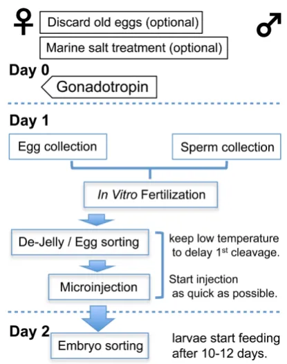
Procedure (diagramed in Fig. 1)
1. Sperm collection(Figure 2 and video “Sperm collection”)
1.1 Transfer some sexually matured male newts (older than 6 months post fertilization) into the anesthetic solution. When the newts are anesthetized, wipe the animals lightly to remove water drops.
1.2 Press lightly on the abdomen of the newts to excrete the urine. When squeezing the sperm strongly with urine in the blad-der, the bladder will break and the newt will die.
1.3 Squeeze the sperm by pressing the abdomen until the sperm seep out of the cloaca. Collect with a pipette and transfer to a microtube containing 300-500 mL of 100% HF. Sperm can be
collected from the same animals every 2–4 weeks.
1.4 Leave the microtube at room temperature for 20-30 min. Then, gently tap the microtube to suspend the sperm. The sperm suspension is slightly cloudy. The sperm will die if mixed vigorously.
1.5 The sperm suspension can be stored for up to 4 hours at room temperature.
Sperm concentration of 2000 sperm/mL is sufficient (Supp. Fig.
3). If the fertilization rate is low, confirm the sperm concentration.
2. Egg collection (Figure 2 and video “Egg collection”)
2.1 (Optional) In order to obtain high quality eggs, keep the female newts (older than 9 months) in an aquarium supplied with 6 g/L of Tetra Marine salt (artificial seawater, provided by Spec-trum Brands Ltd., Atlanta, GA) for more than 2 weeks (Supp. Fig. 4). It may be unnecessary depending on the water quality of the breeding facility.
2.2 (Optional) when the spawning interval is longer than 8 weeks, the female will lay over-maturated eggs. In such a case, discard old
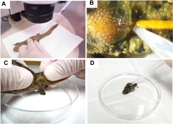
Fig 2. Sperm, egg collection and in vitro insemination. (A) Sperm collection from anesthetized male newts under a
stereomicro-scope. (B) Higher magnification of the cloaca
and sperm sucked up with the micropipette
(also see video “Sperm collection”). (C) Egg
collection from anesthetized female animal. Collect eggs in a dry plastic dish while pushing the abdomen (also see movie “Egg collection”).
(D) The sperm suspension was mixed with the
eggs by administration of 100–200 units of hCG 2 weeks before the injection (Supp. Fig. 4).
2.3 To stimulate ovulation, inject 100–200 units hCG subcuta-neously into the lower jaw of mature females, 14 to 18 hours prior to egg collection. The egg collection should be finished before 24 hours after hCG injection otherwise the quality of the eggs will decline after that period.
2.4 The next day, when the female newts start spawning, anes-thetize the newts in the anesthetic solution. Wipe the newts lightly to remove water drops and then push the abdomens gently and collect the eggs in a dry plastic dish (f100mm). After egg
collec-transfer pipet.
3.5 Soak the fertilized eggs in de-jelly solution for 30-60 sec with gentle shaking and then rinse the eggs 5 times in 25% HF solution. 3.6 Incubate the eggs at 8°C for 20-30 min in 25% HF solution.
During this period, any remaining jelly melts away.
3.7 Sort good quality eggs (Fig. 3 A,B) under a stereomicroscope and transfer them into an injection-dish filled with injection medium.
3.8 Remove the eggshells using fine forceps. The eggs without a shell settle in the well at the bottom (Fig. 3C, video “Eggshell removal”); it helps to hold the eggs during microinjection.
3.9 Store the eggs at 6-8°C until microinjection.
4. Microinjection
4.1 Set up the microinjector and the glass needle filled with injection-solution (DNA, RNA, or proteins) according to the manu-facturer’s protocol. The opening of the glass needle should be 10-30
mm. Adjust the injection volume to between 9-30 nL.
4.2 Take out the injection tray from the incubator and put it under the injector.
4.3 Support gently the egg with the forceps, and prick the animal pole side with the glass needle.
4.4 Inject the injection-solution, and then pull out the needle. 4.5 Leave the injected eggs, and move to the next one. Repeat steps 3-5 until all eggs have been injected. To obtain fully transgenic or full knockout animals, it is important to inject at the single-cell embryo stage. Therefore, the injection is performed as quickly as possible after fertilization.
4.6 Incubate the injected eggs at room temperature overnight. 4.7 (Optional) After injection of DNA, incubate the eggs (em-bryos) at 10–12°C for 4–8 hours to delay the first cleavage, and
then keep at 16°C overnight. This step might facilitate integration
of the injected DNA into the newt genome.
5. Embryo sorting
5.1 Transfer normally developing embryos into petri dishes or plastic cups filled with 25% HF solution using a transfer pipet (Fig. 4 A,B). The criteria for the developing stages were defined by Shi and Boucaut (1996).
5.2 Rinse the embryos 2-3 times in 25% HF solution.
5.3 Incubate the embryos in 25% HF solution for 6–8 days (hatching-stage) and then transfer them into tap water. Start feeding with blain shrimp after 10-12 days post fertilization.
5.4 (Optional) Examination of the expression of the transgene. Expression of a reporter protein such as a fluorescent protein can be confirmed under a fluorescent stereomicroscope.
5.5 Sort intended transgenic or knockout animals by expression of the reporter gene or genotyping.
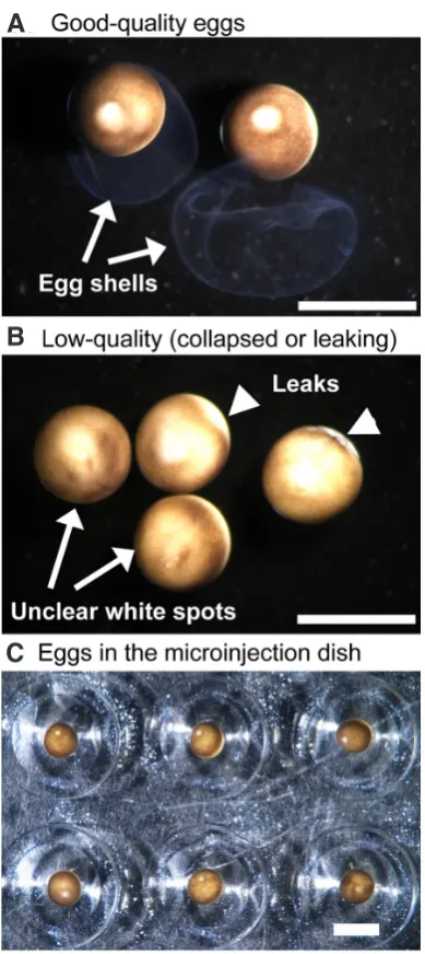
Fig 3. Egg sorting and microinjection. (A) Good quality eggs. The animal pole sides are clearly pigmented, and a white spot on the animal pole was
a sign of good-quality eggs. There was no leak of cytoplasm. (B) On the
other hand, a white spot was blurred or difficult to see on low quality eggs (arrows). These eggs also showed leak of cytoplasm (arrowheads in B).
(C) After eggshell removal, they settled in the well at the bottom of the
injection dish. Scale bars 2 mm.
B
5.6 (Optional) For genotyping, collect the tail tips or limb tips under anesthesia (0.01% ethyl 3-aminobenzoate methanesulfonate is suitable for the larvae).
5.7 Raise these animals to the desired stages. To increase the growth rate of the animals, the optimal temperature range is 24-26°C.
However, they can be maintained in a healthy state at 16-23°C.
Results and Discussion
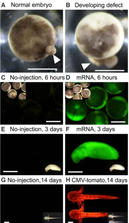
Anticipated results obtained from our injection protocol are shown in Fig. 4. When mRNA is injected, fluorescence can be confirmed after about 6 hours (Fig. 4 C-F). In the case of plasmid DNA, it depends on the enhancer/promoter activity (Fig. 4G and H), but fluorescence can be confirmed at least after 24-36 hours (depend on temperature). Therefore, expression of the transgene was thought to begin after mid-blastula transition.
We examined the conditions of the microinjection method, such as egg preparation (Supp. Fig. 4), de-jelly solution (Supp. Fig. 1), and formulation of injection medium (Supp. Fig. 2). Finally, we have improved the microinjection protocol for a new model animal,
P. waltl newts.
In the new protocol, the survival rate was increased by chang-ing the conditions for the de-jelly treatment (Supp. Fig. 1). Sodium thioglycolate (2% dissolved in 25% HF, 5 min) can be used instead of cysteine solution. After Sodium thioglycolate treatment, the eggshell becomes soft and easier to remove than with cysteine treatment. However, the ratio of normal embryos would decline slightly compared to cysteine solution (0.5% dissolved in 25% HF, 30 sec, see Supp. Fig. 1).
We also applied and optimized the methylcellulose-based injection medium for the P. waltl originally developed for Xenopus (Tanigawa et al., 2009). Since the methylcellulose-injection medium has a low specific gravity and high viscosity, it is easy to perform the injection operation compared to the Ficoll-based injection me-dium. The survival rate in methylcellulose-injection medium was not different from the survival rate in Ficoll-injection medium used in previous protocols (Supp. Fig. 2). In addition, the cost of the methylcellulose-injection medium was 1/10 that of a Ficoll-based medium. Finally, the salt for artificial seawater was a useful option for obtaining high quality eggs (Supp. Fig. 4).
In the present study, we showed the typical results obtained by injection of DNA vectors and mRNA. However, our microinjection protocol can be used for Genome Editing using the CRISPR/Cas9 system (Suzuki et al., 2018).
The P. waltl newt is a very useful laboratory animal for various research fields, such as developmental biology, neurobiology, cancer research, evolution, ethology, toxico-genomics, and regen-eration biology. This microinjection protocol will increase the value of the newts as an experimental model. Furthermore, there is a possibility that this protocol can also be applied to other newts or Urodele amphibians such as axolotls, and Frogs (eg. Xenopus). This protocol will facilitate research through gene manipulation not only in newts but amphibians.
Acknowledgments
We would like to thank Kyorin Corporation (Hyogo, Japan) for kindly providing feed for the newts. This research was supported by MEXT/JSPS KAKENHI (grant numbers, JP16H01254, JP16K08467, and 16H04794).
References
BEETSCHEN J C (1995) The embryonic development of Pleurodeles waltl (Michah). Reappraisal of the Gallien-Durocher stage series. Int J Dev Biol 39: 425-426. BROCKES J P, Kumar A (2002) Plasticity and reprogramming of differentiated cells
in amphibian regeneration. Nat Rev Mol Cell Biol 3: 566-574.
CASCO-ROBLES M, Yamada S, Miura T, Nakamura K, Haynes T, Maki N, DEL
RIO-Fig 4. Embryo sorting and anticipated results. Typical examples of
normally developing embryo, blastula stage (A) and developing defect
(B). The embryo in (B) showed abnormal cleavage. Arrows indicate leaked
cytoplasm. Bars: 1 mm. (C-F) GFP expression in GFP-mRNA (1 ng/egg)
injected embryos (D,F) and un-injected controls (C,E). All embryos were
incubated at 25°C. (C,D) Exposure for 3 seconds, whereas (E,F) were
ex-posed for 300 ms under a green filter. Insets show the bright fields views. Bars: 1 mm. (G,H) RFP expression in CMV driven tdTomato plasmid vector
(110 pg/egg) injected embryos (G) and un-injected controls (H). Photos
HAYASHI T, YOKOTANI N, TANE S, MATSUMOTO A, MYOUGA A, OKAMOTO M, TAKEUCHI T (2013) Molecular genetic system for regenerative studies using newts. Dev Growth Differ 55: 229-236.
GALLIEN L, DUROCHER M (1957) Table chronologique du developpement chez
Pleurodeles waltlii (Michah). Bull Biol Fr Belg 91: 97-114.
INGRAM A J (1972) The lethal and hepatocarcinogenic effects of dimethylnitrosamine injection in the newt Triturus helveticus. Br J Cancer 26: 206-215.
IWAO Y (2012) Egg activation in physiological polyspermy. Reproduction 144: 11-22. MAKITA R, KONDOH H, OKAMOTO M (1995) Transgenesis of newt with exogenous
gene expression facilitated by satellite 2 repeats. Dev Growth and Differ 37: 605-616. SPEMANN H, MANGOLD O (1924) Über Induktion von Embryonalanlagen durch
Implantation artfremder Organisatoren. Arch Mikrosk Anat En 100: 599–638 MATSUNAMI M, SUZUKI M, HARAMOTO Y, FUKUI A, INOUE T, YAMAGUCHI K,
UCHIYAMA I, MORI K, TASHIRO K, ITO Y, TAKEUCHI T, SUZUKI K T, AGATA Kiyokazu A, SHIGENOBU S, HAYASHI (2019) A comprehensive reference tran-scriptome resource for the Iberian ribbed newt Pleurodeles waltl, an emerging model for developmental and regeneration biology. DNA Research, in press. (doi: 10.1093/dnares/dsz003).
regulators. Nature 554: 50-55.
OGINO H, MCCONNELL W B, GRAINGER R M (2006) High-throughput transgenesis in Xenopus using I-SceI meganuclease. Nat Protoc 1: 1703-1710.
OKAMOTO M (1987) Induction of ocular tumor by nickel subsulfide in the Japanese common newt, Cynops pyrrhogaster. Cancer Res 47: 5213-5217.
PICHERAL B (1977) Fertilization in triturus Pleurodeles. Passage through the ovum envelope by spermatozoa. J Ultrastruct Res 60: 106-120.
SHI D L, BOUCAUT J C (1995) The chronological development of the urodele am-phibian Pleurodeles waltl (Michah). Int J Dev Biol 39: 427-441.
SPERRY R W (1943) Effect of 180 degree rotation of the retinal field on visuomotor coordination. J Exp Zool 92: 263-279.
SUZUKIM, HAYASHIT, INOUET, AGATA K, HIRAYAMAM, SUZUKI M, SHIGENOBU S, TAKEUCHI T, YAMAMOTO T, and SUZUKIK T (2018) Cas9 ribonucleoprotein complex allows direct and rapid analysis of coding and noncoding regions of target genes in Pleurodeles waltl development and regeneration. Dev Biol 443: 127-136. TANIGAWA Y, IKENISHI K, and KOMIYA T (2009) A novel method for microinjection
into Xenopus eggs and embryos supported in methylcellulose solution. Int J Dev
What mechanisms drive cell migration and cell interactions in Pleurodeles? J C Boucaut, L Clavilier, T Darribère, M Delarue, J F Riou and D L Shi
Int. J. Dev. Biol. (1996) 40: 675-683
http://www.intjdevbiol.com/web/paper/8877440
Primordial germ cell development: is the urodele pattern closer to mammals than to anurans? M Wakahara
Int. J. Dev. Biol. (1996) 40: 653-659
http://www.intjdevbiol.com/web/paper/8877437
How did urodele embryos come into prominence as a model system? J C Beetschen
Int. J. Dev. Biol. (1996) 40: 629-636
http://www.intjdevbiol.com/web/paper/8877434
What are the key advantages and disadvantages of urodele species compared to anurans as a model system for experimental analysis of early development?
P D Nieuwkoop
Int. J. Dev. Biol. (1996) 40: 617-619
http://www.intjdevbiol.com/web/paper/8877432
The chronological development of the urodele amphibian Pleurodeles waltl (Michah) D L Shi and J C Boucaut
Int. J. Dev. Biol. (1995) 39: 427-441
http://www.intjdevbiol.com/web/paper/7577434
The embryonic development of Pleurodeles waltl (Michah). Reappraisal of the Gallien-Durocher stage series
J C Beetschen
Int. J. Dev. Biol. (1995) 39: 425-426
http://www.intjdevbiol.com/web/paper/7577433
Primordial germ cell proliferation in the salamander Pleurodeles waltl: genetic control before gonadal differentiation
C Dournon, C Demassieux, D Durand and M Lesimple Int. J. Dev. Biol. (1989) 33: 477-485