Paracrine actions of oocytes in the mouse pre-ovulatory follicle
ANTONIETTA SALUSTRI
Department of Public Health and Cell Biology, Faculty of Medicine, University of Rome “Tor Vergata”, Rome, Italy.
ABSTRACT In mammals, ovulation requires a tight control of extracellular matrix modifications, within both the follicle wall and the inner mass of granulosa cells surrounding the oocyte, namely the cumulus cells. During the pre-ovulatory period, mural granulosa cells promote selective degradation of perifollicular matrix, resulting in the formation of a follicle rupture site. Conversely, cumulus cells synthesize a large amount of a muco-elastic matrix that plays an essential role in the extrusion of the oocyte from the follicle and in the subsequent fertilization process. Formation of such matrix by cumulus cells in the pre-ovulatory follicle appears to be controlled by a paracrine influence by the oocyte. We have shown that mouse oocytes modulate the response of cumulus cells to an ovulatory gonadotropin stimulus by promoting the synthesis and preventing the degradation of cumulus matrix. Therefore, although gonadotropins are essential for triggering the complex events involved in ovulation, the oocyte appears to have an active role in this process. In the present review current data and hypotheses concerning molecular mechanisms involved in the organization and synthesis of cumulus matrix are discussed.
KEY WORDS: oocyte, cumulus cells, gonadotropins, ovulation, extracellular matrix.
0214-6282/2000/$20.00
© UBC Press Printed in Spain
www.ehu.es/ijdb
*Address correspondence to: Antonietta Salustri. Dipartimento di Sanità Pubblica e Biologia Cellulare, Università di Roma Tor Vergata, Via Orazio Raimondo, 8, 00173 Roma, Italy. TEL: 39 067 259 6168. FAX: 39 067 259 6172. e-mail: salustri@med.uniroma2.it
Abbreviations used in this paper: COC, cumulus cell-oocyte complex; CS-PG, chondroitin sulfate proteoglycan; dbcAMP, dibutyryl cAMP; FCS, fetal calf serum; FSH, follicle stimulating hormone; hCG, human chorionic gonadotropin; GDF-9, growth differentiation factor-9; HAS2, hyaluronan synthase 2; LH, luteinizing hormone; IαI, inter-alpha-trypsin inhibitor; PαI, pre-alpha-trypsin inhibitor; PMSG, pregnant mare serum gonadotropin; TGF beta1, transforming growth factor beta 1; TSG-6, tumor necrosis factor stimulated gene-6; uPA, urokinase plasminogen activator.
Introduction
In mammalian antral follicles, granulosa cells surrounding the oocyte, namely cumulus cells, and granulosa cells lining the follicle wall, referred to as mural granulosa cells, activate divergent func-tions in response to gonadotropin stimuli. Under follicle stimulating hormone (FSH) action, mural granulosa cells synthesize increas-ing amounts of luteinizincreas-ing hormone (LH) receptors and steroid hormones, while cumulus cells show a limited increase of such functions (Zoller and Weisz, 1979; Lawrence et al., 1980; Channing et al., 1981; Oxberry and Greenwald, 1985; Zlotkin et al., 1986; Ishimura et al., 1989). These two cell populations also differ in growth factor expression: higher mRNA levels of insulin-like growth factor (Oliver et al., 1989), vascular endothelial growth factor (Dissen et al., 1994), inhibin beta A (Braw-Tal, 1994) and mullerian inhibiting substance (Ueno et al, 1989) are reached in cumulus cells in comparison to mural granulosa cells, while the opposite occurs for the c-kit ligand (Manova et al, 1993; Motro and Bernstein 1993). Following an endogenous LH-FSH surge or administration of an ovulatory dose of hCG, mural granulosa cells promote selective degradation of peri-follicular matrix bringing to the formation of a rupture site in the follicle wall. Conversely, cumulus cells synthesize an abundant muco-elastic extracellular matrix promoting detach-ment of cumulus cell-oocyte complex (COC) from the follicle wall and a 20-40 fold increase in the volume of the cumulus mass, a process termed cumulus expansion or mucification (Tsafriri and
Reich, 1999). At ovulation, the expanded COC leaves the follicle through the ruptured wall, while mural granulosa cells remain in the follicle anchored to the basement membrane.
Cumulus cells provide the proper environment for successful oocyte maturation and fertilization. Soluble factors, synthesized and secreted by cumulus cells, improve meiotic progression, fertilizability of oocytes and embryo development (Vanderhyden and Armstromg, 1989; Goud et al., 1998; De La Fuente and Eppig, 1999). The muco-elastic extracellular matrix appears to be essen-tial for oocyte extrusion from the follicle and for pick-up by the fimbria, since when its synthesis is prevented or altered, a signifi-cant lower number of oocytes are released from follicles and in addition, they are not transferred into the oviduct (Chen et al., 1993; Hess et al., 1999; Hizaki et al., 1999). In addition, a function of selective barrier for sperm has been also advanced for this matrix (Carrell et al., 1993; Bedford and Kim, 1993).
producing progesterone required for successful implantation of the embryo and maintenance of the pregnancy. Thus, the acquisition of divergent metabolic activities by the two follicle sub-populations is functional to their different role in follicle development, essen-tially steroidogenic for mural granulosa cells, and oocyte nursing for cumulus cells. Any alteration in the differentiation process of one or the other cell type may compromise fertility. Relative position of cumulus cells and mural granulosa cells within the follicle is essential in determining their response to gonadotropins. Evidence has been accumulating that contact with the basement membrane and soluble products secreted by theca cells, such as androgens and growth factors, co-operate with gonadotropins to promote mural granulosa cell phenotype (Adashi, 1994). In addi-tion, the identification of some components of the expanded extracellular matrix and the study of the regulation of their synthe-sis in mouse COC has highlighted the essential role of oocytes in modulating cumulus cells’ response to gonadotropins and in deter-mining their functional pathway (Salustri et al., 1990b; Buccione et al., 1990). This research field is rapidly evolving and the list of cumulus cell activities affected by the oocyte is lengthening. Here we will focus on the progress of knowledge about the regulation of cumulus expansion which has allowed us to propose a model of the molecular mechanisms involved in the organization and synthesis of cumulus matrix during the pre-ovulatory period.
Structure and molecular components of expanded
cu-mulus matrix
Cumulus expansion depends on synthesis and secretion of hyaluronan by cumulus cells (Fig. 1). Hyaluronan is a glycosaminoglycan with exceptionally high molecular weight (up to a few million Dalton) and large hydrodynamic domains which is synthesized at the plasma membrane, with the elongating chain being extruded into the extracellular space. Its ability to attract
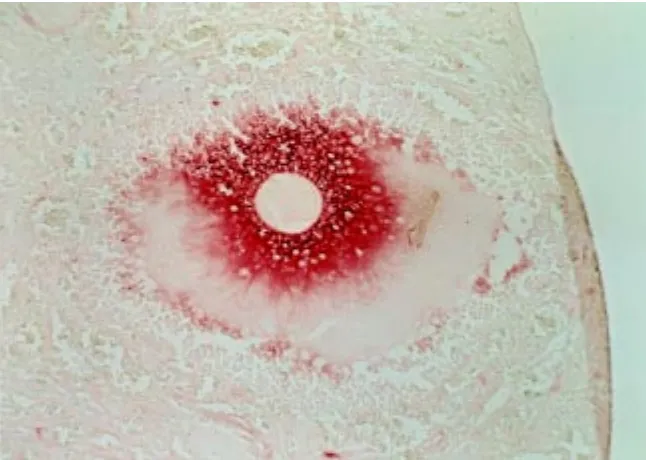
water is most likely responsible for the hydration of COC extracellular matrix and the increase of intercellular space among cumulus cells. When this matrix is analyzed by electron microscopy, the hyaluronan strands appear to be organized into twisted or coiled fibrils which interconnect to each other to form a homogeneous mesh-like network (Yudin et al., 1988). Protease treatment causes dissociation of the fibrils into individual hyaluronan filaments and produces loss of COC integrity (Cherr et al., 1990). Therefore, proteins are required to organize and maintain hyaluronan in such a highly structured gel, conferring on the COC matrix unique physical properties. For in-stance, expanded COCs are only temporarily deformed when subjected to shearing forces. Such elasticity and stability of the matrix may play an essential role in ovulation by promoting the protrusion of the COC and protecting the oocyte from mechanical stress. The hyaluronan-protein fibrillar network is attached on cumulus cell face and penetrates into the zona pellucida sur-rounding the oocyte (Yudin et al., 1988). Most likely these interactions allow cumulus cells and oocyte to remain firmly bound within the matrix, so that they are not dispersed during extrusion. Fig. 1. Localization of hyaluronan in cumulus matrix. The image shows a peri-ovulatory
follicle from a hamster ovarian section stained with biotinylated hyaluronan binding proteins followed by avidin-peroxidase (Salustri et al., 1992).
Evidence obtained in vivo and in vitro suggests that proteins both derived from the serum and synthesized by cumulus cells participate in organizing hyaluronan in the matrix. Mouse COCs in vitro treated with FSH in the presence of fetal calf serum (FCS) undergo expansion (Eppig, 1979). However, when COCs are stimulated with FSH in the absence of serum the elastic-expanded matrix is not formed and cumulus cells lose contact from each other and settle individually on the bottom of the culture dish. In such culture condition, cumulus cells synthesize hyaluronan but they fail to organize it in the intercellular space so that most of hyaluronan diffuses into the culture medium (Eppig, 1980a). The absence of serum does not affect the size of the synthesized hyaluronan suggesting that the observed phenomenon is not due to changes in the physiological properties of this polymer (Salustri et al., 1989). If serum is added at the beginning of culture and removed while COC expansion is ongoing, hyaluronan synthe-sized before the removal is retained in the matrix while that synthesized afterwards is released into the medium (Camaioni et al., 1993). This finding indicates that serum factors must be present while hyaluronan is being synthesized in order to organize it into a matrix. Factors with such functional ability have been isolated from the serum and identified as inter-alpha-trypsin inhibi-tor (IαI) and a related protein pre-alpha-trypsin inhibitor (PαI). Each of these molecules can substitute for serum in retaining hyaluronan among cumulus cells, whereas serum depleted of them is inactive (Chen et al., 1992). Co-localization in vivo of Iα I-related molecules and hyaluronan in the pre-ovulatory follicle suggests their physiological role. It has been proposed that these serum macromolecules can rapidly enter the follicle after a gona-dotropin ovulatory stimulus increases the permeability of the selective blood-follicle barrier (Chen et al., 1992).
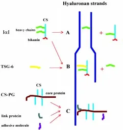
about 16 kDa molecular weight (MW), named bikunin or light chain, and to one (PαI) or two (IαI) additional peptides of about 80 kDa MW, referred to as heavy chains (Salier et al., 1996). The two heavy chains of IαI and the single heavy chain of PαI are encoded by three different genes sharing a high degree of homology. These peptides show the ability both to bind to hyaluronan through stretches rich in positively charged amino acid residues and to covalently link to hyaluronan via displacement of the chondroitin sulfate chain (Chen et al., 1994, 1996). Thus, these interactions can lead to cross bridges between adjacent hyaluronan strands thereby stabilizing cumulus matrix (Chen et al., 1996) (Fig. 2A). Alternatively or in addition, IαI could act as an anti-protease within cumulus matrix. The bikunin peptide of IαI and PαI consists of two tandemly arranged domains, each with proteinase inhibitor activity (Wachter and Hochstrasser, 1979). Its anti-proteolytic activity is increased by tumor necrosis factor-stimulated gene-6 (TSG-6), a protein which can form a stable complex with IαI by displacing a heavy chain (Wisniewski et al., 1994, 1996) (Fig. 2B). The expres-sion of TSG-6 gene is up regulated in cumulus cells during COC expansion and high levels of this messenger are maintained throughout the pre-ovulatory period (Fulop et al., 1997a). Interest-ingly, TSG–6 can also bind to hyaluronan through a highly specific binding site (Lee et al., 1992). Therefore, TSG-6 could activate and specifically localize the anti-protease bikunin on hyaluronan strands
elongating at the cumulus cell membrane, thereby tightly protect-ing from degradation hyaluronan-associated proteins involved in matrix organization. Indeed, proteolytic activity increases in the follicular fluid during the pre-ovulatory period due to the increased production of proteases by mural granulosa cells (Tsafriri and Reich, 1999).
Cumulus cells synthesize two additional hyaluronan binding molecules which are retained within the matrix: a chondroitin sulfate proteoglycan (CS-PG) with a molecular weight larger than 106 Dalton (Camaioni et al., 1996), and the link protein, a
glycopro-tein able to interact with both hyaluronan and a proteoglycan, thus stabilizing their linkage (Kobayashi et al., 1999) (Fig. 2C). Inhibition of chondroitin sulfate chain elongation by b-xyloside treatment apparently does not affect cumulus expansion, implying that the negatively charged glycosaminoglycan chain of large CS-PG do not significantly contribute to the swelling of the matrix (Salustri et al. 1989). The large CS-PG in the COC matrix is more likely involved in matrix organization. For its structural characteristics and immunological affinity it can be considered a member of proteoglycan versican family (Camaioni et al., 1996; Eriksen et al., 1999). The core protein of this type of proteoglycan has a functional N-terminal hyaluronan-binding domain and a C-terminal lectin-binding domain, through which it can bind adhesive molecules (Aspberg et al., 1995). Tenascin C and other adhesive proteins have been found in the extracellular matrix of COC (Familiari et al., 1996) and versican-like PG may serve as cross-linking agent between such molecules and the hyaluronan scaffold (Fig. 2C).
Role of gonadotropins and oocyte in cumulus
expan-sion
When large antral follicles are punctured under a dissecting microscope before gonadotropin surge, cumulus cells remain closely associated with the oocyte, and intact cumulus cell-oocyte complexes (COCs) can be isolated and induced to expand in vitro. In all examined species, gonadotropins are potent inducers of COC expansion in vitro, although cumulus cells are more sensitive to follicle stimulating hormone (FSH) than to luteinizing hormone (LH) or human chorionic gonadotropin (hCG) (Magnusson et al., 1982; Ball et al., 1982; Singh et al., 1993; Armstrong et al., 1996). In fact, highly purified LH and hCG fail to induce expansion of isolated mouse COCs (Eppig, 1979). These findings are consistent with the low or undetectable levels of LH receptors in the cumulus cells in the pre-ovulatory follicle.
The study of hyaluronan synthesis by mouse COCs stimulated to expand in vitro by FSH has allowed molecular mechanisms involved in cumulus expansion to be studied in detail. Hyaluronan synthesis is first detected 2-3 hours after FSH stimulation, in-creases to a maximum rate sustained between 4-10 hours, and then declines and ceases by 12-18 hours, when maximum expan-sion has been reached (Salustri et al., 1989). Several lines of evidence suggest that FSH exerts its influence on hyaluronan synthesis via cAMP. Follicle stimulating hormone stimulates cAMP production by cumulus cells, and dibutyryl cAMP (dbcAMP), a membrane-permeable cAMP analog, shows a potency and tempo-ral pattern of hyaluronan synthesis induction similar to that ob-served with FSH (Tirone et al., 1997). Interestingly, a level of 14 fmol of cAMP/COC has to be reached to obtain maximum hyaluronan synthesis by in vitro FSH-stimulated COCs, and a similar cAMP level is achieved by COCs in vivo 2-4 h after hCG injection into Fig. 2. Models for interaction between hyaluronan, IαI, TSG-6 and
versican-like CS-PG.(A) The heavy chain of IαI can be covalently linked to a hyaluronan through a reaction in which hyaluronan displaces the CS chain of IαI. This polypeptide, tightly linked to hyaluronan can interact with another hyaluronan strand by positive charge residues leading to the formation of a cross-bridge. (B) TSG-6 displaces a heavy chain of IαI forming a stable complex with IαI. TSG-6 increases the anti-proteolytic activity of IαI and it binds to hyaluronan through a specific binding site. (C)
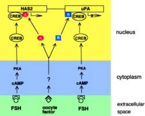
PMSG-primed mice, a time at which hyaluronan synthesis is initiated. Since, as discussed above, mouse cumulus cells do not appear to have LH/hCG receptors, the hCG-dependent cAMP elevation observed in cumulus cells in vivo is likely mediated by mural granulosa either by direct transfer of cAMP via gap junctions or by the synthesis or activation of a molecule that can activate adenylate cyclase in cumulus cells (Schultz et al., 1983; Eppig, 1980b, 1981; Downs and Longo, 1982; Salustri et al., 1985). Based on in vitro results, it is reasonable to hypothesize a primary role for cAMP in regulating hyaluronan synthesis and in promoting cumu-lus expansion in vivo.
Regulation of hyaluronan synthesis in cumulus cells is primarily controlled at the level of hyaluronan synthase-2 (HAS2) gene expression (Fulop et al., 1997b). Hyaluronan synthase-2 mRNA is at very low levels in the compact COCs isolated from PMSG-primed mice, but reaches high levels 2-3 hours after hCG injection into the animals. By the time hyaluronan synthesis ceases, HAS2 mRNA returns to basal levels. Therefore, it is likely that a cAMP dependent protein kinase promotes the activation of a cAMP responsive element binding protein acting as a transcription factor for HAS2 gene.
Although the oocyte does not synthesize hyaluronan, it has an essential role in cumulus expansion in the mouse. When the oocyte is mechanically removed from the COC, isolated cumulus cells exposed to FSH synthesize low levels of hyaluronan, and do not form an expanded matrix (Salustri et al., 1990b; Buccione et al., 1990). A high level of hyaluronan synthesis is restored in FSH-stimulated cumulus cells by adding back either oocytes or oocyte-conditioned medium to the cultures. Therefore, a soluble factor secreted by the oocyte is required for FSH to exert its action on cumulus cells. We have investigated at which intracellular level the oocyte factor synergizes with FSH to induce hyaluronan synthesis. An oocyte influence on FSH binding to its receptor and/or on the generation of an intracellular signal has been excluded by the evidence that equal amounts of cAMP are generated by
hormone-stimulated cumulus cells cultured in the presence or in the absence of oocytes (Buccione et al., 1990) and that dbcAMP fails to increase hyaluronan synthesis by cumulus cells in the absence of oocytes (Salustri et al., 1990b). FSH and the oocyte factor differ in their temporal patterns of action on hyaluronan synthesis (Tirone et al., 1997). Mouse COCs produce maximal hyaluronan synthesis if they are exposed to FSH for only 2 hours, indicating that hormone action is completed within this time. Conversely, the presence of the oocyte is not required during this initial inductive phase, because nearly identical results were obtained when the oocytes were either added with FSH at the beginning of culture or 2 h later. However, continuous presence of the oocyte is required from 2 hours onwards to promote and sustain maximal hyaluronan syn-thesis. Therefore, it is likely that the oocyte factor(s) does not act on FSH signal transduction. The level of HAS2 mRNA significantly increases in cumulus cells stimulated in vitro by FSH in the presence of oocytes while it only slightly increases in cumulus cells cultured with either oocytes or FSH alone, implying that the oocyte factor synergistically acts with FSH to induce HAS2 gene expres-sion (Fulop, Salustri and Hascall, unpublished results). Notewor-thy, if the oocytes are removed from FSH-stimulated COCs at 6 h of culture, when maximum HAS2 transcription and HA synthesis are achieved, hyaluronan synthesis ceases in 2-3 h (Tirone et al., 1997). The same result is obtained if transcription inhibitor actino-mycin D is added at 6 h to FSH-stimulated COC cultures. There-fore, it is likely that removal of the oocyte rapidly down regulates HAS2 transcription and that continuous presence of oocyte factor is required to sustain the expression of this gene.
As reported above, cumulus cells synthesize hyaluronan-bind-ing proteins that maybe are required for matrix assembly and stability. At the present it is not known if the oocyte can influence the synthesis of such proteins by cumulus cells. However, the oocyte inhibits gonadotropin-induced protease synthesis by cumu-lus cells, thereby contributing to protect matrix protein components (Canipari et al., 1995). Cumulus cells show significantly lower levels of urokinase plasminogen activator (uPA) mRNA than mural granulosa cells in mice injected with an ovulatory dose of gonadotropins. Such difference is not observed when isolated cumulus cells and mural granulosa cells are stimulated in vitro with FSH. However, when cumulus cells are cultured with oocytes or oocyte conditioned medium, significantly lower levels of uPA mRNA are achieved under FSH stimulus, mimicking what occurs in vivo. Thus, up-regulation of uPA expression by hormones is suppressed by a soluble oocyte factor and differences in uPA mRNA levels observed in vivo between cumulus and mural granu-losa cells are due to the inhibitory influence of the oocyte.
Based on the similarities in physicochemical characteristics and in pattern of secretion during oogenesis between the oocyte factor regulating hyaluronan synthesis and that regulating uPA synthesis (Vanderhyden et al., 1990; Eppig et al., 1993a, 1993b; Canipari et al., 1995), we have advanced the hypothesis that both activities could be controlled by a single factor produced by the oocyte (Canipari et al., 1995). The results are consistent with the following model: FSH activates a cAMP-responsive element binding (CREB) protein which acts as a transcription factor for both HAS2 and uPA gene. The oocyte factor operates through a separate receptor and signaling pathways to activate other proteins which regulate the activity of the cAMP-dependent transcription factor, positively on HAS2 and negatively on uPA gene (Fig. 3).
Mechanisms limiting oocyte factor action in the
pre-ovulatory follicle
Mural granulosa cells isolated from pre-ovulatory follicles as-sume cumulus cell phenotype when cultured in vitro in the pres-ence of oocytes. For instance, in this condition they increase hyaluronan (Salustri et al., 1990a) and decrease uPA (Canipari et al., 1995) synthesis, as cumulus cells do. In addition, it has been observed that the synthesis of LH receptor mRNA is suppressed in these cells as well as in cumulus cells by a factor secreted by oocytes (Eppig et al., 1997). However, some differences can be observed, such as mural granulosa cells do not require the gona-dotropin stimulus to respond to the oocyte in terms of hyaluronan synthesis (Salustri et al., 1990a). This may depend on the ability of mural granulosa cells to synthesize molecules, such as prostag-landin E and/or epidermal growth factor, which can substitute for FSH and act in an autocrine way synergizing with the oocyte factor (Salustri et al., 1985; Salustri et al., 1990a). Whatever the mecha-nism, results show that even though mural granulosa cells show functional specificity, still they can be modulated in their function by the oocyte. So, how do mural granulosa cells escape from the oocyte control in the follicle? Most likely this goal is accomplished by limited diffusion of oocyte factors. This hypothesis is supported by the evidence that media conditioned by FSH-stimulated COCs, in which the oocyte is enclosed within the cumulus cell mass, have only 10- 20% of the hyaluronan stimulatory activity of media conditioned by an equal number of isolated oocytes when tested in mural granulosa cell cultures. Furthermore, the activity of medium conditioned by isolated oocytes is reduced to ∼30% by pre-incubation with isolated cumulus cells (Salustri et al., 1992). These results suggest that cumulus cells can bind and inactivate the factor, a process that would create a decreasing concentration gradient with increasing distance from the oocyte. In agreement with this hypothesis, the innermost layers of granulosa cells lining the antrum, that are closest to the COC, show an intermediate cell phenotype between cumulus and mural granulosa cells (Salustri et al., 1992) (Fig. 1). In conclusion, limited diffusion of oocyte factor may have an essential role in ensuring that an appropriate number of granulosa cells differentiate and remain within the follicle to form with theca cells a corpus luteum suitable to support embryo implantation.
Identification of oocyte factor
Mural granulosa cells are more abundant and easier to isolate than cumulus cells. For this reason, mural granulosa cell cultures have been used to test a series of known growth factors for ability to stimulate hyaluronan synthesis, as the oocyte factor does. Of growth factors studied, only transforming growth factor (TGF) beta1 resulted to induce hyaluronan synthesis and expansion by FSH-stimulated cumulus cells (Salustri et al., 1990a; 1993). How-ever, neutralizing antibodies against TGF beta1 do not inhibit the oocyte factor activity, indicating that they are different (Salustri et al., 1990a). On the other hand, their effects are addictive at suboptimal doses, and they show identical temporal pattern of induction of hyaluronan synthesis (Tirone et al., 1997). Thus, we have proposed that the oocyte factor could be a member of the TGF beta family, different from TFG beta1 but triggering similar intrac-ellular signals. This hypothesis has been recently confirmed. Recombinant mouse growth differentiation factor-9 (GDF-9)
pro-tein, a member of TGF beta family exclusively expressed by oocytes in mouse ovary (McGrath et al., 1995), is able to increase HAS2 mRNA by mural granulosa cells and to induce cumulus expansion (Elvin et al., 1999). This growth factor also shows the ability to mimic the oocyte factor in decreasing uPA and LH receptor mRNA levels in mural granulosa cells. The receptor and signal transduction pathways utilized by GDF-9 have not been studied yet. However, similarity of action between oocyte condi-tioned medium and TGFbeta1 suggests that GDF-9 could bind to serine/threonine kinase receptors and transduce signals through SMAD proteins, as TGF beta family members do. Following TGF beta binding to receptor, SMADs are phosphorylated and translocaled to the nucleus. These proteins can access target genes by synergistically binding to DNA with cofactors (such as cAMP responsive element binding protein) and can induce or inhibit gene expression by recruiting co-activators or co-repressors (Massague and Chen, 2000). If GDF-9 is signaling through this intracellular pathway in FSH-stimulated cumulus cells, interaction of SMADs with different molecules acting at HAS2, uPA and LH-receptor gene level could explain the opposite effects of this growth factor on the expression of these cAMP-dependent genes. GDF-9 expression has been also demonstrated in rat, ovine, bovine and human oocytes (Jaatinen et al., 1999; Bodensteiner et al., 1999; Aaltonen et al., 1999). In agreement with these findings, it has been shown that porcine, rat and bovine oocytes can substitute for mouse oocytes in inducing cumulus expansion. However, cumulus cells isolated from these species, in contrast with those from the mouse, can be stimulated to expand in vitro by FSH treatment in the absence of oocytes (Prochazka et al., 1991; Singh et al., 1993; Vanderhyden et al., 1993; Ralph et al., 1995). It should be noted , however, that if rat cumulus cell are pre-cultured for 5-6 h in the absence of oocytes, they can be induced to expand thereafter only if oocytes are added to the culture together with FSH (Vanderhyden, 1993). Thus, it is likely that species differences between cumulus cells are related to the duration of GDF9 activity or the stability of cumulus cell response to this factor.
Acknowledgements
I wish to express my sincere thanks to Prof. Gregorio Siracusa, Dr. Vincent C. Hascall and Dr. Antonella Camaioni for a fruitful collaboration over many years. This work was supported by MURST grant for National Project “Development and Differentiation of Germ Cells” and by CNR grant n.98.00512.CT04.
References
AALTONEN, J., LAITINEN, M.P., VUOJOLAINEN, K., JAATINEN, R., HORELLI-KUITUNEN, N., SEPPA, L., LOUHIO, H., TUURI, T., SJOBERG, J., BUTZOW, R., HOVATA, O., DALE, L. and RITVOS, O. (1999). Human growth differentiation factor 9 (GDF-9) and its novel homolog GDF-9B are expressed in oocytes during early folliculogenesis. J. Clin. Endocr. Metab. 84: 2744-2750.
ADASHI, E. (1994). Endocrinology of the ovary. Hum. Reprod. 9: 815-827.
ARMSTRONG, D.T., XIA, P., DE GANNES, G., TEKPETEY, F.R. and KHAMSI, F. (1996). Differential effects of insulin-like growth factor-1 and follicle-stimulating hormone on proliferation and differentiation of bovine cumulus cells and granulosa cells. Biol. Reprod. 54: 331-338.
ASPBERG, A., BINKERT, C. and ROUSLAHTI, E. (1995). The versican C-type lectin domain recognizes the adhesion protein tenascin-R. Proc. Natl. Acad. Sci. USA 92: 10590-10594.
BEDFORD, J. and KIM, H. (1993). Cumulus oophorus as a sperm sequestering device in vivo. J. Exp. Zool. 265: 321-328.
BODENSTEINER, K.J., CLAY, C.M., MOELLER, C.L. and SAWYER, H.R. (1999). Molecular cloning of the ovine Growth/Differentiation factor-9 gene and expres-sion of growth/differentiation factor-9 in ovine and bovine ovaries. Biol. Reprod. 60: 381-386.
BRAW-TAL, R. (1994). Expression of mRNA for follistatin and inhibin/activin subunits during follicular growth and atresia. J. Mol. Endocrinol. 13: 253-264.
BUCCIONE, R., VANDERHYDEN, B.C., CARON, P.J. and EPPIG, J.J. (1990). FSH-induced expansion of the mouse cumulus oophorus in vitro is dependent upon a specific factor(s) secreted by the oocyte. Dev. Biol. 138:16-25.
CAMAIONI, A., HASCALL, V.C., YANAGISHITA, M. and SALUSTRI, A. (1993). Effect of exogenous hyaluronic acid and serum on matrix organization and stability in the mouse cumulus cell-oocyte complex. J. Biol. Chem. 268: 20473-20481.
CAMAIONI, A., SALUSTRI, A., YANAGISHITA, M. and HASCALL V.C. (1996). Proteoglycans and proteins in the extracellular matrix of mouse cumulus cell-oocyte complexes. Arch. Biochem. Biophys. 325: 190-198.
CANIPARI, R., EPIFANO, O., SIRACUSA, G. and SALUSTRI, A. (1995). Mouse oocytes inhibit plasminogen activator production by ovarian cumulus and granu-losa cells. Dev. Biol. 167: 371-378.
CARRELL, D., MIDDLETON, R., PETERSON, C., JONES, K. and URRY, R. (1993). Role of the cumulus in the selection of morphologically normal sperm and induction of the acrosome reaction during human in vitro fertilization. Arch. Andrology 31: 133-137.
CHANNING, C.P., BAE, I.H, STONE, S.L., ANDERSON, L.D., EDELSON, S. and FOWLER, S.C. (1981). Porcine granulosa and cumulus cell properties. LH/hCG receptors, ability to secrete progesterone and ability to respond to LH. Mol. Cell. Endocrinol. 22: 359-70.
CHEN, L., MAO, S.J., MCLEAN, L.R., POWERS, R.W. and LARSEN, W.J. (1994). Proteins of the inter-alpha-trypsin inhibitor family stabilize the cumulus extracel-lular matrix through their direct binding with hyaluronic acid. J. Biol. Chem. 269: 28282-28287.
CHEN, L., RUSSELL, P.T. and LARSEN, W.J. (1993). Functional significance of cumulus expansion in the mouse: roles for the preovulatory synthesis of hyalu-ronic acid within the cumulus mass. Mol. Reprod. Dev. 34: 87-93.
CHEN, L., ZHANG, H., POWERS, R.W., RUSSELL, P.T. and LARSEN, W.J. (1996). Covalent linkage between proteins of the inter-alpha-inhibitor family and hyalu-ronic acid is mediated by a factor produced by granulosa cells. J. Biol. Chem. 271: 19409-19414
CHEN, L.C., MAO, S.J.T. and LARSEN, W.L. (1992). Identification of a factor in fetal bovine serum that stabilizes the cumulus extracellular matrix. J. Biol. Chem. 267: 12380-12386.
CHERR, G.N., YUDIN, A.I. and KATZ, D.F. (1990). Organization of the hamster cumulus extracellular matrix: a hyaluronate-glycoprotein gel, which modulates sperm access to the oocyte. Dev. Growth. Differ. 32: 353-65.
DE LA FUENTE, R., O’BRIEN, M.J. and EPPIG, J.J. (1999). Epidermal growth factor enhances preimplantation development competence of maturing mouse oocytes. Hum. Reprod. 14: 3060-3068.
DISSEN, G.A., LARA, H.E., FAHRENBACH, W.H., COSTA, M.E. and OJEDA, S.R. (1994). Immature rat ovaries become revascularized rapidly after autotrasplantation and show a gonadotropin-dependent increase in angiogenic factor gene expres-sion. Endocrinology 134: 1146-1154.
DOWNS, S.M. and LONGO, F.J. (1982). Effects of indomethacin on preovulatory follicles in immature, superovulated mice. Am. J Anat. 164: 265-274.
ELVIN, J.A., AMANDER, T.C., WANG, P., WOLFMAN N.M. and MATZUK M.M. (1999). Paracrine actions of growth differentiation factor-9 in the mammalian ovary. Mol. Endocrinol.13: 1035-48.
EPPIG, J.J. (1979). FSH stimulates hyaluronic acid synthesis by oocyte-cumulus cell complexes from mouse preovulatory follicles. Nature 281:483-484.
EPPIG, J.J. (1980a). Role of serum in FSH stimulated cumulus expansion by mouse oocyte-cumulus cell complexes in vitro. Biol.Reprod.22: 629-633.
EPPIG, J.J. (1980b). Regulation of cumulus oophorus expansion by gonadotropins in vivo and in vitro. Biol. Reprod. 23:545-552.
EPPIG, JJ. (1981). Prostaglandin E2 stimulates cumulus expansion and hyaluronic acid synthesis by cumuli oophori isolated from mice. Biol. Reprod. 25:191-195.
EPPIG, J.J., PETERS, A.H.F.M., TELFER, E. and WIGGLESWORTH, K. (1993a).
Production of cumulus expansion enabling factor by mouse oocytes grown in vitro: preliminary characterization of the factor. Mol. Reprod. Dev. 34: 450-456.
EPPIG, J.J., WIGGLESWORTH, K. and CHESNEL, F. (1993b). Secretion of cumulus expansion enabling factor by mouse oocytes: relationship to oocyte growth and competence to resume meiosis. Dev. Biol. 158: 400-409.
EPPIG, J.J., WIGGLESWORTH, K., PENDOLA, F. and HIRAO, Y. (1997). Murine oocytes suppress expression of luteinizing hormone receptor messenger ribo-nucleic acid by granulosa cells. Biol. Reprod. 56: 976-984.
ERIKSEN, G.V., CARLSTEDT, I., MORGELIN, M., ULDBJERG, N. and MALMSTROM, A. (1999). Isolation and characterization of proteoglycans from human follicular fluid. Biochem. J. 340: 613-620.
FAMILIARI, G., VERLENGIA, C., NOTTOLA, S.A., RENDA, T., MICARA, G., ARAGONA, C., ZARDI, L. and MOTTA (1996). Heterogeneous distribution of fibronectin, tenascin-C, and laminin immunoreactive material in the cumulus-corona cells surrounding mature human oocytes from IVF-ET protocols—evi-dence that they are composed of different subpopulations: an immunohistochemi-cal study using scanning confoimmunohistochemi-cal laser and fluorescence microscopy. Mol. Reprod. Dev. 43: 392-402.
FULOP, C., KAMATH, R.V., LI, Y., OTTO, J.M., SALUSTRI, A., OLSEN, B.R., GLANT, T.T. and HASCALL, V.C. (1997a). Coding sequence, exon-intron struc-ture and chromosomal localization of murine TNF-stimulated gene 6 that is specifically expressed by expanding cumulus cell-oocyte complexes. Gene 202: 95-102.
FULOP, C., SALUSTRI, A. and HASCALL, V.C. (1997b). Coding sequence of a hyaluronan synthase homologue expressed during expansion of the mouse cumulus-oocyte complex. Arch. Biochem. Biophys. 337: 261-266.
GOUD, P.T., GOUD, A.P., LAVERGE, H., VAN DER ELST, J., DE SUTTER, P. and DHONT, M. (1998). In-vitro maturation of human germinal vesicle stage oocytes: role of cumulus cells and epidermal growth factor in the culture medium. Hum. Reprod. 13: 1638-1644.
HESS, K.A., CHEN, L. and LARSEN, W.J. (1999). Inter-α-inhibitor binding to hyaluronan in the cumulus extracellular matrix is required for optimal ovulation and develop-ment of mouse oocytes. Biol. Reprod. 61: 436-443.
HIZAKI, H., SEGI, E., SUGIMOTO, Y., HIROSE, M., SAJI, T., USHIKUBI F.,MATSUOKA, T., NODA, Y., TANAKA, T., YOSHIDA, N., NARUMIYA, S. and ICHIKAWA, A. (1999). Abortive expansion of cumulus and impaired fertility in mice lacking the prostaglandin E receptor subtype EP(2). Proc. Natl. Acad. Sci. USA 31: 10501-10506.
ISHIMURA, K., YOSHINAGUA-HIRABAYASHI, T., TSURI, H., FUJITA, A. and OSAWA, Y. (1989). Further immunocytochemical study on the localization of aromatase in the ovary of rats and mice. Hystochemistry 90: 413-416.
JAATINEN, R., LAITINEN, MP, VUOJOLAINEN, K., AALTONEN, J., HEIKINHEIMO, K., LEHTONEN, E. and RITVOS, O. (1999). Localization of growth differentiation factor-9 (9) mRNA and protein in rat ovaries and cDNA cloning of rat GDF-9 and its novel homolog GDF-GDF-9B. Mol. Cell Endocrinol. 156: 18GDF-9-1GDF-93.
KOBAYASHI, H., SUN, G.W., HIRASHIMA, Y. and TERAO, T. (1999). Identification of link protein during follicle development and cumulus cell cultures in rats. Endocrinology 140: 3835-3842.
LAWRENCE, T.S., DEKEL, N. and BEERS, W.H. (1980). Binding of human chorionic gonadotropin by rat cumuli oophori and granulosa cells: a comparative study. Endocrinology 106: 1114-8.
LEE, T.H., WISNIEWSKI, H.G. and VILCEK, J. (1992). A novel secretory tumor necrosis factor-inducible protein (TSG-6) is a member of the family of hyaluronate binding proteins, closely related to the adhesion receptor CD44. J. Cell Biol.116: 545-57.
MAGNUSSON, C., BILLING, H., ENEROTH, P., ROOS, P. and HILLENSJO, X. (1982). Comparison between the progestin secretion responsiveness to gonadot-ropins of rat cumulus and mural granulosa cells in vitro. Acta Endocrinol. 101: 611-616.
MANOVA, K., HUANG, E.J., ANGELES, M., DE LEON, V., SANCHEZ, S., PRONOVOST, S.M., BESMER, P. and BACHVAROVA, R.F. (1993). The expres-sion pattern of the c-kit ligand in gonads of mice supports a role for the c-kit receptor in oocyte growth and in proliferation of spermatogonia. Dev. Biol. 157: 85-99.
MASSAGUE, J. and CHEN, YG (2000). Controlling TGF-beta signaling. Genes Dev. 14: 627-644.
MOTRO, B. and BERNSTEIN, A. (1993). Dynamic changes in ovarian c-kit and steel expression during the estrous reproductive cycle. Dev. Dynam. 197: 69-79.
OLIVER, J.E., AITMAN, T.J., POWELL, J.F., WILSON, C.A. and CLAYTON, R.N. (1989). Insulin-like growth factor I gene expression in the rat ovary is confined to the granulosa cells of the developing follicles. Endocrinology 124: 2671-2679.
OXBERRY, B.A. and GREENWALD, G.S. (1982). An autoradiographic study of the binding of 125I-labeled follicle stimulating hormone, human chorionic gonadotropin,
and prolactin to the hamster ovary throughout the estrous cycle. Biol. Reprod. 27: 505-516.
PROCHAZKA, E., NAGYOVA, E., RIMKEVIOVA, T., NAGAI, T., KIKUCHI, K. and MOTLIK, J. (1991). Lack of effect of oocytectomy on expansion of the porcine cumulus. J. Reprod. Fertil. 93:569-576.
RALPH, J.H., TELFER, E.E. and WILMUT, I. (1995). Bovine cumulus cell expansion does not depend on the presence of an oocyte secreted factor. Mol. Reprod. Dev. 42: 248-253.
SALIER, J.P., ROUET, P., RAGUENEZ, G. and DAVEAU, M. (1996). The inter-alpha-inhibitor family: from structure to regulation. Biochem. J. 315: 1-9.
SALUSTRI, A., HASCALL, V.C., CAMAIONI, A. and YANAGISHITA, M. (1993). Oocyte-granulosa cell interactions. In The Ovary, (Eds. E.Y. Adashi and P.C.K. Leung). Raven Press Ltd., New York, pp. 209-225.
SALUSTRI, A., PETRUNGARO, S. and SIRACUSA, G. (1985). Granulosa cells stimulate in vitro the expansion of isolated mouse cumuli oophori: involvement of prostaglandin E2. Biol. Reprod. 33: 229-234.
SALUSTRI, A., ULISSE, S., YANAGISHITA, M. and HASCALL, V.C. (1990 a). Hyaluronic acid synthesis by mural granulosa cells and cumulus cells in vitro is selectively stimulated by a factor produced by oocytes and by transforming growth factor-beta. J. Biol. Chem. 265:19517-19523.
SALUSTRI, A., YANAGISHITA, M. and HASCALL, V.C. (1990 b). Mouse oocytes regulate hyaluronic acid synthesis and mucification by FSH-stimulated cumulus cells. Dev. Biol. 38: 26-32.
SALUSTRI, A., YANAGISHITA, M. and HASCALL, V.C. (1989). Synthesis and accu-mulation of hyaluronic acid and proteoglycans in the mouse cumulus cell-oocyte complex during follicle stimulating hormone-induced mucification. J. Biol. Chem. 264: 13840-13847.
SALUSTRI, A., YANAGISHITA, M., UNDERHILL, C., LAURENT, T.C. and HASCALL, V.C. (1992). Localization and synthesis of hyaluronic acid in the cumulus cells and mural granulosa cells of the preovulatory follicles. Dev. Biol. 151: 541-51.
SCHULTZ, R.M., MONTGOMERY, R.P. and BELANOFF, J.R. (1983). Regulation of mouse oocyte meiotic maturation: implication of a decrease in oocyte cAMP and protein dephosphorylation in commitment to resume meiosis. Dev. Biol. 97: 264-273.
SINGH, B., ZHANG, X. and ARMSTRONG, D.T. (1993). Porcine oocytes release cumulus expansion-enabling activity even though porcine cumulus expansion in
vitro is independent of the oocyte. Endocrinology 132: 1860-1862.
TIRONE, E., D’ALESSANDRIS, C., HASCALL, V.C., SIRACUSA, G. and SALUSTRI, A. (1997). Hyaluronan synthesis by mouse cumulus cells is regulated by interac-tions between follicle-stimulating hormone (or epidermal growth factor) and a soluble oocyte factor (or transforming growth factor beta 1). J. Biol. Chem. 272: 4787-94.
TSAFRIRI, A. and REICH, R. (1999). Molecular aspects of mammalian ovulation. Exp. Clin. Endocrinol. Diabetes 107: 1-11.
UENO, S., TAKAHASHI, M., MANGANARO, T.F., RAGIN, R.C. and DONAHOE, P.K. (1989). Cellular localization of mullerian inhibiting substance in the developing rat ovary. Endocrinology 124: 1000-1006.
VANDERHYDEN, B.C. (1993). Species differences in the regulation of cumulus expansion by an oocyte-secreted factor(s). J. Reprod. Fertil. 98: 219-227.
VANDERHYDEN, B.C. and ARMSTRONG, D.T. (1989). Role of cumulus cells and serum on the in vitro maturation, fertilization, and subsequent development of rat oocytes. Biol. Reprod. 40: 720-728.
VANDERHYDEN, B.C., CARON, P.J., BUCCIONE, R. and EPPIG, J.J. (1990). Developmental pattern of the secretion of cumulus-expansion enabling factor by mouse oocytes and the role of oocytes in promoting granulosa cell differentiation. Dev. Biol. 140: 307-317.
WACHTER, E. and HOCHSTRASSER, K. (1979). Kunitz-type proteinase inhibitors derived by limited proteolysis of the inter-alpha-trypsin inhibitor, III. Sequence of the two Kunitz-type domains inside the native inter-alpha-trypsin inhibitor, its biological aspects and also of its cleavage products. Hoppe. Seylers. Z. Physiol. Chem. 360: 1305-1311.
WISNIEWSKI, H.G., BURGESS, W.H., OPPENHEIM, J.D. and VILCEK, J. (1994). TSG-6, an arthritis-associated hyaluronan binding protein, form a stable complex with the serum protein inter-alpha-trypsin inhibitor. Biochem. 33: 7423-7429.
WISNIEWSKI, H.G., HUA, J.C., POPPERS, D.M., NAIME, D., VILCEK, J. and CRONSTEIN, B.N. (1996). TNF/IL-1 inducible protein TSG-6 potentiates plasmin inhibition by inter-alpha-inhibitor and exerts a strong anti-inflammatory effect in vivo. J. Immunol. 156: 1609-1615.
YUDIN, A.I., CHERR, G.N. and KATZ, D.F. (1988). Structure of the cumulus matrix and zona pellucida in the golden hamster: a new view of sperm interaction with oocyte-associated extracellular matrix. Cell Tissue Res. 251: 555-564.
ZLOTKIN, T., FARKAS, Y. and ORLY, J. (1986). Cell-specific expression if immunore-active cholesterol side-chain cleavage cytochrome P-450 during follicular develop-ment in the rat ovary. Endocrinology 119: 2809-2820.
ZOLLER, L.C. and WEISZ, J. (1979). A quantitative cytochemical study of glucose-6-phosphate dehydrogenase and ∆5-3β-hydroxysteroid dehydrogenase activity in the