Original Article
Cell-matrix interactions and cell-cell junctions during epithelial
histo-morphogenesis in the developing mouse incisor
SANDRINE KIEFFER-COMBEAU, JEAN-MARIE MEYER and HERVÉ LESOT*
INSERM U424, Institut de Biologie Médicale, Faculté de Médecine, Strasbourg, France
ABSTRACT The continuously growing rodentincisor develops mainly along its antero-posterior axis. The labio-lingual asymmetry which characterizes this tooth is initiated at the cap stage and increases furtherduring the cap to bell transition (ED14 to ED16) when histogenesis of the enamel organ proceeds. Histology, transmission electron microscopy (TEM), and immunostaining were used to document the changes in the basement membrane (BM) as well as the modifications of epithelial cell-matrix and cell-cell interactions during this period. The expression of plakoglobin, desmoglein and E-cadherin at ED14 suggested that the main cell-cell junctional complexes were adherens junctions. The expression of desmoglein and TEM observationssuggested a progressive antero-posterior stabilization of the enamel organ by means of desmosomes from ED14 to ED18.
α6 integrin, BP 230 and laminin γ2 chain were all expressed in the developing incisor but were not always co-distributed. Immunostaining and TEM suggested that only primitive type II hemidesmosomes were present. At ED14, cells of the enamel knot (EK) did not show any specific expression for antigens involved in cell-cell interaction. However, strong staining for the laminin γ2 chain characterized the BM in contact with EK cells. The BM in the labial part of the cervical loop demonstrated ultrastructural changes: the presence of loops of the lamina densa in this region preceeded the differential expression of the integrin α6 subunit and that of the laminin γ2 chain in the labial/lingual parts of the cervical loop. Apoptosis was transiently observed in the contiguous mesenchyme. Thisaffected osteoblasts and also nerve cells close to the labial part of the cervical loop.
KEY WORDS:
histo-morphogenesis, desmosome, adherens junction, basement membrane, incisor
0214-6282/2001/$25.00
© UBC Press Printed in Spain
www.ijdb.ehu.es
*Address correspondence to: Dr. Hervé Lesot. INSERM U424, Institut de Biologie Médicale, Faculté de Médecine, 11 rue Humann, 67085 Strasbourg Cedex, France. Fax: +33-390-243-564. e-mail: [email protected]
Abbreviations used in this paper: 3D, three dimensional; BM, basement mem-brane; ED, embryonic day; EK, enamel knot; TEM, transmission electron microscopy
Introduction
The mouse incisor is characterized by continuous growth and a labio-lingual asymmetry where the labial aspect represents the crown-analogue of the tooth and the lingual part, the root-analogue portion. The morphogenesis of the mouse lower incisor has previ-ously been investigated using 3D reconstructions and histological observations. From the cap stage (ED14), the enamel organ grows rapidly and histogenesis of the enamel organ is initiated. The outer dental epithelium (ODE), the inner dental epithelium (IDE) and the stellate reticulum start to differentiate at this stage whereas the stratum intermedium only appears at the bell stage (ED16) on the labial part (Hay, 1961 ; Kieffer et al., 1999).
Cell-matrix interactions and cell-cell junctional complexes play major roles during organogenesis, histo-morphogenesis, and in the regulation of cell migration and proliferation (for review see Gumbiner, 1996; Hata, 1996; Huang and Ingber, 1999). Epithelial cell junctions consist of adherens junctions and desmosomes. Adherens junctions, connecting actin filaments to the plasma membrane, are mediated by
behavior (Kowalczyk et al., 1999). Desmosomes contain two sub-types of the transmembrane glycoprotein superfamily of cadherins: desmogleins and desmocollins. These desmosomal cadherins as-sociate with two additional proteins: plakophilins and desmoplakins.
Desmoplakins, plakoglobin (γ-catenin) and plakophilins reside in the desmosomal zone between the plasma membrane and intermediate filaments (Burdett, 1998).
Hemidesmosomes are specialized junctional complexes con-necting epithelial cells to the basement membrane. Ultrastructur-ally, hemidesmosomes appear as electron-dense structures con-sisting of an inner and outer plaque as well as a sub-basal plate. The inner plaque is composed of the HD1/plectin and BP230 proteins, which are involved in connecting the hemidesmosome to the keratin intermediate filament system. The outer plaque contains transmembrane proteins: α6β4 integrin and BP 180. The α6β4 integrin mediates interactions with the underlying basement membrane by binding laminin-5 (Nievers et al., 1999). Moreover, α6β4 integrin is not only involved in cell adhesion and hemidesmosomes assembly; it may also be implicated in the transduction of signals that modulate or regulate cell proliferation, differentiation, apoptosis and cell migration (Giancotti et al., 1996). The intermediate filaments which terminate in desmo-somes and hemidesmodesmo-somes consist of cytokeratins. Human tooth germs express several cytokeratins (CK) including CK7, 13, 14 and 19 (Kasper et al., 1989; Domingues et al., 2000).
In this paper, we used transmission electron microscopy and indirect immunofluorescence to investigate the changes in cell-cell and cell-matrix junctions that occur when the incisor develops from the cap to bell stage, as already documented in the molar (Palacios et al., 1995; Salmivirta et al., 1996; Fausser et al., 1998; Obara et al., 1998).
Results
3D reconstructions: localization of the EK and apoptosis
At ED14 the lower incisor tooth germ had reached the cap stage (Fig. 1A). The EK was located on the lateral side of the enamel organ close to the tip of the incisor (Fig. 1 A-D). The diameter of the EK gradually decreased antero-posteriorly (Fig. 1 C,D). At ED15, the incisor had extended posteriorly but still remained at the cap stage: wide open on its medial side (Fig. 1 E,F). The EK decreased dramatically in length (Fig. 1 E,G).
In the enamel organ at ED14, apoptosis was restricted to the most anterior part, in the stalk (Fig. 1C) and no apoptosis was observed in the EK (Fig. 1 C,D). At ED15, there was still no apoptosis associated with the EK (Fig. 1 G,H).
Apoptosis was observed in the mesenchyme posteriorly to the labial part of the cervical loop at ED14 (Fig. 1A). At ED15, apoptosis decreased considerably in this region of the mesenchyme, when compared to ED14 (compare Fig. 1E with 1A).
Histology
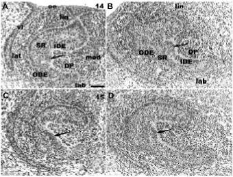
On histological sections at ED14, the EK was formed by con-densed cells and protruded towards the mesenchyme (Fig. 2 A,B). At ED15 the cells of the EK were much less condensed (Fig. 2 C,D) than at ED14 (Fig. 2 A,B).
At ED14, apoptosis in the mesenchyme posterior to the labial part of the CL was observed close to osteoblasts accumulating extracellular matrix (Fig. 3 A,B).
Electron microscopy
Apoptotic figures close to the labial part of the cervical loop could not be characterized from histological sections (Fig. 3A). Trans-Fig. 1. 3D reconstructions of epithelium and enamel knot (EK) of the
lower incisor at ED14 (A-D) and ED15 (E-H). From ED14 to ED15, the EK (represented in green) remains located very close to the tip of the incisor
(A,E), on the lateral side of the tooth (B,F). Red spots represent distribution of metaphases in the epithelium (C,D,G,H). Apoptosis in the epithelium is represented as white spots (C,D,G,H) and as yellow spots when in the mesenchyme (A,E). an, anterior; po, posterior; lab, labial; lin, lingual; lat, lateral; med, medial; oe, oral epithelium. Bar, 100 µm.
A
B
C
D
E
F
mission electron microscopy of this part of the mesenchyme showed that apoptotic figures were observed in close vicinity to nervous cells (Fig. 3 C,D). Degenerate mitochondriae were present in cells very close to nerve fibers (Fig. 3 D,E,F), and also in nerve cells themselves (Fig. 3G).
At ED14, the cells of the EK were rather small, condensed, and in most cells, the nucleus remained distant from the basement membrane (Fig. 4 A,B). These cells thus had a specific shape with a cytoplasmic extension coming in contact with the basement membrane. The number of mitochondriae in EK cells was very high (Fig. 4 A,B,C).
In most parts of the incisor, the basement membrane appeared as a classical lamina densa separated from the apical pole of epithelial cells by the lamina lucida (Fig. 4 D,E). However, the basement membrane in contact with the cells of the EK at ED14 appeared much thicker (compare Fig. 4C with 4D). Indeed, this resulted from an increase in the fibrillar network associated with the lamina densa, towards the lamina fibroreticularis (Fig. 4 B,C). The normal thickness of the plasma membrane of cells in this region indicated that the increase in thickness of the basement membrane was not caused by tangential sectioning.
Loops of lamina densa were observed in association with the epithelial cells in the labial part of the cervical loop at ED14 (Fig. 5A). Similar loops of lamina densa were still observed at ED15 (Fig. 5C) and ED16 (Fig. 5E). At all these stages, the loops remained strictly limited to the labial part of the cervical loop as can be seen from comparison with the lingual side at corresponding stages (Fig. 5 B,D,F).
Immunostaining
Intermediate filaments
At ED14, most of the enamel organ was positive for cytokeratin 14 (CK14) except for some cells in the labial part of the cervical loop
(Fig. 6 A,C). However at ED16 (Fig. 6B) and at ED18 (Fig. 7A), all epithelial cells expressed the antigen.
Antigens associated with cell-cell junctions
At ED14, the vestibular lamina and the oral epithelium reacted strongly with anti-desmoglein antibodies, but the dental epithelium remained unstained (Fig. 6D). At ED16, the expression of desmoglein by dental epithelial cells increased strongly and the antigen was intensely expressed by cells of the stratum intermedium on the labial side of the incisor (Fig. 6 E and F). On the lingual side, epithelial cells in contact with the IDE were also positive. The staining for the IDE itself remained very weak (Fig. 6 E,F). At ED18, the dental epithelial cells in contact with the BM expressed strongly desmoglein on the labial and lingual sides of the tooth (Fig. 7D). At this stage, the staining was not restricted to the anterior part of the tooth as at ED16 but extended very much posteriorly (compare Fig. 7D to Fig. 6 E,F). The lingual part of the CL was positive (Fig. 7F), whereas the labial part was negative (Fig. 7E).
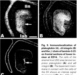
Anti-plakoglobin antibodies stained the dental epithelium at ED14, although much less in the region of the cervical loop than in the more anterior part of the incisor (Fig. 6G). On frontal sections, the EK area expressed plakoglobin although EK cells in contact with the basement membrane were less intensely labelled (Fig. 9A). At the early bell stage, (ED16), the stratum intermedium was intensily stained for plakoglobin (Fig. 6 H, I) and the labial part of the cervical loop showed less staining than the lingual part (Fig. 6H). At ED18, anti-plakoglobin antibodies strongly stained the stratum intermedium and the epithelial cells in contact with the lingual IDE (Fig. 7 G,H). Plakoglobin was not detected in the cells of the CL (Fig. 7 G,I).
The expression of E-cadherin was homogeneous in all the dental epithelium at ED14 (Fig. 6J), and decreased in the labial portion of the cervical loop at ED16 (Fig. 6 K,L) and at ED18 (Fig. 7 J-L).
Fig. 2.Histological sections of the lower inci-sor at ED14 (A,B) and ED15 (C,D). Frontal (A)
and parasagittal (B) sections in the enamel knot (EK) area at ED14. The arrows indicate the con-densed cells of the inner dental epithelium (IDE) forming the EK on the lateral side of the incisor. Frontal section (C) and sagittal section (D) at ED15 in the EK area. The EK at ED15 is less protruding in the dental papilla than at ED14 (A,B). oe, oral epithelium; DP, dental papilla; ODE, outer dental epithelium; SR, stellate reticulum; lin, lin-gual; lab, labial; lat, lateral; med, medial; vl, vesti-bular lamina. Bar, 50 µm.
A
B
Antigens involved in cell-matrix interactions
At ED14, in the enamel organ, α6 integrin was not homoge-neously distributed : the staining of cells in the stalk and that of the prospective ODE were less intense (Figs. 8A, 9B). The dental mesenchyme also reacted with antibodies to α6 integrin (Figs. 8A, 9B). The use of frontal sections showed that the antigen was intensely expressed outside the EK (Fig. 9B). At ED16, the staining
for the integrin α6 remained diffuse in cells of the dental epithelium. The staining was less intense in the labial than in the lingual part of the cervical loop (Fig. 8 B,C). At ED16 and ED18, the staining started to be restricted to cells in contact with the basement membrane in the labial part of the incisor. At ED18, the lingual part was negative for α6 integrin (Fig. 7 M,O). In the dental papilla, staining for integrin α6 was no longer visible at ED16 and ED18 (Fig. 7 M-O and Fig. 8 B,C), except for blood vessels which remained positive at ED16 (Fig. 8 B,C).
Immunostaining for the laminin γ2 chain demon-strated that at ED14 the antigen was present in the basement membrane in contact with the ODE and IDE (Fig. 8D). The staining of the basement mem-brane was weaker in the lingual part of the cervical loop (Fig. 8D). In the labial part of the cervical loop, all epithelial cells expressed the γ2 chain of laminin (Fig. 8D). The staining of the BM for the γ2 chain of laminin was more intense in contact with the EK than in other regions (Fig. 9C). At the early bell stage and still at ED18, the staining for the laminin γ2 chain tended to decrease in the basement membrane associated with the IDE except for the most anterior part of the incisor (Fig. 7P and Fig. 8E). At ED16, the labial part of the cervical loop weakly expressed the γ2 chain whereas no staining was visible on the lingual side (Fig. 8 E,F). At ED18, the γ2 chain of laminin was detected in the BM in contact with the ODE but only on the labial side of the incisor (Fig. 7 P,Q). The lingual part of the tooth as well as the labial part of the CL were negative (Fig. 7 P-R).
Immunostaining for BP230 showed that at ED14 the cells of the dental epithelium expressed the antigen except for the IDE (Fig. 8 G,I). At ED16, the same pattern of expression was observed: the IDE remained unstained (Fig. 8H). At the cap and bell stage, staining for BP230 was much more intense in the oral epithelium or the vestibulum than in the dental epithelium and this difference increased with time (Fig. 8 G,H). At ED18, the staining for BP230 was strong in the anterior part and in the lingual portion of the tooth (Fig. 7 S,U) whereas staining in the labial part remained very weak and negative in the region of the cervical loop (Fig. 7 S,T).
Discussion
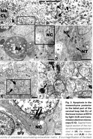
The cap stage in the mouse incisor lasts for about 36 hours. During this period, the enamel organ remains open on the medial side of the tooth. At the same time, the incisor elongates posteriorly, histogenesis Fig. 3. Apoptosis in the
mesenchyme posterior to the labial part of the cervical loop (lab CL) of the lower incisor at ED14 by light (A,B) and trans-mission electron micros-copy (C-G). Sagittal histo-logical section of the inci-sor showing apoptosis (ar-row) in (A) the mesen-chyme, and (A,B) in the vicinity of osteoblasts accumulating extracellular matrix; B is a magnification of the black area boxed in A. Bar, 50 µm.The mesenchyme area delimited by dotted line in A was observed by (C-G) transmission electron microscopy. (C) An apoptotic body(Ap) is detected in the vicinity of nervous cells (NC) and capillary (cap). Bar, 2 µm. (D) Neurofilaments (NF) and neurotubules (NT) are visible in the nervous cell. D is a magnification of the area boxed in C). Bar, 2 µm. In this zone of the mesenchyme, (E,F) degenerative mitochondriae (arrows) are visible among a nervous plexus; F is a magnification of the area boxed in E). Bar, 1 µm.
(G) Mitochondriae showing degeneration (arrow) as well as glycogen patches (gly) are also detected in cells adjacent to the cytoplasmic process of a nervous cell (NC). Bar, 1 µm.
of the enamel organ progresses in the same direction and the EK disappears. The cap to bell transition then occurs within about 12 hours (Kieffer et al., 1999; Miard et al., 1999). From ED14 to ED18, all the epithelial cells in contact with the basement membrane in the cervical loop area divide, mediating the posterior growth of the incisor. The labial and lingual parts of the cervical loop extend at the same rate, but the fate of the cells on each side is different: only the
A
B
C
D
E
F
cells of the IDE on the labial side of the incisor will give rise to functional ameloblasts (Smith & Warshawsky 1975). Cell-cell and cell-matrix interactions are involved in the histo-morphogenesis of the enamel organ, and in the regulation of cell migration and proliferation. Immunohistological approaches were combined with 3D-reconstructions, histology and transmission electron micros-copy to study the localization of desmosomes, adherens junctions and hemidesmosomes in the mouse lower incisor from the cap to bell stages.
Anterior part of the incisor
Histogenesis of the enamel organ is initiated in the anterior part of the developing incisor at ED14. The IDE and ODE as well as the stellate reticulum started to differentiate (Kieffer et al., 1999). Although only very faint staining for desmoglein was observed at ED14, two days later, the antigen became strongly expressed by the cells of the stratum intermedium on the labial side and by cells in contact with the IDE on the lingual side. This expression of desmoglein at ED16 is in agreement with TEM observations showing the presence of desmosomes in the stratum intermedium (not shown) and with the histological observations showing that the stratum intermedium, visible at ED16 was not yet present at ED14 (Kieffer et al., 1999). Sasaki et al., (1984) also showed that cells of the stratum intermedium were connected to each other and to the stellate reticulum cells and ameloblasts by desmosomes in human teeth. This suggested that the stratum intermedium might be very
important in stabilizing the differentiating labial IDE. However, similar cell-cell junctions also exist in epithelial cells in contact with the IDE on the lingual part of the developing incisor where the stratum intermedium does not differentiate. The stabilizing role of the epithelial cells in contact with the IDE should thus be correlated with histogenesis of the enamel organ and not the cytodifferentia-tion of ameloblasts. From ED16 to ED18, the expression of desmoglein progressed posteriorly suggesting a stabilization of this region by desmosomes.
Adherens junctions were visualized using antibodies to E-cadherin and desmosomes with antibodies to desmoglein. Plakoglobin is present in both the adherens junctions and desmo-somes. Comparison of the three patterns of expression in the tooth at ED14 suggested that the main cell-cell junctional complexes were adherens junctions. E-cadherin has been proposed to play a key role in the formation of pre-adherens-junctions (Vasioukhin et al., 2000).
To analyze epithelial cell-matrix interactions, three antigens have been immunolocalized: the γ2 chain of laminin-5, α6 integrin and the BP230. Laminin-5, a component of the basal lamina, is a heterotrimeric protein composed of α3, β3 and γ2 subunits. The expression of laminin-5 subunits during embryonic mouse tooth development has been well documented (Salmivirta et al., 1997; Fig. 4. Transmission electron micrographs of the lower incisor at
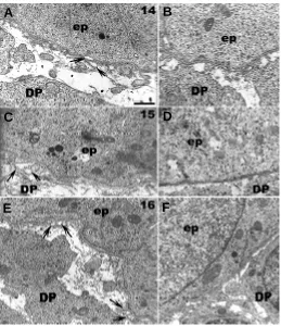
ED14.(A) A semi-thin transverse section through the enamel knot (EK) shows the position of the nuclei of the EK epithelial cells distantfrom the basement membrane. Dense granules (arrows) accumulate in these cells facing the dental papilla (DP). Bar, 10µm. (B) An ultrathin section in the same region shows that these granules correspond to mitochondriae. Bar, 5 µm. (B,C) Fibrillar material is condensed under the basal lamina (bl) in contact with cells of the EK. Bar, 1 µm. At a distance from the EK, the aspect of the basement membrane in contact with cells of the (D) inner dental epithelium (IDE) and of the (E) outer dental epithelium (ODE) is different: fibrillar material is no longer associated with the lamina densa. Bar, 1 µm. ep, epithelium; PDM, peridental mesenchyme.
Fig. 5. Ultrastructural aspects of the epithelio-mesenchymal junction in the labial (A,C,E) and the lingual part (B,D,F) of the cervical loop at ED14 (A,B), ED15 (C,D) and ED16 (E,F). The basement membrane (lamina fibroreticularis) shows more fibrillar material on the labial (A,C,E) than on the lingual (B,D,F) side of the cervical loop. On the labial side the lamina densa is duplicated and makes loops (arrows), which become more frequent from␣ ED14 to ED16 (A,C,E). On the lingual side, no loops are apparent (B,D,F). Bar, 1µm. DP, dental papilla; ep, epithelium.
A
B
C
D
E
A
B
C
D
Yoshiba et al., 1998a, 1998b, 2000). Laminin-5 is involved in the anchorage/motility of epithelial cells through integrins α6β4, α6β1 and α3β1 (Delwel and Sonnenberg, 1996). The BP230 was used as a marker of hemidesmosomes (Nievers et al., 1999). The γ2 subunit of laminin-5, the α6 chain of integrin as well as the BP230 were all expressed in the developing incisor but did not always co-distribute. For example, at ED14 the α6 subunit of integrin had a rather ubiquitous distribution in the enamel organ while the γ2 chain of laminin was restricted to the BM in the anterior part of the incisor (i.e. where the histogenesis was more advanced). At this stage, most of the epithelial cells in contact with the dental papilla remained negative after staining for BP230. Furthermore, observa-tions by transmission electron microscopy did not allow detection of hemidesmosomes. Such an apparent discrepancy and the weak staining for BP230 have already been described in the developing molar where it was suggested that only primitive type II hemidesmosomes may be present (Fausser et al., 1998). In other models such as lens cell differentiation, α6β4 integrin is also expressed but in the absence of hemidesmosomes (Walker and Menko, 1999).
From ED14 to ED18 in the incisor, changes occurred in the distribution of BP230, α6 integrin, and the laminin γ2 chain. The staining for BP230 decreased and appeared very weak when compared to the staining in the vestibular lamina or in the oral epithelium. At ED18, the staining for the γ2 chain of laminin-5 became restricted to the BM and the BM in contact with the cells of the ODE was more heavily stained. In the developing molar, the
temporo-spatial expression of laminin-5 subunits appeared to be differentially controlled by the dental/peri-dental mesenchyme (Yoshiba et al., 1998a) so a similar situation may be expected in the incisor.
Enamel knot
Another aspect of the histo-morphogenesis of the enamel organ is the formation and disappearance of the EK. From ED14 to ED15, the EK is transiently detected in the antero-lateral part of the incisor. It progressively disappears although apoptosis is not involved and probably occurs as a result of histological reorganization (Kieffer et al., 1999). Immunostaining for antigens associated with desmosomes or adherens junctions was similar for EK cells when compared to neighbouring epithelial cells. However, the BM in contact with the cells of the EK demonstrated a specific ultrastructural appearance. Using transmission elec-tron microscopy, we observed an increase in the fibrillar network associated to the lamina densa, towards the lamina fibroreticularis underlying EK cells in the incisor. Very similar observations have been made when looking at the basement membrane in contact with EK cells in the molar (Lesot et al., 1999). In this region, the lamina densa itself did not change, either in the incisor, or in the molar. However, the BM underlying the EK cells was intensely stained by antibodies to the γ2 chain of laminin-5 at ED14 in the incisor but not in the molar (Yoshiba et al., 1998a). The proteolytic processing of laminin-5 by matrix metalloproteinases (MPPs) has been suggested to influence cell migration as well as Fig. 6. Immunolocalization of cytokeratin 14 (CK 14) (A-C), desmoglein (D-F), plakoglobin (G-I) and E-cadherin (J-L) on sagittal sections of the lower incisor at ED14 (A,C,D,G,J) and at ED16 (B,E,F,H,I,K,L). At ED14, the enamel organ shows ubiquitous staining for CK14 (A), plakoglobin (G)
hemidesmosome formation (Gianelli et al., 1997; Goldfinger et al., 1998). In addition to providing stable adhesion laminin-5, by means of proteolytic cleavage, could also serve as a motility factor (Kikkawa et al., 1996). Furthermore, the EK cells in the molar expressed less α6 integrin than other epithelial cells (Salmivirta et al., 1996), which was not the case in the incisor. These differences in EK cell-basement membrane interactions in the inci-sor and molar might thus have to be correlated with the different ability of these cells to migrate in the two teeth. Indeed, EK cells were shown to migrate and segregate in the molar but not in the incisor (Coin et al., 1999; 2000).
Posterior part of the incisor
The labio-lingual asymmetry in the cervical loop was already visible at ED14 after staining for CK14 or the γ2 chain of laminin-5. CK14 was present in most cells of the enamel organ at ED14 except for some cells on the labial part of the cervical loop. All epithelial cells of the labial part of the cervical loop still expressed the γ2 chain of laminin-5 although the antigen became restricted to the BM in all other parts of the developing incisor. This transition char-acterizes the maturation of epithelial tissues and has already been reported for the molar (Yoshiba et al., 1998a; 2000). This maturation is not achieved in the incisor at birth (Yoshiba et al., 1998b).
At ED16, the staining for α6 integrin was stronger in the lingual part of the cervical loop than in the labial part where it tended to concentrate at the pole of cells in contact with the BM. The very low level of expres-sion of α6 integrin and the absence of BP230 in the labial part of the cervical loop confirmed that in this part of the tooth, the cell-matrix interactions appeared to be weaker compared to the IDE in more anterior part of the incisor. The α6β4 integrin might be impli-cated in the transduction of signals that modulate or regulate cell proliferation and migration (Giancotti et al., 1996). Such a potential role and the differential expression of the molecule in the lingual and labial portions of the cervical loop would have to be taken into account to better understand the different cell behaviour in the two regions. From the cap to bell stage, loops of the lamina densa in contact with the labial part of the cervical loop were observed by transmission electron microscopy as originally de-scribed at ED16 (Meyer et al., 1995). These loops of the lamina densa on the labial part of the cervical loop were already present at ED14, before the differential expression of the α6 integrin subunit. This suggests that other yet unidentified molecules should show earlier asymmetrical changes in their expression during incisor development.
In the labial and lingual parts of the cervical loop, the absence of desmoglein and the very faint
growth of the embryonic incisor. Previous investigations on the developing molar also showed very poor staining of the cervical loop area until the stratum intermedium differentiated in this Fig. 7. Immunolocalization of cytokeratin 14 (CK14) (A-C), desmoglein (D-F), plakoglobin (G-I), E-cadherin (J-L), α6 integrin (M-O), γ2 chain of laminin-5 (P-R) and BP230 (S-U) on sagittal sections of lower incisor at ED18. All cells of the enamel organ express cytokeratin 14 (A-C) and E-cadherin (J-L). However the staining for CK14 decreases in the inner dental epithelium (IDE) (A) and part of the cervical loop (B) on the labial side. This region of the cervical loop also shows less staining for E-cadherin (J,K). The staining for desmoglein (D-F) and plakoglobin (G-I) is detected in the epithelial cells in contact with the IDE (D,F,G,H) except in the most posterior part of the labial cervical loop (E,G).α6 integrin is expressed only in the labial side of the incisor (M-O). The γ2 chain of laminin-5 is present in the basement membrane in contact with the outer dental epithelium in the anterior and labial parts of the tooth (P-R). The basement membrane in contact with the IDE is negative for the γ2 chain of laminin-5 except in the labial anterior region (P). BP230 is strongly expressed in the anterior and in the lingual regions of the incisor (S-U).lin, lingual; lab, labial. Bar, 100 µm.
region at ED 19 (Fausser et al., 1998). At ED16, the IDE differen-tiated but the staining for antigens associated with adherens junctions (plakoglobin and E-cadherin) was much weaker in the labial part of the cervical loop. In this region, the cellular dynamics are expected to be very high: the incorporation of BrdU was much greater in the labial portion of the cervical loop than in the lingual one (Coin et al., 2000). This compartment is also supposed to contain stem cells allowing the continuous growth of the incisor in
rodents (Smith and Warshawsky, 1975; 1976). These cells would then not only have specific abilities to proliferate and to migrate, but also have different developmental potentialities.
Close to the labial part of the cervical loop, but in the mesen-chyme, apoptotic cells and bodies accumulated at ED13,5-14 (Kieffer et al., 1999; Miard et al., 1999). Apoptosis in this area tended to disappear from ED15 and thus was not related to the posterior growth of the incisor, which had only started. These apoptotic figures were located outside the dental sac, but were too close to the incisor to be related to the disappearance of potential dental mesenchymal cells from the diastema. Ultrastructural observations showed that these apoptotic figures were indeed very closely or even directly associated with nerve cells. During development, immature neuronal cells disappear by apoptosis. This could result from competition for limited access to neu-rotrophic factors and involve active signalling through death receptors (Raoul et al., 2000). Nerve growth factor (NGF) and its low affinity p75 neurotrophin receptor (p75NTR) are involved in death signalling (Casaccia-Bonnefil et al., 1998; 1999; Barrett, 2000). NGF and p75NTR are expressed by the developing incisor (Mitsiadis et al., 1993). However, young stages have not been investigated. Thus, the potential role of such factors in the elimination of nerve cells posterior to the labial part of the incisor at ED14 remains unclear. Experimental approaches have dem-onstrated that neurturin, another neurotrophic factor, neither stimulates cell proliferation nor prevents apoptotic cell death in isolated dental mesenchyme (Luuko et al., 1998). These authors suggested that FGF4 might prevent apoptosis in the dental mesenchyme. However, the expression of this growth factor and the related receptors in the region posterior to the developing lower incisor from ED13 to ED16 has not been investigated yet. If involved, the antagonist effects of FGF4/BMPs would more prob-ably play a role in controlling apoptosis (Buckland et al., 1998).
Fig. 8. Immunolocalization of α6 integrin (A-C), γ2 chain of laminin-5 (D-F) and BP230 (G-I) on sagittal sections of lower incisor at ED14 (A, D, G, I) and at ED16 (B, C, E, H).
From ED14 (A) to ED16 (B,C) [C is a magni-fication of B], the staining for α6 integrin decreases in the mesenchyme and in the epithelial cells of the labial part of the cervical loop. The staining for laminin 5 is less intense from ED14 (D) to ED16 (E,F).The basement membrane in contact with cells of the inner dental epithelium (IDE) on the labial and lin-gual sides is no more stained for laminin 5 at ED16 (E,F) [F is a magnification of E]. From ED14 to ED16, the cells of the IDE never express BP230 (G,H,I) [I is a magnification of G]. lin, lingual; lab, labial; oe, oral epithelium; vl, vestibular lamina. Bar, 100 µm.
Materials and Methods
Histology
Laboratory inbred ICR mice were mated overnight and the midnight before the morning detection of the vaginal plug was determined as embryonic day (ED) 0.0. The embryos were harvested at ED14, ED15. For histological sections and 3D reconstructions, the developmental stage of specimens of the same chronological age was specified in more detail by the wet body weight of embryos before fixation (Peterková et al., 1993). The embryos were fixed in Bouin-Hollande fluid and their heads processed for
histology. 5 µm frontal serial sections from paraffin embedded heads were
stained with Mallory or alcian blue-hematoxylin-eosin.
3D reconstructions
The contours of the mandibular dental and adjacent oral epithelium as well as the delimitation of the enamel knot cells, were drawn from serial
frontal histological sections (5 µm intervals) using a Leica DMRB
micro-scope equipped with a drawing chamber at a magnification of 320x. Apoptoses were recorded in the epithelium and mesenchyme on the basis of morphological criteria (Kerr et al., 1995, Turecková et al., 1996); their nature has previously been confirmed using the TUNEL method (Turecková et al., 1996). The digitalization of the serial drawings and correlation of successive images (Olivo et al., 1993) have been previously described (Lesot et al., 1996). Software packages allowing image acquisition and treatment were developed and adapted to this work. Three-dimensional images were generated using a volume rendering program (Sun Voxel, Sun Microsystems).
Immunohistochemistry
Heads from ED14, ED16 and ED18 ICR mouse embryos were removed, washed in Hanks’ balanced salt (GibcoBRL, Life Technologies) and frozen
in dry ice cooled 2 methyl butane and stored at -20°C. Serial 7-8 µm thick
sections were prepared using a Jung CM 3000 cryostat.
After washing with Tris buffered saline pH 7.4 (TBS: Tris HCl 50mM, NaCl 150mM), sections were permeabilized with Triton X-100 (0.1% in TBS for 5 min) prior to saturation with Bovine Serum Albumin (BSA, 1% in TBS for 10 min) and incubated with primary antibody (30 min). After three washing steps in TBS, BSA 1%/TBS and TBS (5 min each), sections were incubated with secondary antibody (30 min), washed for 5 min in TBS then 5 min in Triton X-100 (0.1% in TBS) and finally 5 min in TBS and then mounted in a solution of p-phenylene diamine in glycerol.
The immunostaining for laminin γ2 chain was performed as described by
Yoshiba et al. (1998a,b).
Controls were performed with omission of the primary antibody. Obser-vations were made using a Nikon microphot-FXA fluorescence micro-scope.
Antibodies
Mouse monoclonal antibodies against human E-cadherin (clone 36)
and plakoglobin (γ catenin, clone 15) (Transduction Laboratories,
Lexing-ton, KY, USA) were used at a 1/50 dilution. Desmoglein was detected with a mouse monoclonal antibody used at a 1/20 dilution, which reacts with desmoglein isoforms Dsg-1 and Dsg-2 (Schaëfer et al., 1996) (clone DG 3.10; Progen, Heidelberg, Germany). Hemidesmosomes were stained with human antibody 5E-Hy-4B specifically directed against BP230 (diluted 1/ 160). The BP230 antiserum was generously provided by Dr G. Meneguzzi and Dr. D. Aberdam (INSERM U385, Nice, France). The affinity-purified
rabbit polyclonal antibody SE144 (diluted 1:200) specific for laminin γ2,
(Aberdam et al., 1994) was used to follow the expression of laminin-5. A rat
monoclonal antibody against integrin α6 (clone GoH3, Serotec, Oxford,
England) was used at a 1/50 dilution. Cytokeratin 14 was detected with a mouse monoclonal anti human CK 14 (clone CKB1, Sigma, France) diluted at 1/40.
CYtm 3 goat anti-mouse and anti-rabbit secondary antibodies (Jackson
Immunoresearch Laboratories, Inc., West Grove, PA, USA) were used at
a 1/400 dilution, CYtm 3 goat anti-rat, and anti-human secondary antibodies
(Jackson Immunoresearch Laboratories, Inc., West Grove, PA, USA) were used at a 1/300 dilution.
Transmission electron microscopy
Tooth germs of ED14, ED15 and ED16 mouse embryos were immersed in a solution containing 2% glutaraldehyde in 0.1 M cacodylate buffer for 1
h, rinsed in cacodylate buffer and post-fixed for 1 h in a 1% OsO4 solution
in the same buffer. After dehydration, the specimens were embedded in Epon 812. Semithin sections were stained with toluidine blue. Ultrathin sections, contrasted with uranyl acetate and lead citrate, were examined in a Siemens Elmiskop 102.
Acknowledgements
We are grateful to Dr. G. Meneguzzi and Dr. D. Aberdam for the gift of
BP230 and laminin γ2 chain antiserum. We thank Dr R. Peterková for kindly
providing the ICR mouse histological serial sections and Dr. A.J. Smith and Prof. J.V. Ruch for critical reading of this manuscript. Sandrine Kieffer-Combeau was financed by Le ministère de l’Education Nationale, de la Recherche et de la Technologie (Grant 97-5-33546).
References
ABERDAM, D., AGUZZI, A., BAUDOIN, C., GALLIANO, M.-F., ORTONNE, J.-P., and MENEGUZZI, G. (1994). Developmental expression of nicein adhesion protein (laminin-5) subunits suggests multiple morphogenic roles. Cell Adhes. Commun. 2: 115-129.
ABERLE, H., SCHWARTZ, H. and KEMLER, R. (1996). Cadherin-catenin complex: protein interactions and their implications for cadherin function. J. Cell Biochem. 61(4): 514-523.
BARRETT, G.L. (2000). The p75 neurotrophin receptor and neuronal apoptosis. Prog. Neurobiol. 61(2) : 205-29.
BUCKLAND, R.A., COLLINSON, J.M., GRAHAM, E., DAVIDSON, D.R. and HILL, R.E. (1998). Antagonistic effects of FGF4 on BMP induction of apoptosis and chondrogenesis in the chick limb bud. Mech. Dev. 71(1-2): 143-150.
BURDETT, I.D. (1998). Aspects of the structure and assembly of desmosomes. Micron. 29 (4): 309-328.
CASACCIA-BONNEFIL, P., GU, C. and CHAO, M.V. (1999). Neurotrophins in cell survival/death decisions. Adv. Exp. Med. Biol. 468: 275-282.
CASACCIA-BONNEFIL, P., KONG, H. and CHAO, M.V. (1998). Neurotrophins: the biological paradox of survival factors eliciting apoptosis. Cell Death Differ. 5(5) : 357-364.
COIN, R., LESOT, H., VONESCH, J.L., HAIKEL, Y. and RUCH, J.V. (1999). Aspects of cell proliferation kinetics of the inner dental epithelium during mouse molar and incisor morphogenesis: a reappraisal of the role of the enamel knot area. Int. J. Dev. Biol. 43(3): 261-267.
COIN, R., KIEFFER, S., LESOT, H., VONESCH, J.L. and RUCH, J.V. (2000). Inhibition of apoptosis in the primary enamel knot does not affect specific tooth crown morphogenesis in the mouse. Int. J. Dev. Biol. 44 : 389-396.
DELWEL, G.O. and SONNENBERG, A. (1996). Laminin isoforms and their integrin receptors. In: Horton, M.A. (Eds.), Adhesion receptors as therapeutic targets. CRC press, Boca Raton, pp. 9-36.
DOMINGUES, M.G., JAEGER, M.M., ARAUJO, V.C. and ARAUJO, N.S. (2000). Expression of cytokeratins in human enamel organ. Eur. J. Oral Sci. 108(1): 43-47.
FAUSSER, J.L., SCHLEPP, O., ABERDAM, D., MENEGUZZI, G., RUCH, J.V. and LESOT, H. (1998) Localization of antigens associated with adherens junctions, desmosomes, and hemidesmosomes during murine molar morphogenesis. Dif-ferentiation. 63(1): 1-11.
GIANCOTTI, F.G.(1996). Signal transduction by the α6β4 integrin: charting the path between laminin binding and nuclear events. J. Cell Sci. 109: 1165-1172.
GIANELLI, G., FALK-MARZILLIER, J., SCHIRALDI, O., STETLER-STEVENSON, W.G. and QUARANTA, V. (1997). Induction of cell migration by matrix metalloprotease-2 cleavage of laminin-5. Science. 277: 225-228.
5 and its functional consequences: role of plasmin and tissue-type plasminogen activator. J. Cell Biol. 144: 255-265.
GUMBINER, B.M.(1996). Cell adhesion: the molecular basis of tissue architecture and morphogenesis. Cell. 84(3):345-57.
HATA, R.I. (1996). Where am I? How a cell recognizes its positional information during morphogenesis. Cell Biol Int. 20(1):59-65.
HAY, M. F. (1961). The development in vivo and in vitro of the lower incisor and molars of the mouse. Arch. Oral Biol. 3 : 86-109.
HUANG, S. and INGBER, D.E. (1999). The structural and mechanical complexity of cell-growth control. Nat Cell Biol. 1(5):E131-8.
KASPER, M., KARSTEN, U., STOSIEK, P. and MOLL, R. (1989). Distribution of intermediate-filament proteins in the human enamel organ: unusually complex pattern of coexpression of cytokeratin polypeptides and vimentin. Differentiation. 40(3): 207-14.
KERR, J.F.R., GOBE, G.C., WINTERFORD, C.M., and HARMON, B.V. (1995). Anatomical methods in cell death. In Methods in Cell Biology (Eds. L.M. Schwartz and B.A. Osborne). Academic Press, London, pp. 1-27.
KIEFFER, S., PETERKOVA, R., VONESCH, J.L., RUCH, J.V., PETERKA, M. and LESOT, H. (1999). Morphogenesis of the lower incisor in the mouse from the bud to early bell stage. Int. J. Dev. Biol. 43(6): 531-539.
KIKKAWA, Y., AKAOGI, K., MIZUSHIMA, H., YAMANAKA, N;, UMEDA, M. and MIYAZAKI, K. (1996). Stimulation of epithelial cell migration in culture by ladsin, a laminin-5-like cell adhesion protein. In Vitro Cell Dev. Biol. Anim. 32: 46-52.
KOWALCZYK, A.P., BORNSLAEGER, E.A., NORVELL, S.M., PALKA, H.L. and GREEN, K.J. (1999). Desmosomes: intercellular adhesive junctions specialized for attachment of intermediate filaments. Int. Rev. Cytol. 185: 237-302.
LESOT, H., PETERKOVA, R., SCHMITT, R., MEYER, J.M., VIRIOT, L., VONESCH, J.L., SENGER, B., PETERKA, M. and RUCH, J.V. (1999). Initial features of the inner dental epithelium histo-morphogenesis in the first lower molar in mouse. Int. J. Dev. Biol. 43(3): 245-254.
LESOT, H., VONESCH, J.L., PETERKA, M., TURECKOVA, J., PETERKOVA, R. and RUCH, J.V. (1996). Mouse molar morphogenesis revisited by three-dimensional reconstruction. II. Spatial distribution of mitoses and apoptosis in cap to bell staged first and second upper molar teeth. Int. J. Dev. Biol. 40(5): 1017-1031.
LUUKKO, K., SAARMA, M. and THESLEFF, I. (1998). Neurturin mRNA expression suggests roles in trigeminal innervation of the first branchial arch and in tooth formation. Dev. Dyn. 213(2): 207-219.
MEYER, J.M., RUCH, J.V, KUBLER, M.D, KUPFERLE, C. and LESOT, H. (1995). Cultured incisors display major modifications in basal lamina deposition without further effect on odontoblast differentiation. Cell Tissue Res. 279(1): 135-147.
MIARD, S., PETERKOVA, R., VONESCH, J.L., PETERKA, M., RUCH, J.V. and LESOT, H. (1999). Alterations in the incisor development in the Tabby mouse. Int. J. Dev. Biol. 43(6): 517-529.
MITSIADIS, T.A., COUBLE, P., DICOU, E., RUDKIN, B.B. and MAGLOIRE, H. (1993). Patterns of nerve growth factor (NGF), proNGF, and p75 NGF receptor expression in the rat incisor: comparison with expression in the molar. Differentiation. 54(3): 161-175.
NIEVERS, M.G, SCHAAPVELD, R.Q.J. and SONNENBERG A. (1999). Biology and function of hemidesmosomes. Matrix Biology. 18 : 5-17.
OBARA, N., SUZUKI, Y., NAGAI, Y. and TAKEDA, M. (1998). Expression of E- and P-cadherin during tooth morphogenesis and cytodifferentiation of ameloblasts. Anat Embryol (Berl). 197(6): 469-475.
OLIVO, J.C., IZPISUA-BELMONTE, J.C., TICKLE, C., BOULIN, C. and DUBOULE, D. (1993). Reconstruction from serial sections: a tool for developmental biology.
Application to Hox genes expression in chicken wing buds. Bioimaging 1: 151-158.
PALACIOS, J., BENITO, N., BERRAQUERO, R., PIZARRO, A., CANO, A. and GAMALLO, C. (1995). Differential spatiotemporal expression of E- and P-cadherin during mouse tooth development. Int. J. Dev. Biol. 39(4): 663-666.
PETERKOVA, R., PETERKA, M., VONESCH, J.L. and RUCH, J.V. (1993). Multiple developmental origin of the upper incisor in mouse: histological and computer assisted 3-D-reconstruction studies. Int. J. Dev. Biol. 37: 581-588.
RAOUL, C., PETTMANN, B. and HENDERSON, C.E. (2000). Active killing of neurons during development and following stress: a role for p75(NTR) and Fas? Curr. Opin. Neurobiol. 10(1): 111-117.
SALMIVIRTA, K., GULLBERG, D., HIRSCH, E., ALTRUDA, F. and EKBLOM, P. (1996). Integrin subunit expression associated with epithelial-mesenchymal inter-actions during murine tooth development. Dev. Dyn. 205(2): 104-113.
SALMIVIRTA, K., SOROKIN, L.M. and EKBLOM, P. (1997). Differential expression of laminin alpha chains during murine tooth development. Dev. Dyn. 210(3): 206-215.
SASAKI, T., SEGAWA, K., TAKIGUCHI, R. and HIGASHI, S. (1984). Intercellular junctions in the cells of the human enamel organ as revealed by freeze-fracture. Arch. Oral Biol. 29(4) : 275-286.
SCHAËFER, S., STUMPE, S. and FRANKE, W.W. (1996). Immunological identifica-tion and characterizaidentifica-tion of the desmosomal cadherin Dsg2 in coupled and uncoupled epithelial cells and in human tissues. Differentiation. 60: 99-108.
SMITH, C.E. and WARSHAWSKY, H. (1975). Histological and three dimensional organization of the odontogenic organ in the lower incisor of 100 gram rats. Am J Anat 142:403-430
SMITH, C.E. and WARSHAWSKY, H. (1976). Movement of entire cell populations during renewal of the rat incisor as shown by radoioautography after labeling with 3H-thymidine. The concept of a continuously differentiating cross-sectional seg-ment. Am. J. Anat. 145(2): 225-259.
TURECKOVA, J., LESOT, H., VONESCH, J.L., PETERKA, M., PETERKOVA, R. and RUCH, J.V. (1996). Apoptosis is involved in disappearance of the diastemal dental primordia in mouse embryo. Int. J. Dev. Biol. 40: 483-489.
VASIOUKHIN, V., BAUER, C., YIN, M. and FUCHS, E. (2000). Directed actin polymerization is the driving force for epithelial cell-cell adhesion. Cell. 100(2): 209-219.
WALKER, J.L. and MENKO, A.S. (1999). alpha6 Integrin is regulated with lens cell differentiation by linkage to the cytoskeleton and isoform switching. Dev. Biol. 210(2): 497-511.
YOSHIBA, K., YOSHIBA, N., ABERDAM, D., MENEGUZZI, G., PERRIN-SCHMITT, F., STOETZEL, C., RUCH, J.V. and LESOT, H. (1998a). Expression and localiza-tion of laminin-5 subunits during mouse tooth development. Dev. Dyn. 211(2): 164-176.
YOSHIBA, N., YOSHIBA, K., ABERDAM, D., MENEGUZZI, G., PERRIN-SCHMITT, F., STOETZEL, C., RUCH, J.V. and LESOT, H. (1998b). Expression and localiza-tion of laminin-5 subunits in the mouse incisor. Cell Tissue Res. 292(1): 143-149.
YOSHIBA, K., YOSHIBA, N., ABERDAM, D., MENEGUZZI, G., PERRIN-SCHMITT, F., STOETZEL, C., RUCH,J.V. and LESOT, H. (2000). Differential expression of laminin-5 subunits during incisor and molar development in the mouse. Int. J. Dev. Biol. 44: 337-340.

![Fig. 6. Immunolocalization of cytokeratin 14 (CK14) (A-C), desmoglein (D-F), plakoglobin (G-I)and E-cadherin ubiquitous staining for CK14 tion of A] and plakoglobin (G) are expressed less](https://thumb-us.123doks.com/thumbv2/123dok_us/1024302.1127262/6.609.44.382.83.424/immunolocalization-cytokeratin-desmoglein-plakoglobin-ubiquitous-staining-plakoglobin-expressed.webp)