Cnidarians as a Model System for Understanding
Evolution and Regeneration
BRIGITTE GALLIOT
1and VOLKER SCHMID*
,21Department of Zoology and Animal Biology, University of Geneva,
Switzerland and 2Institut of Zoology, Biozentrum/Pharmazentrum, Basel, Switzerland
0214-6282/2002/$25.00
© UBC Press Printed in Spain www.ijdb.ehu.es
*Address correspondence to: Dr. Volker Schmid. Institut of Zoology, Biozentrum/Pharmazentrum, Klingelbergstrasse 50, CH-4056 Basel, Switzerland. Fax: +41-6-1267-1627. e-mail: [email protected]
The Developmental Interest of Cnidarians
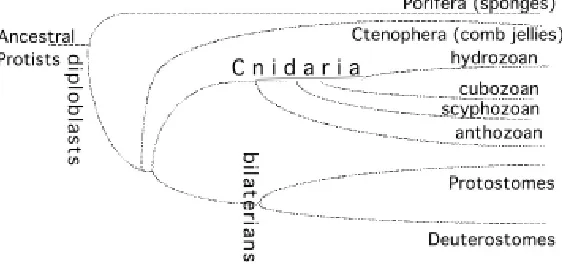
Cnidarians are simple animals that display either a simple tube-shape form, called the polyp, or a more sophisticated morphology named medusa. Cnidarians are made up of two multifunctional epithelial muscle layers separated by an extra-cellular substance named the mesoglea. Additionally the tissues contain nerve cells, nematocytes, interstitial cells and when appropriate, gametes. Cnidaria differentiate structures along their body axis that perform specific functions, like the head at the apical pole, responsible for the active feeding behavior, and in the medusa, monofunctional tissues like striated muscle or complex sense organs, like lens eyes or statocysts (Bouillon, 1994). The Cnidaria phylum, which together with the Ctenophora (comb jellies) and the Porifera (sponges) represent the only surviving diploblast species (Fig. 1), is supposed to predate the protostome / deuterostome divergence, representing thus a sister group to the bilaterian species. The cnidarian species distribute among four distinct classes, the Anthozoa, Hydrozoa, Scyphozoa and Cubozoa. From the molecu-lar data accumulated during the past ten years, it appears that among the four cnidarian classes, the anthozoans (sea anemone, coral) arose first (Bridge et al., 1992; Bridge et al., 1995; Odorico and Miller, 1997; Schuchert, 1993). However, the cellular and molecular complexity of the medusa organisation (see below) remains enigmatic (Boero et al., 1998).
More than 99% of the cnidarians are sea-water animals, and according to their class, live as polyps exclusively (all anthozoans, some hydrozoans like Hydra or Hydractinia) or alternatively differ-entiate both forms (many hydrozoans, all scyphozoans and cubozoans, see Fig. 2). The polyp can bud, either to reproduce asexually as in hydra, or to develop the parental form, the medusa that will complete the sexual cycle. In addition to budding and sexual reproduction, all polyp species and many medusa types can regenerate. Additionally when dissociated into small tissue frag-ments or single cells, reaggregation and regeneration occurs in all species and life stages, especially good results are observed with polyps. These later events prove that the developmental programs can be reactivated whatever the age of the animals. In general polyp forms are regarded as immortal whereas the medusa dies after liberation of gametes, with one exception, Turritopsis, where all animals transforms into polyps again (Piraino et al., 1996).
For developmental purposes, different cnidarian species dis-play complementary advantages: the hydrozoan freshwater hydra
Abbreviations used in this paper: bHLH, basic helix-loop-helix; BMP, bone
is probably the most well know cnidarian because of its regenera-tive possibilities. It is easily maintained in the laboratory where grafting, budding, regeneration and reaggregation are amenable to experimentation. In addition, a collection of different strains and mutants is available. However, its sexual cycle is most often seasonal and not adapted for extensive manipulations (Fig. 2, Left). In contrast, the light-inducible and short sexual development of the marine hydrozoan species, Hydractinia and Podocoryne, make them suitable for genetic purposes, e.g. the study of axis formation in early development. Additionally Podocoryne has a full life cycle including medusa development, the life stage when formation of monotypical tissues like the striated muscle or the subumbrellar plate, complex nerve systems and sense organs, can be observed (Fig. 2 Right). None of these structures differen-tiates in the polyp form. In anthozoans, the corals display an annual sexual cycle that cannot take place under laboratory conditions; this, however, is possible with sea anemones.
History and Contribution of the Cnidarian Model
Sys-tem over the Last 260 Years
Abraham Trembley, a gentleman from Geneva, incidentally discovered hydra regeneration at the time he was teaching the children of a Dutch prince in the Netherlands (Trembley, 1744; Lenhoff and Lenhoff, 1986; see Fig. 1 of article by Buscaglia and Duboule in this issue, pp. 6). His approach was very original, because he was the first scientist to perform systematic animal experimentation in order to understand and describe the process he had discovered. The fact that the original question was: is hydra a plant or an animal, might have helped him to apply strategies that were so far reserved to plants. Whereas experimental cnidarian research was not continued in Switzerland after Trembley, it became an important study subject at the turn between the XIXth and the XXth centuries, when most developmental biologists were fascinated by the regeneration potential of cnidarians. The prob-lem of axially polarized regeneration was already recognized and studied. Morgan investigating head regeneration in Tubularia proposed in 1905 that “a gradient of material is regulating the hydranth (young polyp) forming material, which decreases from the apical towards the basal end” (Morgan, 1905). In 1907, Child concluded from his studies on Tubularia: “we may regard polarity as an axial difference in the character and energy of reactions
hydra reacts to light stimulus (Tardent and Frei, 1969; Borner and Tardent, 1971). His interest in marine hydrozo-ans included the histology, development and regeneration of the eye of the Cladonema medusa (Weber, 1981) and the development (Frey, 1968; Schmid and Tardent, 1969; Brändli, 1971; Schmid, 1972), regeneration (Schmid and Tardent, 1971; Schmid et al., 1976) and transdifferentiation (Frey, 1968; Schmid, 1972) of medusae.
Pierre Tardent influenced cnidarian research in many ways. Firstly, his own students (Volker Schmid, Robert Stidwill) and postdocs (B. Marcum, Lynne Littlefield, Tho-mas Holstein) carried on with projects they had initiated in his laboratory. Secondly, the knowledge accumulated by the group of Pierre Tardent on cnidarians stimulated the formation of new cnidarian groups, especially that of Alfred Gierer who, in the early 70s, decided to use hydra as a model system for understanding pattern formation Fig. 1. Four distinct classes form the Cnidaria phylum. Both Hydra and
Podocoryne carnea belong to the hydrozoan class.
resulting from part physiological relations” (Child, 1907). At the same time, Elena Browne by inducing the formation of extra-heads upon grafting small pieces of hydra tissue, discovered the phenom-ena of tissue induction (Browne, 1909), a discovery that took place 18 years before that of Speeman’s on vertebrates.
processes. Together with his collaborators in Tübingen (Ger-many), Gierer discovered that cells obtained from dissociated hydra could reaggregate and reform a complete normal animal within a relatively short period of time (Gierer et al., 1972).
Finally, as a contribution of the cnidarian model system to our current view of developmental mechanisms, hydra was the first species where conservation over evolution of molecules regulating cell differentiation and developmental processes could be demon-strated. Early investigations in the late 50s already proved the presence of substances which, once extracted from hydra and applied on regenerating animals, would promote the regeneration of head structures (Burnett, 1965). In the 70s, similar properties were demonstrated for the neuropeptide Head Activator (Schaller, 1973; Schaller et al., 1979; Schaller et al., 1989). This peptide was biochemically purified and sequenced from both sea anemone (Schaller and Bodenmüller, 1981) and bovine (Bodenmuller and Schaller, 1981) showing a strikingly identical sequence that proved a perfect conservation from cnidarians to vertebrates.
Genetic Conservation from Cnidarians to Bilaterians
The recent years were an active period for cloning evolutionarily conserved genes from cnidarian species. It is now clear that those species make use of highly evolutionarily-conserved genes that encode functional domains, involved in gene regulation, transla-tional control, signal transduction, apoptosis, extra-cellular signal-ling, specification of the myogenic cell lineage and cell/extra-cellular matrix (ECM) interactions (reviewed in (Galliot, 2000). Furthermore migration of nematocytes seem to be controlled through RGD dependent ligand-receptor complexes (Ziegler and Stidwill, 1992) and the entire Wnt/wingless signalling pathway is conserved in hydra (Hobmayer et al., 2000). These data prove first, that most if not all of the gene families do have representatives in
cnidarians, and second, that their diversification in many cases occurred prior to the Cnidaria divergence.
Moreover, the conservation from cnidarians to chordates is remarkable. For example, the sequence of the mesoderm specifi-cation factor Twist in the bHLH domain is significantly closer to vertebrate than drosophila or nematode twist cognate sequences (Spring et al., 2000). Cnidarians have actually retained gene families, like the Syk protein-tyrosine kinase (Steele et al., 1999), the Not, Hex (Gauchat et al., 2000) and Pax-3/7 (Miller et al., 2000; Gröger et al., 2000) homeobox genes, that have been lost in some bilaterian species, because they were not found in the nematode and/or Drosophila genomes. In addition, genomic data show the conservation of introns at fixed positions within functional domains (Galliot et al., 1995). All together, this high level of gene conserva-tion strengthens the validity of the cnidarian model systems for studying basic developmental processes shared by eumetazoans. Despite the lack of genetic tools currently available in cnidarian species so far, functional assays such as antisense (Yan et al., 2000a) or dsRNA interference (Lohmann et al., 1999) should rapidly highlight the developmental functions of these genes.
Apical and Axial Patterning in Cnidarians
Since the discovery of the conservation of the homeobox motif between Drosophila and vertebrates genes (McGinnis et al., 1984; Scott and Weiner, 1984; Carrasco et al., 1984), it became more and more obvious that pieces of evolutionarily-conserved develop-mental pathways were recruited for similar developdevelop-mental tasks by protostomes and deuterostomes (Duboule and Wilkins, 1998). Such functional conservation implies that these developmental pathways were already present in their common ancestor. Thus, the cnidarians are the best candidates for investigating a non-bilaterian representation of these ancestral developmental path-Fig.2. Life cycle patterns in the Hydroidea (Hydrozoa, Cnidaria). (Left) Sexual cycle of the freshwater hydrozoan, Hydra vulgaris, a strain obtained from Pierre Tardent which constitutively produces gametes (Grassi et al., 1995). Testis are visible in (A,C), ovary in (B), fertilized eggs still attached to the parental polyp in C, detached embryos in (D). After a variable period from 2 weeks to several months, a young hydra will hatch (E,F). (Right) The life cycle of the marine hydrozoan Podocoryne carnea.
A
B
C
D
E
ways. Those are supposed to trigger basic developmental func-tions, as neurogenesis, head patterning and positional information along the axis, that make possible an active feeding behavior and an active locomotion.
Recently, expression analyses performed at the quantitative and qualitative levels were made available from different cnidarian species (Hydra, Podocoryne, Hydractinia, coral) and permitted to distinguish genes that might exert a developmental function. Several of these evolutionarily conserved genes clearly show an expression that is regulated in time and in place where develop-mental events take place (Fig. 4). These molecular approaches have already brought some light on the conservation or the divergence of molecular mechanisms that support similar
develop-mental events in cnidarians and bilaterians. For example, apical patterning in hydrozoans on one hand and anterior patterning in chordates on the other, use structurally-related genes like forkhead (Martinez et al., 1997), emx (Mokady et al., 1998), paired-like (Gauchat et al., 1998; Broun et al., 1999) genes. This suggests some conservation of ancestral basic mechanisms involved in head organizer activity (Galliot and Miller, 2000). However, axis formation from a pool of cells that will differentiate both an apical and a basal pole, as it occurs during budding, regeneration and reaggregation, also likely requires the activity of Hox-related (Schummer et al., 1992; Gauchat et al., 2000) as well as that of the brachyury (Technau and Bode, 1999) and the Wnt-pathway (Hobmayer et al., 2000; Technau et al., 2000) genes. Conse-quently, the apico-basal axis of the cnidarian polyp cannot be considered as a primitive bilaterian anterior-posterior or dorso-ventral axis (Gauchat et al., 2000; Galliot, 2000). Axis formation is nevertheless a difficult question in cnidarians. As described above, the axis can be established in three different contexts in cnidarians: i) during sexual development, from the egg to the swimming planula (the larva that will metamorphoses into the polyp), ii) in the polyp at the time it buds or regenerates a new polyp (asexual reproduction), or iii) in the medusa at the time it buds from the polyp. Moreover, the larva and the polyp axis are often considered as inverted: the anterior larval pole will adhere to the substrate while the posterior larval pole will become the polyp mouth with ten-tacles. Are there common molecular mechanisms underlying these different processes? Which of these axis-forming contexts reflect at best the mechanisms at work in the ancestor common to cnidarians and bilaterians? Extensive comparative data are needed before we can answer to these questions.
Molecular Markers of the Podocoryne carnea Life Cycle
Although all essential elements and the diversity of the cnidarian life cycles are well established (reviewed in Bouillon, 1994), recent experimental and molecular data add new aspects in how the different life cycle stages can be viewed. The developmental Fig. 3. Pierre Tardent,
at a conference (ca. 1970).
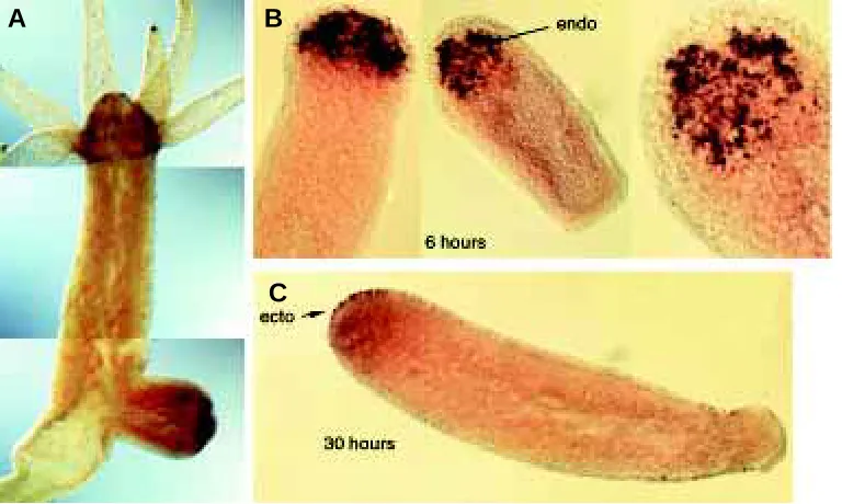
Fig. 4. The regulation of the homeobox paired-like gene prdl-a possibly reflects an ancestral function in apical patterning which was conserved and recruited for anterior patterning in bilaterians (Gauchat et al., 1998; Galliot and Miller, 2000). (A) In adult polyps, prdl-a displays an expression which is restricted to the nerve cell lineage of the head. (B,C) During regeneration, prdl-a shows a sequential expression, first in endodermal cells of head-regenerating stumps 6 hours after bisection (B) and later in the ectodermal cells of the regenerating head (C). Prdl-a shows a similar biphasic mode of expression during budding. In (A) expression of the prdl-b gene was simultaneously detected along the body column with a fluorescein-labelled probe (red dots). ecto, ectoderm; endo, endoderm.
A
B
program of the full hydrozoan life cycle has two distinctive and separate stages: first, the formation of the planula larva and its transformation to the polyp; second, the formation of the medusa from polyp tissues (Fig. 5). It appears that all cell types and the bilayered body structure of the polyp have already differentiated in the young Podocoryne larva 25-36 hrs postfertilization (Gröger and Schmid, 2001). Whereas the differentiating smooth muscles, RFamide positive nerve cells and most nematocytes express no axial polarity, the tyrosine-tubulin positive nervous system devel-ops gradually in repetitive patterns from anterior to posterior. In contrary, the large tyrosin-tubulin positive nematoctes are found only at the posterior end of the larva. This is paralleled, spatially and temporally, by the anterior expression of the Podocoryne genes Cnox2-Pc (an orphan Hox) in the ectoderm (Masuda-Nakagawa et al., 2000), and Gsx (paraHox) in the endoderm (Yanze et al., 2001). The posterior pole of the larva seems to be defined by Cnox4-Pc, an other orphan Hox gene, which is expressed as a maternal message in the egg at the site where first cleavage is initiated. At subsequent stages, Cnox4-Pc transcripts are exclusively detected in the few blastomere cells that localize to the future posterior pole (Yanze et al., 2001), the site of gastrulation (Freeman, 1981). Formation of the polyp occurs by settlement of the swimming larva and rearrangment of the cell types and tissues (Tardent, 1978). This process is not investi-gated yet at the molecular level.
In contrast to polyp formation, the development of the medusa is a de novo developmental process as the medusa differs in body structure and cell types from that of the polyp. The early medusa are formed from dedifferentiating polyp cells which proliferate intensively (Brändli, 1971; Bölsterli, 1977; Spring et al., 2000). Later in medusa development a third tissue layer separates from the ectoderm (Kühn, 1910; Bölsterli, 1977) forming the medusa bell that contains several non-myoepithelial cell types and sense organs (Tardent, 1978). The development of the third layer, called
entocodon, and the fact that it cavitates and produces the smooth and striated muscle of the medusa bell has put forward the idea that hydrozoans are ancestral and derived triploblasts (Boero et al., 1998). This idea is supported by molecular data, which suggest that the structural genes (myosin heavy chain (Schuchert et al., 1993) or tropomyosin (Gröger et al., 1999)), the genes that specify the myogenic linage (Twist (Spring et al., 2000), Brachyury, Snail and Mef (Spring et al., submitted)) and those regulating the muscle differentiation (the HLH family genes JellyD or Id (Müller et al., in preparation)) are structurally and functionally conserved in Podocoryne.
The Hydra Regenerating Stump: a Paradigm for
Orga-nizer Activity
In Hydra, grafting experiments have provided the first hints about the regions where organizer activity distributes along the axis (Browne, 1909; Webster, 1966; Wolpert et al., 1971) and when this organizer activity appears in the regenerating stump (Berking, 1979; MacWilliams, 1983a; MacWilliams, 1983b). Two pairs of gradients, one each for the head and the foot, display activation and inhibition parallel graded activities along the body axis with the maxima in the head region for the head activation/ head inhibition, and in opposite directions for the foot activation/ foot inhibition. After amputation, a rapid and long-lasting drop of head inhibition was immediately observed, consistently with its major source of production in the head (MacWilliams, 1983a), while a delayed head activation (nowadays named head organizer activity) was progres-sively appearing in the tip of the regenerating-stump, reaching a plateau level about 10 hours after mid-gastric section (MacWilliams, 1983b) (Fig. 6A). At the cellular level, the restoration of lost structures occurs initially without cell proliferation but rather, by the direct differentiation of the stem cells and precursor cells present in the body column (Park et al., 1970; Holstein et al., 1991). Thus, Fig. 5. Life cycle stages of Podocoryne carnea. (A) Planula larva, the animal shows the graded anterior(a)-posterior(p) expression pattern of Cnox1-Pc, an ante-rior Hox-like gene. (B) Cnox1-Pc expres-sion in the gastrozooid and the gonozooid. Staining is preferentially strong in the developing striated muscle tissue of the medusa buds. (C) Medusa stained with BrdU antibody which detects all nuclei, that have undergone DNA replication dur-ing the incubation time. Growth zones are located in the manubrium and at the periphery of the bell. (D) Podocoryne carnea egg. Nucleus and nucleolus(n) are located in the animal half.
A
D
unlike axolotls, planarians or medusa, Hydra “bypasses” blastema formation and goes directly from wound healing to regeneration proper, a process named morphallaxis.
After Hydra bisection, gene regulation is detectable immedi-ately (within 1 hour), early (within 10 hours), early-late (from 15 to 36 hours) or late (after 40 hours) (Fig. 6B). These “immediate”, “early”, “early-late” and “late” successive waves of gene regulation are concomitant with the wound healing phase, the establishment of organizer activity and the differentiation of head structures, respectively. In most cases, the “immediate” and very “early” gene modulations are observed in both head- and foot-regeneration stumps, implying that some components are shared at this stage. Recent work has demonstrated that most of the early regulatory genes can be detected in the endodermal cells of the regenerating stump (Galliot, 2000). In addition, when documented the expres-sion of these “early” genes like brachyury (Technau and Bode, 1999) or wnt (Hobmayer et al., 2000) are altered in the regenera-tion-deficient mutant reg-16. Taken together, these observations suggest that endodermal cells of the stump establish organizer activity in Hydra thanks to these “immediate/early” regulatory genes. At the “early-late” and “late” stages, genes are most often expressed as broad domains in the layer, either ectodermal or endodermal, that corresponds to the future adult expression do-main. During the third day of head regeneration, these expression domains get restricted to reach the adult pattern.
How amputation activates or represses the expression of these
developmentally regulated genes, however, remains a mystery. Amputation induces the release of “messenger” molecules, which lead to immediate modifications in DNA-binding activity (Galliot et al., 1995) and transiently target regeneration-specific pathways (Hampe et al., 1999), among which the PKC (Hassel et al., 1998) and the CREB pathways (Kaloulis et al., submitted). Such regula-tion may be mediated by peptides (Schaller et al., 1996; Grens et al., 1999; Hampe et al., 1999; Lohmann and Bosch, 2000), low-molecular weight substances like fatty acids and their derivatives (HETEs) (Hassel et al., 1996), ubiquitously distributed factors like hydroperoxides (Jantzen et al., 1998), evolutionarily-conserved signals (Hobmayer et al., 2000), specific metalloproteinases whose down-regulation prevents either head or foot regeneration (Leontovich et al., 2000; Yan et al., 2000a; Yan et al., 2000b). These substances likely translate the physical stimulus of amputa-tion into cascades of gene expression. The first stage in regenera-tion after amputaregenera-tion is the closure of the wound by cell and tissue migration.
The comparison of the expression patterns observed in the adult polyps with those detected in budding, regenerating or reaggregating animals implies that patterning is actively main-tained in the adult animal by genetic networks that are extensively reprogrammed when the animal starts forming a new head, a new foot or a new axis. At the initial phase of budding, a specific genetic program that involves the Otx (Smith et al., 1999) and the wnt / b-catenin (Hobmayer et al., 2000) genes, takes place at the position along the body column where the bud will emerge. At the subse-quent stages, genetic regulations are highly similar to that ob-served during regeneration of the head and the foot. Thus, except the initiation phase, budding and regeneration appear as the two faces of the same coin.
Instability of the Differentiated State
One of the central issue addressed by regeneration is that of the mechanisms that maintain the developmental program accessible in those species whatever the age of the animal. In principal the regenerate can be formed either by morphallactic rearrangement of the remaining tissues (sponges, some cnidaria), or by recruit-ment of undifferentiated cells (neoblasts in planaria), or by activa-tion of differentiated cells through dedifferentiaactiva-tion and
ming of their cellular committment, a process called transdifferentiation. In general, cellular commitment and the differ-entiated state are stably controlled and in many cell types seem to be irreversibly fixed. Although transdifferentiation was mainly investigated in the context of regeneration (reviewed in Okada, 1991), more recent reports demonstrate that it can be also part of normal development (Bölsterli, 1977; Bode et al., 1986; Schmid, 1992), even in vertebrates (Mochii et al., 1998; Patapoutian et al., 1995).
Cnidarians seem to be well suited to analyse the mechanisms of transdifferentiation. They regenerate well, consist of a small number of cell types and the mayor families of regulatory genes have less members than in higher phyla. Because it can be easily isolated as a monotypical tissue and cultured, the isolated striated muscle of medusae was intensively investigated for its transdifferentiation potential (reviewed in Schmid, 1992). When isolated by microsurgery together with portions of adhering ECM and cultured in artificial seawater without further treatment, the differentiated state of the striated muscle is maintained until the isolated muscle fragments disintegrate after 3-4 weeks. When activated for transdifferentiation, the striated muscle cells start to
dedifferentiate and DNA replication is induced after 24 to 48 hours (Fig. 7). In all activated isolates flagella are formed de novo and smooth muscle cells and FMRFamide positive nerve cells develop. Occasionally striated muscle isolates are able to regenerate even the feeding and sexual organ (manubrium) and tentacles. In this case the striated muscle cells are able to transdifferentiate into smooth muscle cells, nerve cells, nematocytes. Whereas forma-tion of smooth muscle cells needs no DNA replicaforma-tion, transdifferentiation to nerve cells requires one cell cycle, and all the other cell types two (Alder and Schmid, 1987). Transdifferentiation of isolated striated muscle can be induced by treating this tissue with ECM degrading en-zymes, by drugs activating the PKC such as TPA, mezerein or diacylglycerol, or by grafting muscle isolates onto pieces of ECM (reviewed in Schmid and Reber-Muller, 1995). Induction was also obtained with a mAb specific for ECM-specific carbohydrate moieties (Reber-Muller et al., 1995). Taken together these results demonstrate that the stability of the differentiated state of striated muscle is controlled by the cell-substrate complex and thus confirms in vivo obser-vations (Schmid et al., 1999).
Since regeneration starts with wound closure that re-quires cell and tissue migration, the effect of change in cell substrate adhesion on gene expression was investigated by grafting isolated striated muscle on stretched ECM. The cells quickly adhere on the host ECM and migrate onto it until the muscle tissue is completly stretched (12-24 hrs). Surprisingly expression of striated muscle specific regula-tory genes like the homeobox genes Otx and Cnox1-Pc (Hox1-like) and of the structural genes MHC (myosin heavy chain) and tropomyosin (Tpm2) are downregulated in the migrating cells whereas expression of the ubiqui-tously expressed homeobox gene Cnox3-Pc (Msx-like) is maintained (Yanze et al., 1999). The fact that dedifferentia-tion is not initiated during migradedifferentia-tion indicates that addidedifferentia-tional activating pathways, possibly those stimulating and con-trolling cell cycle activity, are needed for the reprogram-ming of the genome and for the fixation of the newly determined state.
The investigations on gene regulation in the transdifferentiation process demonstrated that the genes specific for the striated muscle (see above) are turned off within hours postactivation (Cnox1-Pc) or 1-3 days (Otx, unpublished), others like Pax-B are permanently (Gröger et al., 2000) or transiently, like BMP2-8 (Reber-Müller, in preparation) turned on. Interestingly Twist which is required to make striated and smooth muscle in medusa devel-opment, is not expressed throughout the transdifferentiation pro-cess (Spring et al., 2000), indicating that regeneration not neces-sarily copies ontogeny.
Conclusion
The question of the conservation of the molecular cascades that underly regeneration in different phyla, either invertebrate or vertebrate, remains open (Sánchez Alvarado, 2000). Identification of molecular markers at work during Hydra regeneration and Podocoryne transdifferentiation will allow the characterization in a close future of the key signaling cascades involved in cnidarian Fig. 7. Experimental conditions leading to transdifferentiation of striated
regeneration. Present work suggests that most of these compo-nents are evolutionarily conserved, then deciphering their regula-tion could open the way to understand how a developmental program can be reactivated, especially in species where this program is locked very rapidly during development.
Summary
Hydra and Podocoryne are two cnidarian animals which provide complementary advantages for analysing developmental mecha-nisms possibly reflecting the basic developmental processes shared by most bilaterians. Interestingly, these mechanisms remain ac-cessible all along the life of these animals, which bud and regen-erate, whatever their age. The Hydra polyp permits a direct study of the molecular cascades linking amputation to regeneration. Podocoryne displays a complete life cycle, polyp and medusa stages with a fast and inducible sexual cycle and an unparalleled in vitro transdifferentiation potential. In both cases, a large number of evolutionarily conserved molecular markers are available, and analysis of their regulation highlights the molecular mechanisms which underly pattern formation in these two species.
KEY WORDS:
cnidarians, hydra, podocoryne, regeneration,
bud-ding, transdifferentiation, evolution, apical/head patterning, axial
patterning, extra-cellular matrix, striated muscle
Acknowledgements
The work performed in BG’s laboratory is supported by the Swiss National Science Foundation, the Canton of Geneva, the Georges et Antoine Claraz Fondation. The work performed in VS’s laboratory is supported by the Swiss National Science Foundation and the Fondation Suisse de Recherche sur les Maladies Musculaires. The authors thank their collaborateurs for the data reproduced in this review.
References
AERNE, B., L., STIDWILL, R.P., TARDENT, P. (1991). Nematocyte discharge in Hydra does not require the presence of nerve cells. J. Exp. Zool. 258: 137-141.
ALDER, H., SCHMID, V. (1987). Cell cycles and in vitro transdifferentiation and regeneration of isolated, striated muscle of jellyfish. Dev. Biol. 124: 358-369.
BERKING, S. (1979). Analysis of head and foot formation in Hydra by means of an endogenous inhibitor. Roux’s Arch. Dev. Biol. 186: 189-210.
BODE, H.R., DUNNE, J., HEIMFELD, S., HUANG, L., JAVOIS, L., KOIZUMI, O., WESTERFIELD, J., YAROSS, M. (1986). Transdifferentiation occurs continu-ously in a adult hydra. Curr. Topics Dev. Biol. 20: 257-280.
BODENMULLER, H., SCHALLER, H.C. (1981). Conserved amino acid sequence of a neuropeptide, the head activator, from coelenterates to humans. Nature 293: 579-580.
BOERO, F., BOUILLON, J., PIRAINO, S., SCHMID, V. (1998). Diversity of hydrozoan life cycles: ecological implications and evolutionary patterns. Ital. J. Zool. 65: 5-9.
BÖLSTERLI, U. (1977). An electron mircoscopic study of early developmental stages, myogenesis, oogenesis and cnidogenesis in the anthomedusa, Podocoryne carnea M. Sars. J. Morph. 154: 259-289.
BORNER, M., TARDENT, P. (1971). Der Einfluss von Licht auf die Sponatnaktivität von Hydra attentuata Pall. Rev. Suisse Zool. 78: 697-704.
BOUILLON, J. (1994). Embranchement des cnidaires (Cnidaria). In Traité de Zoologie. Cnidaires, Cténaires (Ed. P.P. Grassé). Masson, Paris, pp 1-28.
BRÄNDLI, E. (1971). Bedeutung der kolonialen Komponenten für die Bildung und Differenzierung der medusen von Podocoryne carnea M. Sars. Roux’Archiv. Dev. Biol. 166: 254-286.
BRIDGE, D., CUNNINGHAM, C.W., DESALLE, R., BUSS, L.W. (1995). Class-level relationships in the phylum Cnidaria: molecular and morphological evidence. Mol.
Biol. Evol. 12: 679-689.
BRIDGE, D., CUNNINGHAM, C.W., SCHIERWATER, B., DESALLE, R., BUSS, L.W. (1992). Class-level relationships in the phylum Cnidaria: evidence from mitochon-drial genome structure. Proc. Natl. Acad. Sci. USA 89: 8750-8753.
BRIDGE, D.M., STOVER, N.A., STEELE, R.E. (2000). Expression of a novel receptor tyrosine kinase gene and a paired-like homeobox gene provides evidence of differences in patterning at the oral and aboral ends of hydra. Dev. Biol. 220: 253-262.
BROUN, M., SOKOL, S., BODE, H.R. (1999). Cngsc, a homologue of goosecoid, participates in the patterning of the head, and is expressed in the organizer region of Hydra. Development 126: 5245-5254.
BROWNE, E.N. (1909). The production of new hydranths in hydra by the insertion of small grafts. J. Exp. Zool. 7: 1-37.
BURNETT, A. (1965). The acquisition, maintenance, and lability of the differentiated state in Hydra. In The acquisition, maintenance, and lability of the differentiated state in Hydra (Eds. W. Beemann, J. Reinert, H. Ursprung). Springer Verlag, Heidelberg, pp 109-127.
CARRASCO, A. E., MCGINNIS, W., GEHRING, W. J., De ROBERTIS, E. M. (1984). Cloning of an X. laevis gene expressed during early embryogenesis coding for a peptide region homologous to Drosophila homeotic genes. Cell 37: 409-414.
CHILD, C.M. (1907). An analysis of form regulation in Tubularia I. Arch. Entw. Mechan. 23: 396-414.
DUBOULE, D., WILKINS, A.S. (1998). The evolution of ‘bricolage’. Trends Genet. 14: 54-59.
EPP, L., SMID, J., TARDENT, P. (1986). Synthesis of the mesoglea by ectoderm and endoderm in reassembled Hydra. J. Morphol. 189: 271-279.
EPP, L., TARDENT, P. (1978). The distribution of nerve cells in Hydra attentuata Pall. Roux’s Arch. Dev. Biol. 185: 185-193.
FREEMAN, G. (1981). The cleavage initiation site establishes the posterior pole of the hydrozoan embryo. Roux‘ Arch. Dev. Biol. 190: 123-125.
FREY, J.R.A.-Ω. (1968). Die Entwicklungsleistungen der Medusenknospen und Medusen von Podocoryne carnea M. Sars nach Isolation und Dissoziation. Roux’s Arch. Dev. Biol. 160: 428-468.
GALLIOT, B. (2000). Conserved and divergent genes in apex and axis development in cnidarians. Curr. Op. Genet. Dev. 10: 629-637.
GALLIOT, B., MILLER, D. (2000). Origin of anterior patterning. How old is our head? Trends Genet. 16: 1-5.
GALLIOT, B., WELSCHOF, M., SCHUCKERT, O., HOFFMEISTER, S., SCHALLER, H.C. (1995). The cAMP response element binding protein is involved in hydra regeneration. Development 121: 1205-1216.
GAUCHAT, D., KREGER, S., HOLSTEIN, T., GALLIOT, B. (1998). prdl-a, a gene marker for hydra apical differentiation related to triploblastic paired-like head-specific genes. Development 125: 1637-1645.
GAUCHAT, D., MAZET, F., BERNEY, C., SCHUMMER, M., KREGER, S., PAWLOWSKI, J., GALLIOT, B. (2000). Evolution of Antp-class genes and differential expression of Hydra Hox/paraHox genes in anterior patterning. Proc. Natl. Acad. Sci. USA 97: 4493-4498.
GIERER, A., BERKING, S., BODE, H., DAVID, C.N., FLICK, K., HANSMANN, G., SCHALLER, H., TRENKNER, E. (1972). Regeneration of hydra from reaggre-gated cells. Nature New Biol 239: 98-101.
GRASSI, M., TARDENT, R., TARDENT, P. (1995). Quantitative data about gameto-genesis and embryonic development in Hydra vulgaris Pall. (Cnidaria, Hydrozoa). Invertebrate Reproduction and Development 27: 219-232.
GRENS, A., GEE, L., FISHER, D.A., BODE, H.R. (1996). CnNK-2, an NK-2 homeobox gene, has a role in patterning the basal end of the axis in hydra. Dev. Biol. 180: 473-488.
GRENS, A., SHIMIZU, H., HOFFMEISTER, S.A., BODE, H.R., FUJISAWA, T. (1999). The novel signal peptides, pedibin and Hym-346, lower positional value thereby enhancing foot formation in hydra. Development 126: 517-524.
GRÖGER, H., CALLAERTS, P., GEHRING, W.J., SCHMID, V. (1999). Gene duplica-tion and recruitment of a specific tropomyosin into striated muscle cells in the jellyfish Podocoryne carnea. J. Exp. Zool. 285: 378-386.
GRÖGER, H., SCHMID, V. (2001). Larval development in Cnidaria. A connection to Bilateria. Genesis 29: 110-114.
HAMPE, W., URNY, J., FRANKE, I., HOFFMEISTER-ULLERICH, S.A., HERRMANN, D., PETERSEN, C.M., LOHMANN, J., SCHALLER, H.C. (1999). A head-activator binding protein is present in hydra in a soluble and a membrane-anchored form. Development 126: 4077-4086.
HASSEL, M. (1998). Upregulation of a Hydra vulgaris cPKC gene is tightly coupled to the differentiation of head structures. Dev. Genes Evol. 207: 489-501.
HASSEL, M., BRIDGE, D.M., STOVER, N.A., KLEINHOLZ, H., STEELE, R.E. (1998). The level of expression of a protein kinase C gene may be an important component of the patterning process in Hydra. Dev. Genes Evol. 207: 502-514.
HASSEL, M., LEITZ, T., MULLER, W.A. (1996). Signals and signal-transduction systems in the control of development in Hydra and Hydractinia. Int. J. Dev. Biol. 40: 323-330.
HOBMAYER, B., RENTZSCH, F., KUHN, K., HAPPEL, C., CRAMER VON LAUE, C., SNYDER, P., ROTHBÄCHER, U., HOLSTEIN, T. (2000). WNT signaling molecules act in axis formation in the diploblastic metazoan Hydra. Nature 407: 186-189.
HOLSTEIN, T., TARDENT, P. (1984). An ultrahigh-speed analysis of exocytosis: nematocyst discharge. Science 223: 830-833.
HOLSTEIN, T.W., HOBMAYER, E., DAVID, C.N. (1991). Pattern of epithelial cell cycling in hydra. Dev. Biol. 148: 602-611.
JANTZEN, H., HASSEL, M., SCHULZE, I. (1998). Hydroperoxides mediate lithium effects on regeneration in Hydra. Comp. Biochem. Physiol. C Pharmacol. Toxicol. Endocrinol. 119: 165-175.
KÜHN, A. (1910). Die Entwicklungsgeschichte der Geschlechtsindividuen der Hydromedusen. Zool. Jahrb. 30: 145-164.
LENHOFF, S.G., LENHOFF, H.M. (1986). Mémoires, Pour Servir à L’histoire d’un Genre De Polypes d’eau Douce, à Bras en Forme de Cornes. The Boxwood Press. Pacific Grove.
LEONTOVICH, A.A., ZHANG, J., SHIMOKAWA, K., NAGASE, H., SARRAS, M.P., JR. (2000). A novel hydra matrix metalloproteinase (HMMP) functions in extracellular matrix degradation, morphogenesis and the maintenance of differentiated cells in the foot process. Development 127: 907-920.
LITTLEFIELD, C.L. (1984). Interstitial cells control the sexual phenotype of hetero-sexual chimeras of hydra. Dev. Biol. 102: 426-432.
LOHMANN, J.U., BOSCH, T.C. (2000). The novel peptide HEADY specifies apical fate in a simple radially symmetric metazoan. Genes Dev. 14: 2771-2777.
LOHMANN, J.U., ENDL, I., BOSCH, T.C. (1999). Silencing of developmental genes in Hydra. Dev. Biol. 214: 211-214.
MACWILLIAMS, H.K. (1983a). Hydra transplantation phenomena and the mechanism of hydra head regeneration. I. Properties of the head inhibition. Dev. Biol. 96: 217-238.
MACWILLIAMS, H.K. (1983b). Hydra transplantation phenomena and the mechanism of Hydra head regeneration. II. Properties of the head activation. Dev. Biol. 96: 239-257.
MARTIN, R., TARDENT, P. (1962). Kultur von Hydroiden-Zellen in vitro. Rev. Suisse Zool. 70: 312-316.
MARTINEZ, D.E., DIRKSEN, M.L., BODE, P.M., JAMRICH, M., STEELE, R.E., BODE, H.R. (1997). Budhead, a fork head/HNF-3 homologue, is expressed during axis formation and head specification in hydra. Dev. Biol. 192: 523-536.
MASUDA-NAKAGAWA, L.M., GRÖGER, H., AERNE, B.L., SCHMID, V. (2000). The HOX-like gene Cnox2-Pc is expressed at the anterior region in all life cycle stages of the jellyfish Podocoryne carnea. Dev. Genes Evol. 210: 151-156.
MCGINNIS, W., HART, C.P., GEHRING, W.J., RUDDLE, F.H. (1984). Molecular cloning and chromosome mapping of a mouse DNA sequence homologous to homeotic genes of Drosophila. Cell 38: 675-680.
MILLER, D.J., HAYWARD, D.C., REECE-HOYES, J.S., SCHOLTEN, I., CATMULL, J., GEHRING, W.J., CALLAERTS, P., LARSEN, J.E., BALL, E.E. (2000). Pax gene diversity in the basal cnidarian Acropora millepora (Cnidaria, Anthozoa): implica-tions for the evolution of the Pax gene family. Proc. Natl. Acad. Sci. U S A 97: 4475-4480.
MOCHII, M., ONO, T., MATSUBARA, Y., EGUCHI, G. (1998). Spontaneous transdifferentiation of quail pigmented epithelial cell is accompanied by mutation in the Mitf gene. Dev. Biol. 196: 145-159.
MOKADY, O., DICK, M.H., LACKSCHEWITZ, D., SCHIERWATER, B., BUSS, L.W. (1998). Over one-half billion years of head conservation? Expression of an ems
class gene in Hydractinia symbiolongicarpus (Cnidaria: Hydrozoa). Proc. Natl. Acad. Sci. USA 95: 3673-3678.
MORGAN, T.H. (1905). An attempt to analyse the phenomenon of polarity in Tubularia. J. Exp. Zool. 2: 495-.
ODORICO, D.M., MILLER, D.J. (1997). Internal and external relationships of the Cnidaria: implications of primary and predicted secondary structure of the 5'-end of the 23S-like rDNA. Proc. R. Soc. Lond. B. Biol. Sci. 264: 77-82.
OKADA, T.S. (1991). Transdifferentiation. Claredon Press. Oxford.
PARK, H.D., ORTMEYER, A.B., BLANKENBAKER, D.P. (1970). Cell division during regeneration in Hydra. Nature 227: 617-619.
PATAPOUTIAN, A., WOLD, B.J., WAGNER, R.A. (1995). Evidence for developmen-tally programmed transdifferentiation in mouse esophagal muscle. Science 270: 1818-1821.
PIRAINO, S., BOERO, F., AESCHBACH, B., SCHMID, V. (1996). Reversing the life cycle: Medusae transforming into polyps and cell transdifferentiation in Turritopsis nutricula (Cnidaria, Hydrozoa). Biol. Bull. 190: 302-312.
REBER-MULLER, S., SPISSINGER, T., SCHUCHERT, P., SPRING, J., SCHMID, V. (1995). An extracellular matrix protein of jellyfish homologous to mammalian fibrillins forms different fibrils depending on the life stage of the animal. Dev. Biol. 169: 662-672.
RICH, F., TARDENT, P. (1969). Studies of differentiation of nematocytes in Hydra attenuata Pall. Rev. Suisse Zool. 76: 779-787.
SÁNCHEZ ALVARADO, A. (2000). Regeneration in the Metazoans: Why does it happen? BioEssays 22: 578-590.
SCHALLER, H.C. (1973). Isolation and characterization of a low-molecular-weight substance activating head and bud formation in hydra. J. Embryol. Exp. Morphol. 29: 27-38.
SCHALLER, H.C., BODENMÜLLER, H. (1981). Isolation and amino acid sequence of a morphogenic peptide in hydra. Proc. Natl. Acad. Sci. USA 78: 7000-7004.
SCHALLER, H.C., HERMANS-BORGMEYER, I., HOFFMEISTER, S.A. (1996). Neu-ronal control of development in hydra. Int. J. Dev. Biol. 40: 339-344.
SCHALLER, H.C., HOFFMEISTER, S.A., DUBEL, S. (1989). Role of the neuropeptide head activator for growth and development in hydra and mammals. Development 107: 99-107.
SCHALLER, H.C., SCHMIDT, T., GRIMMELIKHUIJZEN, C.J.P. (1979). Separation and specificity of action of four morphogens from hydra. Roux´ Arch. Dev. Biol. 186: 139-149.
SCHMID, V. (1972). Untersuchungen über Dedifferenzierungsvorgänge bei Medusenknospen und Medusen von Podocoryne carnea M. Sars. Wilhelm Roux’s Arch. Dev. Biol. 169: 281-307.
SCHMID, V. (1992). Transdifferentiation in Medusae. Intl. Rev. Cyt. 142: 213-261.
SCHMID, V., ONO, S.I., REBER-MULLER, S. (1999). Cell-substrate interactions in cnidaria. Microsc Res Tech 44: 254-268.
SCHMID, V., REBER-MULLER, S. (1995). Transdifferentiation of isolated striated muscle of jellyfish in vitro: the initiation process. Semin. Cell Biol. 6: 109-116.
SCHMID, V., SCHMID, B., SCHNEIDER, B., STIDWILL, R., BAKER, G. (1976). Factors affecting manubrium regeneration in Hydromedusae. Roux’s Arch. Dev. Biol. 179: 41-56.
SCHMID, V., TARDENT, P. (1969). Zur Gametogenese von Podocoryne carnea M. Sars. Rev. Suisse Zool. 76: 1071-1078.
SCHMID, V., TARDENT, P. (1971). The reconstitutional performances of the leptomedusa Campanularia johnstoni. Mar. Biol. 8: 99-104.
SCHUCHERT, P. (1993). Phylogenetic analysis of the Cnidaria. Z. zool. Syst. Evolut.-forsch. 31: 161-173.
SCHUCHERT, P., REBER-MULLER, S., SCHMID, V. (1993). Life stage specific expression of a myosin heavy chain in the hydrozoan Podocoryne carnea. Differentiation 54: 11-18.
SCHUMMER, M., SCHEURLEN, I., SCHALLER, C., GALLIOT, B. (1992). HOM/HOX homeobox genes are present in hydra (Chlorohydra viridissima) and are differen-tially expressed during regeneration. EMBO J. 11: 1815-1823.
SCOTT, M.P., WEINER, A.J. (1984). Structural relationships among genes that control development: sequence homology between the Antennapedia, Ultra-bithorax, and fushi tarazu loci of Drosophila. Proc. Natl. Acad. Sci. USA 81: 4115-4119.
SMID, I., TARDENT, P. (1984). Migration of I-cells from ectoderm to endoderm in Hydra attenuata Pall (Cnidaria, Hydrozoa) and their subsequent differentiation. Dev. Biol. 106: 469-477.
SMITH, K.M., GEE, L., BLITZ, I.L., BODE, H.R. (1999). CnOtx, a member of the Otx gene family, has a role in cell movement in hydra. Dev. Biol. 212: 392-404.
SPRING, J., YANZE, N., MIDDEL, A.M., STIERWALD, M., GRÖGER, H., SCHMID, V. (2000). The mesoderm specification factor Twist in the life cycle of jellyfish. Dev. Biol. 228: 363-375.
SPRING, J., YANZE, N., JÖSCH, C.H.,MIDDEL, A.M., WINNIGET, B. and SCHMID, V. (2002). Conservation of Brachyury, Metz and Snail in the myogenic lineage of jellyfish: a connection to the mesoderm of Bilateria. Dev. Biol. (submitted).
STEELE, R.E., STOVER, N.A., SAKAGUCHI, M. (1999). Appearance and disappear-ance of Syk family protein-tyrosine kinase genes during metazoan evolution. Gene 239: 91-97.
STIDWILL, R. (1981). Interspezifische Inkompatibilitäten zwischen Arten der gattung Hydra (Hydrozoa, Cnidaria). PhD Thesis. University of Zürich. Zürich.
TARDENT, P. (1952). Ueber Anordnung und Eigenschaften der institiellen Zellen bei Hydra und Tubularia. Rev. Suisse Zool. 59: 247-253.
TARDENT, P. (1954). Axiale Verteilungs. Gradienten der interstitielle Zellen bei Hydra und Tubularia und ihre Bedeutung für die Regeneration. Arch. Entw. Mech. 146: 593-649.
TARDENT, P. (1966). Experimente zur Frage der Geschlechtsbestimmung bei Hydra attentuata Pall. Rev. Suisse Zool. 73.
TARDENT, P. (1968). Experiments about sex determination in Hydra attenuata Pall. Dev. Biol. 17: 483-511.
TARDENT, P. (1978). Coelenterata. Cnidaria. VEB Gustav Fischer. Jena.
TARDENT, P. (1985). The differentiation of germ cells in Cnidaria. The origin and the evolution of sex. (Eds. A. Monroy, H. Halvorson). Alan R. Riss, New York, pp 163-197.
TARDENT, P. (1995). The cnidarian cnidocyte, a high-tech cellular weaponry. BioEssays, 17: 351-362.
TARDENT, P., BUHRER, M. (1982). Intraspecific tissue incompatibilities in the metagenetical Podocoryne carnea M. Sars (Cnidaria, Hydrozoa). Prog. Clin. Biol. Res. 85: 295-308.
TARDENT, P., FREI, E. (1969). Reaction patterns of dark- and light-adapted Hydra to light stimuli. Experientia 25: 265-267.
TARDENT, P., HOLSTEIN, T. (1982). Morphology and morphodynamics of the stenotele nematocyst of Hydra attenuata Pall. (Hydrozoa, Cnidaria). Cell Tissue Res. 224: 269-290.
TARDENT, P., HOLSTEIN, T., WEBER, J., KLUG, M. (1985). The morphodynamics and actions of stenotele nematocysts in Hydra. Arch. Sc. Genève. 38: 401-418.
TARDENT, P., MORGENTHALER, U. (1966). Autoradiographic studies on the problem of cell migration in Hydra attenuate Pall. Rev. Suisse Zool. 73: 468-480.
TARDENT, P., WEBER, C. (1976). A qualitative and quantitative inventory of nervous cells in Hydra attentuata Pall. In Coelenetrate Ecology and Behaviour (Ed. G.O. Mackie) Plenum Press, New York, pp 501-512.
TECHNAU, U., BODE, H.R. (1999). HyBra1, a Brachyury homologue, acts during head formation in Hydra. Development 126: 999-1010.