Title:
The efficacy of bio-aerosol reducing procedures used in dentistry: a systematic review
Authors and Institutions:
*Lakshman Perera Samaranayake1,2 Kausar Sadia Fakhruddin1
Borvornwut Buranawat3
Chamila Panduwawala1
1 Departments of Preventive and Restorative Dentistry and Oral and Craniofacial Health Sciences, University of Sharjah, UAE
2 Faculty of Dentistry, The University of Hong Kong, Hong Kong Special Administrative Region, China
3 Faculty of Dentistry, Department of Periodontics and Implant Dentistry, Faculty of Dentistry, Thammasat University, Pathum Thani, Thailand
Running Title: Bio-aerosol reduction in dentistry
Key words: bio-aerosol, reduction, procedures, dentistry, review
Corresponding author
*Professor Lakshman Samaranayake M28-125, College of Dental Medicine,
University of Sharjah, 27272, United Arab Emirates
E-mail: [email protected]; [email protected]
Abstract
Background and objective:
Microbe laden air particles, known as bio-aerosols, are routinely generated, in clinical dentistry due to the operative instrumentation within a milieu rich in salivary organisms. As the major mode of transmission of SARS-CoV-2 appears to be airborne aerosols and droplets, there has been an intense focus on such aerosol generating procedures (AGP). As there has been no
systematic reviews on the efficacy of bio-aerosol reducing measure in dentistry, the objective of this systematic review was to evaluate the literature on three major AGPs, rubber dam
application, pre-procedural oral rinse, and high-volume evacuation (HVE) aimed at reducing dental bio-aerosols.
Method:
PubMed via Ovid MEDLINE, EBSCO host, Cochrane Library, and Web of Science databases between January 01, 1985, and April 30, 2020, were searched.
Results:
A total of 156 records in the English language literature were identified, of which 17 clinical studies with 724 patients were included in the final analyses. The eligible reviewed articles revealed the inadequacy of the afore mentioned three principal AGPs used in contemporary dental practice to minimise bio-aerosols. HVE appears to be the most efficacious method, although no single approach provides total elimination of bio-aerosols.
Conclusion:
This, the first systematic review on methods of controlling bio-aerosols in dental operatory settings, indicates that employing combination strategies of rubber dam, with a pre-procedural antimicrobial oral rinse, and HVE can significantly minimize bio-aerosols. As the quality of the currently available data on dental bio-aerosols are rather poor, further, controlled, multi-centre studies are essential to address this critical issue.
Introduction:
An aerosol can be defined as a suspension of fine solid particles or liquid droplets in air or another gas. Aerosols can be either natural, such as fog, mist, dust, or, anthropogenic - created by humans or animals when they speak, sneeze, or cough, for instance. On the other hand, bio-aerosols are bio-aerosols comprising particles/droplets with live microorganisms [1, 2]. The characteristics of bio-aerosols and their spread vary considerably depending on numerous factors, as discussed below.
Although there are conflicting reports in the literature on the size of the aerosols and how long they are airborne, early researchers have classified particles <100 μm in diameter as aerosols, and those > 100 μm as droplets or `spatter/splatter`[1]. The latter, usually falls on to the ground immediately, as and when they are expelled. The former, aerosols, on the contrary, may be entrained or suspended in the air for considerable periods depending on the humidity, airflow, and temperature of the environment into which they are expelled, for example, a dental clinic operatory or a hospital ward. Similarly, the large diameter droplets with the offending agents can contaminate surfaces in the immediate vicinity and spread a few meters, yet again depending on the ambient conditions such as the airflow [3].
As mentioned, bio-aerosols can be generated either by humans or animals (anthropogenic) or by various mechanical devices. Humans produce bio-aerosols by talking, breathing, sneezing, or coughing [1, 4, 5], depending on the infectious state of a person, and these may contain fungi, bacteria, or viruses. On the other hand, mechanical devices such as clinic/hospital ventilation systems, air conditioners, air rotors with coolant water spray used in dentistry may spread bio-aerosols equally efficiently, and engineering strategies such as high efficiency particulate filters (HEPA) need to be inatlled to minimize or eliminate such spread [6, 7]. It is also noteworthy, in this context, that a number of microbial factors such as their virulence, the number of infectious particles, and the pathogenicity of the offending microbes, as well as host factors including their immune status, determine the susceptibility of acquiring an infectious agent via a bio-aerosol [8-10].
where HCWs have disproportionately succumbed to the disease [12]. Although thus far, there has been no mortality amongst dental HCWs due to COVID-19, they are considered to be the professional group that has the highest likelihood of acquiring the disease [13].
SARS-CoV-2- and bio-aerosols:
Despite the intricate anatomical configuration of the respiratory system that efficiently filters extraneous pathogenic agents, bio-aerosols have the ability to penetrate deep into the human alveolar sacs [3]. The human nose, for instance, is an efficient filter for particles of
approximately 1 µm or more [2] but SARS-CoV-2 with a diameter of 50–200 nanometres can effectively escape this sentinel mechanism. It has been shown in some studies that aerosolized particles ranging from 0.25 to 0.5 µm [8, 14], can enter the bloodstream, and then the target organs. Once inhaled, SARS-CoV-2 uses the angiotensin-converting enzyme-1 (ACE2 ) present
on eukaryotic cell surfaces, including oral epithelia, as a keyhole for systemic entry [10, 15] making the oral cavity a potential portal for viral entry and subsequent systemic dissemination.
The fact that ACE2 receptors are present in the aqueous humor of the human eye [16], also
implies that the bio-aerosols may take the ophthalmic route to cause upper respiratory tract infections, as the offending pathogen, such as SARS-CoV-2, may has a ready passage via the nasolacrimal duct to reach the nasopharynx and the upper and lower respiratory system A laboratory study has demonstrated a high rate of trans-ocular transmission in subjects, following exposure to aerosols containing live attenuated influenza viruses [17]. is strongly suggests that respiratory virions,
Viral load in the oro-pharynx region:
Bio-aerosol generating dental procedures and implications for dentistry:
Many interventional procedures are known to aerosolize respiratory secretions in healthcare settings [6, 20, 21]. In dentistry, viral particles may be aerosolized by the high-speed handpiece and the accompanying air jet, ultrasonic scaling, air polishing, and air/water syringes [21]. Unless judiciously controlled, these bio-aerosol generating procedures appear to be one of many intrinsic hazards the profession faces, that has been brought into focus by the current COVID-19 pandemic [6].
Thus, in a very early laboratory study, Miller et al. [22] observed that aerosolized microbes generated by high-powered dental drills and periodontal scalers could transmit to around 200 cm distance in the operatory. More recently, van Doremalen and colleagues [23] noted that the SARS-CoV-2 virus could remain aerosolized for up to 3 hours [23]. They reported that the viability of SARS-CoV-2 virions is relatively high compared with their counterparts, and hence viable and infectious virions may be suspended in bio-aerosols for hours, and for several days within a shed inoculum, on solid surfaces such as steel and card board [23]. These findings have direct implications in dentistry as the threat of airborne transmission of the virus in dental bio-aerosols, as well as their deposition on the clinic surfaces, have to be appropriately addressed for safe dental practice. Indeed, Li and colleagues [24], during the post-SARS era, suggested additional measures for bio-aerosol reduction, and concomitant safe dental practice. These include manual scaling, chemo-chemical caries removal, atraumatic restorative technique, open debridement for periodontal surgeries, rubber dam isolation, use of pre-procedure oral rinses, general ventilation, air filtration [13, 24]. Most of these have been currently adopted by various dental jurisdictions during the current pandemic [13].
Although the post-SARS era literature provides useful guidance on bio-aerosol reducing
procedures in dentistry, there has been, to our knowledge, no contemporary systematic review on this subject during the current COVID-19 pandemic. Thus, we aimed to systematically to gather evidence of standard precautionary measures which attempt to reduce bio-aerosol transmission in dentistry, and reviewed the contemporaneous data on three such major strategies, i)
Methods:
Data sources:
An electronic data search of English language manuscripts using Ovid MEDLINE, Web of Science, EBSCO host, and Cochrane Library databases was performed by two investigators (LPS and KSF). Published clinical reports were accessed between January 01, 1985, and April 30, 2020. Afterscreening different electronic databases, a total of seventeen clinical studies (rubber dam and bio-aerosols (x4 studies); HVE and bio-aerosols (x6 ); and pre-procedural oral rinse and bio-aerosols (x7) were identified.
A precise review question was formulated using the PICO framework as follows. Does pre-procedural antimicrobial mouth rinse, rubber dam application, and high volume evacuation (HVE) (I), compared with placebo, no mouth antimicrobial mouth rinse/rubber dam
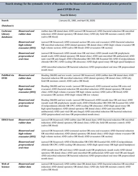
application/use of high volume evacuation (C), results in effective microbial reduction disseminated via aerosol generated during dental procedures (O), in dental patients (male and female) undergoing the dental procedure? (P). The search keywords and combination of the keywords were systematized according to the PICO model (Table 1).
Outcome: The key finding of the present review was the systematic assessment of the efficacy of preprocedural mouth rinse, rubber dam application, and use of HVE in reducing bio-aerosols generated via dental procedures.
Study selection:
Inclusion criteria: Pre-determined inclusion criteria were 1) English language articles; 2) clinical trials; 3) patients undergoing dental procedures (use of high-speed rotary
instruments/ultrasonic scalers/air polishers/triple syringe); 4) pediatric and adult patients; 5) country or date enforced no limitations.
Electronic data search and analysis: To ensure of systematic and comprehensive method approached, we trailed through the PRISMA (Preferred Reporting Items for Systematic Reviews and Meta-Analyses) guidelines.[25, 26] The search approach employed, and results generated, are presented in Fig. 1, and search terms and limits are shown in Table 1.
A three-staged, electronic data search and analysis was carried out. In stage one: the titles and abstracts of all relevant clinical trials meeting our set inclusion criteria were screened. In step- two: a full-text review of all the pertinent articles was completed, which yielded a
comprehensive account of the data. Through the full-text review of the retrieved literature, the investigators used spreadsheets, ensuring that eligibility criteria were met and the reported outcomes were in alignment with the set study objectives. References of the included clinical trials were checked as a backward-search. In stage-three, the reviewers extracted and assessed the data.
Following the full-text review, specific points linked to the characteristics of each included clinical trial were charted using the Cochrane model. This enabled in classifying the setting, study design, and the country. Additionally, sample size, assessment methods, evaluation time, and study conclusions were comprehensively examined. The identified articles were compiled using a bibliographic software tool, Endnote version 9 (Clarivate Analytics, USA).
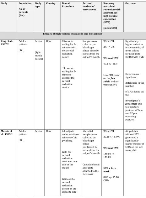
Summary of the characteristics of included clinical trials and the reported results, including the microbial counts generated during dental procedures, are provided in Tables 2, 3, and 4.
Quality Assessment and Overall Risk of bias:
Results and Discussion:
Efficacy of rubber dam in reducing bio-aerosol generated during dental procedures:
In total, we assessed data from 108 participants (40 pediatric and 68 adults) enrolled in four clinical trials evaluating the efficacy of rubber dam in reducing bio-aerosols. In almost all experimental settings [28-31], bio-aerosols were generated using high and low-speed rotary dental devices during restorative dental procedures. The adjunct use of an air-water syringe and high-speed rotary instrumentation, either with or without a rubber dam, was also examined by Cochran et al. in one study [28]. Their data showed up to 90-98% efficacy of the rubber dam in preventing the spread of bio-aerosol during dental procedures (Table 2). Samaranayake and team [29] also observed an up to70 percent reduction in airborne particles, in and around a 1-meter diameter of the operational area, consequential to the use of rubber dam (in comparison to control procedures without the rubber dam); this effect was zero, on cabinetry surfaces located 3 meters from the operative site.
In one study, Tag El din and colleagues [30] compared the efficacy of rubber dam with the addition of an oral antimicrobial rinse before the rubber dam application. They reported no significant difference in the organisms reaching a culture plate, placed at a 1meter distance from the operative site, irrespective of the pre-procedural rinse. The reduction in CFUs was 98.8% and 99.4% in the rubber dam group and the antiseptic plus rubber dam group, respectively. Apart from the above, Al Amad and colleagues [31] reported a significant bacterial reduction due to rubber dam use, although the information on colony counts was not presented in their report.
Efficacy of High-volume evacuator (HVE) in reducing bio-aerosol generated during dental
procedures:
Many dental procedures ranging from routine prophylactic ultrasonic scaling and subgingival restorations to periodontal and oral surgical procedures generate copious bio-aerosols [28, 32, 33]. This has been a major concern, as these procedures are executed in an intraoral milieu with voluminous amounts of microbe laden saliva and blood. Hence, HVE is highly recommended to extract the bio-contaminants originating from the operative site to obviate bio-aerosols released into the operatory environment.
We evaluated data from 128 patients from four experimental settings [32, 34-36], that assessed the efficacy of HVE in reducing bio-aerosol generation; all four studies were conducted when restorative dental procedures were performed. All the included reports [32, 34, 35], except for Desarda et al. [36], noted a significant reduction in CFU when HVE was used during the several dental procedures. The latter group [36] attributed this divergent observation to the reduced efficacy of the evacuation system they used. In another experimental setting where only two patients were used, Bentley et al. [37] observed an efficient reduction of bio-aerosols when HVE was used in a patient who flossed and brushed before ultrasonic scaling. However, the data quality of this study was weak and anecdotal due to the low number of subjects enrolled.
The use of high-speed rotary instruments is common during minor oral surgical procedures, with the simultaneous production of splatter and aerosols [33, 38]. In an elegant study Ishihama et al. have demonstrated `blood mist` carrying potential infectious pathogens in the air of the operatory [38] and the efficacy of an extra-oral evacuator close to the surgical site, that leads to an
immediate reduction of this air plume. In one of the most extensive studies, to date, Yamada and colleagues [39] reviewed data from 281 patients who underwent oral surgery as well as
therapeutic and prophylactic procedures. They tested HVE efficacy, using filters at two differing distances (50 cm and 100 cm) from the patients' mouth. Not surprisingly, they observed that combined use of two extraoral evacuators was more effective than a single evacuator, in reducing bio-aerosol, particularly during third molar surgeries.
close to the surgical site, was significantly better in obviating visible saliva, blood, and water sprays and spatter produced during dental procedures.
The foregoing conclusively demonstrates the efficacy of HVE in reducing bio-aerosols in the clinic environment, and their efficiency is determined by the suction force of the appliance, the proximity of the HVE to the operating site, and the number of evacuators used.
Efficacy of a pre-procedural oral rinse in reducing bio-aerosol generated during dental
procedures:
Although a rubber dam can be applied for many operative procedures, it not a realistic option for some treatment modalities such as crown preparations, subgingival restorations, and full mouth prophylaxis with ultrasonic scaling. The latter method in particular, is notorious in the literature as an intense, bio-aerosol, and spatter producing intervention [40, 41]. Hence it is not startling that a vast majority of the clinical investigations we reviewed appertained to this interventional procedure [42-48]. In total, the database comprised 128 patients, and the bacterial content of the dental aerosols generated through ultrasonic scaling and air polishing procedures, either with or without pre-procedural oral rinsing.
Three major and most popular antiseptics used for pre-procedural oral rinses are chlorhexidine gluconate (CHX), cetyl pyridinium chloride (CPC), and essential oils, and this practice has been shown to reduce bio-aerosols significantly [42-47]. Indeed, chlorhexidine and essential oils are effective antiseptics in reducing the load of both the planktonic organisms suspended in saliva, and those residing within biofilms, either on mucosal or tooth surfaces [6, 49-51].
These randomized controlled trials (RCT) unequivocally imply that a preprocedural
antimicrobial oral rinse efficaciously reduces the number of viable microbes during substantial -bio-aerosol generating procedures such as ultrasonic scaling [42-47]. Nevertheless, a few of these reports had inherent weaknesses. For instance, we noted imprecise information on the allocation concealment in one study , which may have led to inflated estimates of the treatment effect [52]. Moreover, for valid estimates of the effect size of the intervention in RCTs, blinding of the participants and assessors is crucial [53, 54]. Yet, the foregoing information was presented only by Fine et al., in their two studies [42, 43].
Which bio-aerosol mitigating method is superior?
Infective pathogens suspended and entrained in air can be a source of many infectious diseases [1, 38]. Researchers in almost all of the preceding reviewed studies determined that a significant reduction in bacteria-laden aerosols could be achieved by the three major interventional
procedures discussed, viz. rubber dam application, HVE, and preprocedural oral rinses. Additionally, almost all of these workers arrived at this conclusion using the traditional
bacteriological culture plate exposure detection methods, which paints an incomplete picture of the airborne microbial load. There is, for instance, virtually no data in the literature on the aerosol dissemination of other constituents of the oral microbiome such as fungi, and above all viruses. It is hoped that the advent of the novel molecular analytical techniques such as next-generation sequencing (NGS) could redress the situation and shed some light on the all-important viral dissemination that may be associated with dental interventional procedures.
Conclusions
Bioaerosols are generated in clinical dentistry during multiple interventional procedures. The current review summarizes three major approaches used in contemporary dental practice to minimise such bio-aerosols, rubber dam application, pre-procedural oral rinse, and HVE. Altogether our review of a total of 17 clinical studies indicate that HVE is an obligatory
requirement to reduce bio-aerosols in dentistry, while rubber dam application and pre-procedural oral rinses must be utilised when opportune, since all three procedures significantly reduce bio-aerosols. As the quality of the currently available data on bio-aerosols in dentistry are rather scanty, and wanting, further, rigorously controlled, multi-centre studies are urgently needed to address this important issue of containing contagious viral infections that appear to pose a constant infection transmission threat in the dental operatory.
Acknowledgements
Professor Samaranayake gratefully acknowledges the Thammasat University of Thailand for the award of a Bualuang ASEAN Chair Professorship to support this research.
Contributions
LPS, together with KSF and CP performed data collation analysis and drafting the manuscript. BB assisted in data evaluation, manuscript writing and editing. All four authors reviewed, and agreed on the final version of the review to be published, and to be responsible for all aspects of the work.
References:
[1] Samaranayake L. Essential Microbiology for Dentistry. 5th Edition, Elsevier, 2018.:1-400.
[2] Gradon L, Yu CP. Diffusional Particle Deposition in the Human Nose and Mouth. Aerosol Science and Technology 1989;11:213-20.
[4] Bourouiba L. Turbulent Gas Clouds and Respiratory Pathogen Emissions: Potential Implications for Reducing Transmission of COVID-19. JAMA 2020;323:1837-8.
[5] Scharfman B, Techet A, Bush J, et al. Visualization of sneeze ejecta: steps of fluid fragmentation leading to respiratory droplets. Experiments in Fluids 2016;57:24.
[6] Harrel SK, Molinari J. Aerosols and splatter in dentistry: A brief review of the literature and infection control implications. The Journal of the American Dental Association 2004;135:429-37. [7] Chen C, Zhao B, Cui W, et al. The effectiveness of an air cleaner in controlling droplet/aerosol particle dispersion emitted from a patient's mouth in the indoor environment of dental clinics. J R Soc Interface 2010;7:1105-18.
[8] Liu Y, Ning Z, Chen Y, et al. Aerodynamic characteristics and RNA concentration of SARS-CoV-2 aerosol in Wuhan hospitals during COVID-19 outbreak. BioRxiv 2020.
[9] Bennett AM, Fulford MR, Walker JT, et al. Microbial aerosols in general dental practice. British Dental Journal 2000;189:664-7.
[10] Xu H, Zhong L, Deng J, et al. High expression of ACE2 receptor of 2019-nCoV on the epithelial cells of oral mucosa. International journal of oral science 2020;12:8.
[11] Samaranayake LP, Peiris M. Severe acute respiratory syndrome and dentistry: a retrospective view. Journal of the American Dental Association (1939) 2004;135:1292-302.
[12] Wang J, Zhou M, Liu F. Reasons for healthcare workers becoming infected with novel coronavirus disease 2019 (COVID-19) in China. The Journal of hospital infection 2020;105:100-1. [13] Jamal M, Shah M, Almarzooqi SH, et al. Overview of transnational recommendations for COVID-19 transmission control in dental care settings. Oral diseases 2020;n/a.
[14] Dietz L, Horve PF, Coil DA, et al. 2019 Novel Coronavirus (COVID-19) Pandemic: Built Environment Considerations To Reduce Transmission. mSystems 2020;5:e00245-20.
[15] Ciaglia E, Vecchione C, Puca AA. COVID-19 Infection and Circulating ACE2 Levels: Protective Role in Women and Children. Front Pediatr 2020;8:206.
[16] Holappa M, Valjakka J, Vaajanen A. Angiotensin(1-7) and ACE2, "The Hot Spots" of Renin-Angiotensin System, Detected in the Human Aqueous Humor. Open Ophthalmol J 2015;9:28-32. [17] Bischoff WE, Reid T, Russell GB, et al. Transocular entry of seasonal influenza-attenuated virus aerosols and the efficacy of n95 respirators, surgical masks, and eye protection in humans. J Infect Dis 2011;204:193-9.
[18] To KK-W, Tsang OT-Y, Leung W-S, et al. Temporal profiles of viral load in posterior oropharyngeal saliva samples and serum antibody responses during infection by SARS-CoV-2: an observational cohort study. The Lancet Infectious Diseases 2020;20:565-74.
[19] Wang W, Xu Y, Gao R, et al. Detection of SARS-CoV-2 in Different Types of Clinical Specimens. JAMA 2020;323:1843-4.
[20] Weissman DN, de Perio MA, Radonovich LJ, Jr. COVID-19 and Risks Posed to Personnel During Endotracheal Intubation. JAMA 2020;323:2027-8.
[21] Coulthard P. Dentistry and coronavirus (COVID-19) - moral decision-making. British Dental Journal 2020;228:503-5.
[22] Miller RL. Characteristics of blood-containing aerosols generated by common powered dental instruments. Am Ind Hyg Assoc J 1995;56:670-6.
[23] van Doremalen N, Bushmaker T, Morris DH, et al. Aerosol and Surface Stability of SARS-CoV-2 as Compared with SARS-CoV-1. New England Journal of Medicine 2020;382:1564-7.
[24] Li RWK, Leung KWC, Sun FCS, et al. Severe Acute Respiratory Syndrome (SARS) and the GDP. Part II: Implications for GDPs. British Dental Journal 2004;197:130-4.
[26] Liberati A, Altman DG, Tetzlaff J, et al. The PRISMA statement for reporting systematic reviews and meta-analyses of studies that evaluate healthcare interventions: explanation and elaboration. Bmj 2009;339:b2700.
[27] Higgins JP, Altman DG, Gotzsche PC, et al. The Cochrane Collaboration's tool for assessing risk of bias in randomised trials. Bmj 2011;343:d5928.
[28] Cochran MA, Miller CH, Sheldrake MA. The efficacy of the rubber dam as a barrier to the spread of microorganisms during dental treatment. Journal of the American Dental Association (1939)
1989;119:141-4.
[29] Samaranayake LP, Reid J, Evans D. The efficacy of rubber dam isolation in reducing atmospheric bacterial contamination. ASDC journal of dentistry for children 1989;56:442-4.
[30] Tag El-Din AM, NA E-HG. Efficacy of rubber dam isolation as an infection control procedure in paediatric dentistry Eastern Mediterranean Health Journal 1997;3.
[31] Al-Amad SH, Awad MA, Edher FM, et al. The effect of rubber dam on atmospheric bacterial aerosols during restorative dentistry. J Infect Public Health 2017;10:195-200.
[32] Timmerman MF, Menso L, Steinfort J, et al. Atmospheric contamination during ultrasonic scaling. J Clin Periodontol 2004;31:458-62.
[33] Ishihama K, Iida S, Koizumi H, et al. High incidence of blood exposure due to imperceptible contaminated splatters during oral surgery. Journal of oral and maxillofacial surgery 2008;66:704-10. [34] King TB, Muzzin KB, Berry CW, et al. The effectiveness of an aerosol reduction device for ultrasonic scalers. J Periodontol 1997;68:45-9.
[35] Muzzin KB, King TB, Berry CW. Assessing the clinical effectiveness of an aerosol reduction device for the air polisher. Journal of the American Dental Association (1939) 1999;130:1354-9.
[36] Desarda H, Gurav A, Dharmadhikari C, et al. Efficacy of High-volume Evacuator in Aerosol Reduction: Truth or Myth? A Clinical and Microbiological Study. J Dent Res Dent Clin Dent Prospects 2014;8:176-9.
[37] Bentley CD, Burkhart NW, Crawford JJ. Evaluating spatter and aerosol contamination during dental procedures. Journal of the American Dental Association (1939) 1994;125:579-84.
[38] Ishihama K, Koizumi H, Wada T, et al. Evidence of aerosolised floating blood mist during oral surgery. Journal of Hospital Infection 2009;71:359-64.
[39] Yamada H, Ishihama K, Yasuda K, et al. Aerial dispersal of blood-contaminated aerosols during dental procedures. Quintessence Int 2011;42:399-405.
[40] Holbrook WP, Muir KF, Macphee IT, et al. Bacteriological investigation of the aerosol from ultrasonic scalers. Br Dent J 1978;144:245-7.
[41] Gross KB, Overman PR, Cobb C, et al. Aerosol generation by two ultrasonic scalers and one sonic scaler. A comparative study. J Dent Hyg 1992;66:314-8.
[42] Fine DH, Mendieta C, Barnett ML, et al. Efficacy of Preprocedural Rinsing With an Antiseptic in Reducing Viable Bacteria in Dental Aerosols. Journal of Periodontology 1992;63:821-4.
[43] Fine DH, Yip J, Furgang D, et al. Reducing Bacteria in Dental Aerosols: Pre Procedural Use of an Antiseptic Mouthrinse. The Journal of the American Dental Association 1993;124:56-8.
[44] Logothetis DD, Martinez-Welles JM. Reducing Bacterial Aerosol Contamination With A Chlorhexidine Gluconate Pre-Rinse. The Journal of the American Dental Association 1995;126:1634-9. [45] Klyn SL, Cummings DE, Richardson BW, et al. Reduction of bacteria-containing spray produced during ultrasonic scaling. General dentistry 2001;49:648-52.
[46] Feres M, Figueiredo LC, Faveri M, et al. The Effectiveness of a Preprocedural Mouthrinse Containing Cetylpyridinium Chloride in Reducing Bacteria in the Dental Office. The Journal of the American Dental Association 2010;141:415-22.
[48] Dawson M, Soro V, Dymock D, et al. Microbiological assessment of aerosol generated during debond of fixed orthodontic appliances. American Journal of Orthodontics and Dentofacial Orthopedics 2016;150:831-8.
[49] Haffajee AD, Yaskell T, Socransky SS. Antimicrobial effectiveness of an herbal mouthrinse compared with an essential oil and a chlorhexidine mouthrinse. Journal of the American Dental Association (1939) 2008;139:606-11.
[50] Cortelli SC, Cortelli JR, Holzhausen M, et al. Essential oils in one-stage full-mouth disinfection: double-blind, randomized clinical trial of long-term clinical, microbial and salivary effects. J Clin Periodontol 2009;36:333-42.
[51] Meiller TF, Silva A, Ferreira SM, et al. Efficacy of Listerine Antiseptic in reducing viral contamination of saliva. J Clin Periodontol 2005;32:341-6.
[52] Schulz KF, Grimes DA. Allocation concealment in randomised trials: defending against deciphering. The Lancet 2002;359:614-8.
[53] Jüni P, Altman DG, Egger M. Assessing the quality of controlled clinical trials. Bmj 2001;323:42-6. [54] Penić A, Begić D, Balajić K, et al. Definitions of blinding in randomised controlled trials of
Table 1. Employed search-terms and limits
Search strategy for the systematic review of literature on the bioaerosols and standard dental practice protocol in
post-COVID-19 era
Search history
Search
(Database/s)
( January 01, 1985, and April 30, 2020)
Cochrane Library databases
Bioaerosol and rubber dam
(rubber dam OR dental dam) AND (aerosol OR bioaerosol) AND (bacterial reduction OR microbial reduction) AND (dental operatory OR dental clinic) AND (dry field OR moisture control) AND (saliva OR blood)
Bioaerosol and high-volume evacuator (HVE)
(aerosol OR bioaerosol) AND (extraoral suction OR extra oral evacuator) AND (bacterial reduction OR microbial reduction) AND (dental operatory OR dental clinic) AND (high volume evacuator OR high volume suction) AND (saliva OR blood) AND (evacuators OR suction)
Bioaerosol and preprocedural oral rinse
(aerosol OR bioaerosol) AND (mouth rinse OR oral rinse) AND (mouth wash OR prophylactic mouth wash) AND (dental operatory OR dental clinic) AND (anti-microbial OR antibacterial) AND (anti-viral OR anti-fungal) AND (Chlorhexidine OR CHX OR Essential Oil) AND (Cetylpyridinium chloride OR CPC) AND (scaling OR ultrasonic) AND (high-speed rotary OR high-speed handpiece) AND (microbial reduction OR bacterial reduction) AND (preprocedural oral rinse OR preprocedural mouth rinse)
PubMed via OVID
Bioaerosol and rubber dam
Heading (MeSH) and text words: (aerosol OR bioaerosol) AND (rubber dam OR dental dam) AND (bacterial reduction OR microbial reduction) AND (dental operatory OR dental clinic) AND (dry field OR moisture control) AND (saliva OR blood)
Bioaerosol and high-volume evacuator (HVE)
Heading (MeSH) and text words: (aerosol OR bioaerosol) AND (extraoral suction OR extra oral evacuator) AND (bacterial reduction OR microbial reduction) AND (dental operatory OR dental clinic) AND (high volume evacuator OR high volume suction) AND (saliva OR blood) AND (evacuators OR suction) AND (high volume OR low volume)
Bioaerosol and preprocedural oral rinse
Heading (MeSH) and text words: (aerosol OR bioaerosol) AND (mouth rinse OR oral rinse) AND (mouth wash OR prophylactic mouth wash) AND (Chlorhexidine OR CHX OR Essential Oil) AND (Cetylpyridinium chloride OR CPC) AND (scaling OR ultrasonic) AND (high-speed rotary OR high-speed handpiece) AND (dental operatory OR dental clinic) AND (anti-microbial OR antibacterial) AND (anti-viral OR anti-fungal) AND (microbial reduction OR bacterial reduction) AND (preprocedural oral rinse OR preprocedural mouth rinse)
EBSCO host Bioaerosol and rubber dam
(aerosol OR bioaerosol) AND (rubber dam OR dental dam) AND (bacterial reduction OR microbial reduction) AND (dental operatory OR dental clinic) AND (dry field OR moisture control) AND (saliva OR blood)
Bioaerosol and high-volume evacuator (HVE)
(aerosol OR bioaerosol) AND (extraoral suction OR extra oral evacuator) AND (bacterial reduction OR microbial reduction) AND (dental operatory OR dental clinic) AND (high volume evacuator OR high volume suction) AND (saliva OR blood) AND (evacuators OR suction)
Bioaerosol and preprocedural oral rinse
(aerosol OR bioaerosol) AND (Chlorhexidine OR CHX OR Essential Oil) AND (Cetylpyridinium chloride OR CPC) AND (scaling OR ultrasonic) AND (high-speed rotary OR high-speed handpiece) AND (mouth rinse OR oral rinse) AND (mouth wash OR prophylactic mouth wash) AND (dental operatory OR dental clinic) AND (anti-microbial OR antibacterial) AND (anti-viral OR anti-fungal) AND (microbial reduction OR bacterial reduction) AND (preprocedural oral rinse OR preprocedural mouth rinse)
Web of Science
Bioaerosol and rubber dam
Bioaerosol and high-volume evacuator (HVE)
(aerosol OR bioaerosol) AND (extraoral suction OR extra oral evacuator) AND (bacterial reduction OR microbial reduction) AND (dental operatory OR dental clinic) AND (high volume evacuator OR high volume suction) AND (saliva OR blood) AND (evacuators OR suction)
Bioaerosol and preprocedural oral rinse
Table 2: Included Studies (Efficacy of rubber dam isolation and bio-aerosol)
Study Population
No. of patients (No.) Study type Country Dental Procedure Aerosol-method of assessment Summary microbial reduction with and without rubber dam (mean CFU) Outcome
Cochran et al., 1989 28
Adults patients
(16)
Case control
USA 1. The rotary dental instrument with and without rubber dam. during restorative procedures (placement of amalgam and composite resin restorations).
2. Rotary dental instrument and air-water syringe with and without rubber dam. The microbial collection was done during handpiece and air-water syringe spray Case: Microbial collection on the four culture dishes that were attached to the dental operating light positioned perpendicular to 24 inches away from the patients' mouth
Another petri dish containing the same kind of agar placed on the patients' napkin 6-7 inches in front of the patients' chin.
Controls: consisted of sets of four dishes attached to the dental light.
A petri dish on the bracket table, all exposed to the air With the rubber dam: 0.3 ±0.2 (98%) Without the rubber dam: 13 ±0.3 1. Statistically significant reduction in microorganisms with the use of the rubber dam--70% to 88%
2. Statistically significant decrease in microbes with the use of the rubber dam-95% to 99%
Overall, 90% to 98% of all data combined
Samaranayake et al., 1989 29
Pediatric patients
(20)
RCT UK The rotary dental instrument with and without rubber dam
Blood agar plate positioned at 1m, 2m and 3m distances near the headrest area
With the rubber dam:
88% reduction at - 1 m from the headrest
72% reduction at 2 m from the headrest A highly significant (p= 0.001) reduction in bacterial contamination with rubber dam isolation
0 % - No reduction at 3 m from the headrest
m from the headrest
Tag El-Din et al., 1997 30
Pediatric patients
(20)
RCT Egypt Rotary instrument (air-turbine-driven handpiece) Standard restorative procedures performed under rubber dam isolation Standard procedures without rubber dam isolation Use of chlorhexidine mouth rinse 30 minutes before the procedure Use of chlorhexidine mouth rinse before
application of the rubber dam
Four blood agar culture plates placed equidistantly from the child's head
One each on the chest left and right sides, and behind the patient.
Another two plates placed at 1 and 2 meters from the headrest of the dental chair, respectively With the rubber dam: 7.9 ±2.8 Rubber dam + Antiseptic mouth rinse: 5.9±2.0 Without the rubber dam:
19.5 ± 5.8
The bacterial reduction was 98.8% at 1 meter when the rubber dam was used.
The bacterial reduction increased when antiseptic mouth rinse was used before rubber dam application
The reduction in CFUs at one meter was 98.8%, 73.8%, and 99.4% in the rubber dam group, the antiseptic group, and the antiseptic with rubber dam group,
respectively.
The highest bacterial contamination was on the agar plates positioned on the patient's chest
Al- Amad et al., 2017 31
Adult patients
(52)
Table 3: Included Studies related to high-volume evacuation(HVE) and bio-aerosols
Study Population
No. of patients (No.)
Study type
Country Dental Procedure Aerosol-method of assessment Summary microbial reduction with and without high volume evacuation (HVE) (mean CFU) Outcome
Efficacy of High-volume evacuation and bio-aerosol
King et al., 199734 Adults patients (12) In vivo (Split mouth design)
USA Ultrasonic scaling for 5-minutes with the aerosol reduction device
Ultrasonic scaling for 5- minutes without the aerosol reduction device Samples were collected on blood agar plates placed 6 inches from the subject's mouth
With HVE
2.6 +/- 3.6
Without HVE
45.1 +/- 28.9
Low CFU count on the face shield with or without HVE
Significantly higher reduction in the quantity of mean colony forming units (CFUs) with HVE
However, no significant
differences in the number
of CFUs found on the
investigator's
face shield due to operators' position at 9 am and 12 pm operating position
Muzzin et al., 199935
Adults patients
(30)
In vivo USA All subjects
underwent two minutes of air polishing.
With the aerosol reduction device on one side of the mouth
Without the aerosol reduction device on the opposite side Microbial samples were collected on blood agar plates positioned 12 inches from the subject's mouth
One plate blood agar plate attached to the face mask
With HVE
20.10 +/- 53.90
Without HVE
148.00 +/- 145.00
HVE + Face mask
8.80 +/- 15.10 CFUs
Without HVE + Face mask
40.90 +/- 33.80 CFUs
Timmerman et al., 200432
Adults patients
(06)
In vivo Netherland Ultrasonic scaler with either high-volume evacuation (HVE) or conventional dental suction (CDS) 17 treatment sessions, consisting of a 40-min episode
Two plates (blood agar) placed at 40 cm for 5 min After 20 min, the procedure was repeated.
Two plates (blood agar) placed at 150 cm for 20 min. This was followed by exposure to two new Petri dishes for the rest of the session.
The plates were cultured aerobically and anaerobically for 3 and 7 days, respectively.
Mean CFU before treatment never exceeded 0.6 colonies per plate.
At 40 cm, the mean CFU, at 40 mins, was 8.0 for HVE and 17.0 for CDS.
The mean CFU at 150 cm at 40 mins was 8.1 with HVE and 10.3 with the CDS
The use of a
high‐volume
evacuator may, however, help to minimize risks of microbial air contamination HVE Mean Aerobic microbes 0.9 (1.3) Mean Anaerobic microbes 1.1 (1.2) CDS (conventional dental suction) Mean Aerobic microbes 1.0 (1.2) Mean Anaerobic microbes 3.3 (2.7) Desarda et al., 2014 36
Adults patients
(80)
In vivo India Piezoelectric
ultrasonic scaling with or without high-volume evacuator.
Nutrient agar plate placed on patient's chest at 20 inches and another plate was set at 12 inches on the dental assistant side
Scaling was carried out for 10 minutes
Nutrient agar plates (4) were exposed for 20 minutes for microbial sampling
With HVE:
12.14 ± 1.93
Without HVE:
11.08 ± 2.25
There found no statistically significant differences in colony-forming units (CFU) with and without high-volume suction placed at 12 and 20- inches from the oral cavity
Bentley et al., 1994 37
Adult patient (2)
In vivo USA Restorative
procedure using handpiece and high-volume evacuator for 30 minutes Ultrasonic scaling with conventional salivary ejector for 30 minutes
Blood agar plates were placed with on the six spokes of the headrest extension device at 12 and 24 inches from patients' mouth Also, on operators face mask, disposable gowns, head caps. Colonies of alpha-hemolytic streptococci High-volume evacuation during all the restorative procedures shows negligible bacterial counts reaching plates at 24 inches from the mouth
Higher bacterial counts inpatient, who did not brush, or floss for 24 hours compared to the second patient who had brushed and flossed before treatment High-volume evacuation and preoperative toothbrushing and flossing may reduce bacterial contamination and
dissemination
Efficacy of High-volume evacuation and bio-aerosol
Yamada et al., 2011 39
Adult patients (281)
At 50 cm single evacuator
(n = 102)
At 100 cm (n =124)
At 100 cm double evacuator
(n=55)
In vivo Japan At 50 cm and
100 cm from the mouth of the patient with single HVE: Third molar surgery Full-crown preparation, Inlay cavity (Black Class II) preparation,
Scaling with an ultrasonic scaler
At 100 cm from the mouth of the
Test filter At 50 cm from patients' mouth (n=102) with single HVE: Third molar surgery 92% (12/13) Full-crown preparation 70% (21/30) Inlay cavity (Black Class II) preparation
35% (9/26)
Ultrasonic
scaling 33% (11/33)
patient with two HVE:
Third molar surgery
with single HVE:
Third molar surgery 90% (35/39)
Full-crown preparation, 48% (15/31)
Inlay cavity (Black Class II) 29% (6/21),
Ultrasonic scaling 12% (4/33)
At 100 cm from the patient's mouth (n=55) with two HVE:
Table 4: Pre-procedural oral rinse and bio-aerosols
Study Populatio
n
No. of patients (No.)
Study
type Country Dental Procedur e Aerosol-method of assessmen t Microbial
Assay Summary microbial reduction with and without pre-oral rinse Outcome Fine et al., 1992 42 Adults patients (18) Double-blind, controlle d, cross-over, clinical study
USA A 10-minute
ultrasonic scaling
Antiseptic mouthwash
(EO) or a control (20 ml) for 30 seconds Aerosolized bacteria were collected on a sterile filter. Filter was Incubated on trypticase soy agar, aerobically at 370C for
24 to 72 hours. Counting the colony-forming units (CFU) Counting of total CFU with a dissecting microscope EO: reduction of 1.23 CFU (log-transformed ) Placebo: reduction of 0.18 CFU (log transformed ) Difference between groups:
EO: reduced 1.05 more CFU (log-transformed ) Rinsing with the antiseptic mouthwash (EO) produced a 94.1% reduction in CFUs Fine et al., 1993 43 Adults patients (18) Double-blind, controlle d, cross-over, clinical study
USA Full-mouth dental prophylaxis with
ultrasonic scaler for 5 min
Antiseptic mouthwash
(EO) or a control Aerosolized bacteria were collected on a sterile filter. positioned in front of the
participant’s
mouth at a distance of 2 inches
Counting the
forming units (CFU)
Difference between groups:
EO: reduced 1.02 more CFU (log-transformed )
Logotheti s et al., 1995 44
Adults patients
(18)
RCT USA Air polish
device for 3 min
Antiseptic mouthwash
(EO) and (CHX) or a control
Mask of the operator and at 2, 3, 5, 6, and 9 feet from a reference point (patient’s head) Culture grown on eight blood agar plates Counting the colony-forming units (CFU) Anaerobic culture Counting of total CFU with colony counter
CHX versus control,
93.10% reduction
EO versus control, 1%
reduction
Pre-rinse with CHX can effectively reduce most of the bacterial aerosols generated via the use of the air-polishing device,
Pre-rinse reduces Aerosol as far as 9 feet from the patients’ head Klyn et al., 2001 45 Adults patients (15)
RCT USA Full-mouth dental prophylaxis with
ultrasonic scaler for 5 min Antiseptic mouthwash (CHX vs. control) Bio-aerosols were collected on four blood agar plates. Three agar plates were placed at 6 inches from the oral cavity,
One agar plate was placed 2 feet from the oral cavity
Counting of CFU
CHX versus control,
51.43% reduction
The use of preoperative
CHX
mouth rinse reduces the disseminatio n of bacteria
Feres et al., 201046 Adults patients (60)
RCT Brazil Full-mouth dental prophylaxis with ultrasonic Bio-aerosols were collected on Checkerboar d DNA-DNA hybridizatio
CHX versus water, 70% microbial reduction Mouth rinses containing 0.12% CHX
scaler for 10 minutes
Antiseptic mouthwash
(CHX) and (CPC) or a control
five blood agar plates:
Three on the support board,
One on the
participant’s
chest
One on the
clinician’s
forehead
n (40 species) Anaerobic culture: counting of CFU with colony counter
CPC versus water, 68% microbial reduction
CHX versus no-rinse, 78% microbial
reduction
CPC versus no-rinse, 77% microbial reduction equally effective in reducing the levels of spatter containing microbes generated during ultrasonic scaling Dawson et al., 201648 Adults patients (18)
RCT UK Low-speed handpiece
Antiseptic mouthwash
(CHX) and a control (water) Petri dish with anaerobe agar The extension tube was positioned at the level of
the patient’s
mouth at a distance of 30 centimeters Anaerobic culture Polymerase chain reaction (PCR) using universal primer) for total bacterial count
CXH versus no-rinse, a 77% increase
CHX versus water, a 25.3% increase
The use of preprocedur al 0.2% CHX
mouth rinse increases in the numbers and diversity of airborne microbes Retamel -Valdez et al., 201747 Adults patients (60)
RCT Brazil Full-mouth dental prophylaxis using
ultrasonic scaler for 10 min Bio-aerosols were collected on five agar plates:
Three on the support board,
One on the
participant’s
chest, and
One on the
clinician’s forehead Anaerobic culture: counting of CFU with colony counter Checkerboar d DNA-DNA hybridizatio n (40 species)
CXH versus no-rinse: 77% reduction CPCversus no-rinse: 70% reduction CHXversus water: 70% reduction CPCversus water, 61%
reduction
Preprocedur al mouth rinse with
CHX and CPC
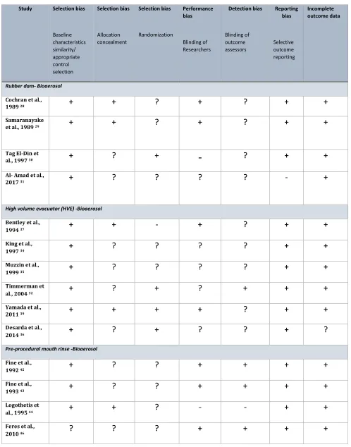
Table 5. Risk of Bias of the included studies
Study Selection bias
Baseline characteristics similarity/ appropriate control selection
Selection bias
Allocation concealment
Selection bias
Randomization
Performance bias
Blinding of Researchers
Detection bias
Blinding of outcome assessors
Reporting bias
Selective outcome reporting
Incomplete outcome data
Rubber dam- Bioaerosol
Cochran et al.,
1989 28 + + ? + ? + +
Samaranayake
et al., 1989 29 + + ? + ? + +
Tag El-Din et
al., 1997 30 + ? +
-
? + +Al- Amad et al.,
2017 31 + ? ? ? ? - +
High volume evacuator (HVE) -Bioaerosol
Bentley et al.,
1994 37 + + - + ? + +
King et al.,
1997 34 + ? ? ? ? + +
Muzzin et al.,
1999 35 + ? ? ? ? + +
Timmerman et
al., 2004 32 + ? + ? + + +
Yamada et al.,
2011 39 + + + + ? + +
Desarda et al.,
2014 36 + ? + ? ? + ?
Pre-procedural mouth rinse -Bioaerosol
Fine et al.,
1992 42 + ? ? + + + +
Fine et al.,
1993 43 + ? ? + + + +
Logothetis et
al., 1995 44 + + ? - - + +
Feres et al.,
Dawson et al.,
2016 48 - ? - + + ? ?
Retamel -Valdez et al., 2017 47
+ ? + + + + ?