Original Article
Clinical efficacy of laparoscopic hepatectomy
and its effects on cellular immune function
Haiying Sun*, Ju Liu*, Yongpeng Liu, Mingmin Wang, Zirong Wen
Department of Liver Diseases, Hospital of Infectious Disease, Qingdao, Shandong, China. *Equal contributors and
co-first authors.
Received April 23, 2017; Accepted May 24, 2017; Epub July 15, 2017; Published July 30, 2017
Abstract: Objective: To investigate the clinical efficacy of laparoscopic hepatectomy and its effects on organism
cellular immune function. Methods: Two hundred liver cancer patients cured in our hospital from January 2013 to December 2016 were enrolled in this study and randomly divided into observation group and control group, with 100 cases in each group. The patients in the observation group were treated with laparoscopic hepatectomy, while patients in the control group were treated with open hepatectomy. Various intraoperative indexes, postoperative
short-term and long-term efficacy, occurrence of complications, ratio of CD3+ T lymphocytes and their subsets (CD4+ and CD8+) at different time points before and after operation, as well as expression levels of serum interleukin-6
(IL-6) and tumor necrosis factor-α (TNF-α) in two groups of patients were compared. Results: The operation time of the
observation group was longer than that of the control group; total bleeding volume and blood occlusion rate were less than those of the control group; incision length was shorter than that of the control group. Compared with the control group, the indwelling time of the drainage tube, time to start eating and postoperative hospital stays of the
patients in the observation group were significantly less than those in the control group. Compared with the control
group, 24 h and 72 h after operation, the indexes of AST, ALT and TBIL of the patients in the observation group
markedly reduced, while ALB level obviously increased (P<0.05), which was statistically significant. There were no
statistical differences of postoperative metastasis rate, relapse rate and mortality between the two groups, but the
incidence of complications in the observation group was significantly lower than that in the control group (P<0.05).
At postoperative 72 h, the ratio of CD3+ T lymphocytes and their subsets (CD4+ and CD8+) cell populations in the observation group was basically recovered to the preoperative level (P>0.05), while those in the control group were remarkably lower than the preoperative level (P<0.05). Compared with the preoperative condition, the levels of IL-6
and TNF-α in the control group increased 24 h and 72 h after operation (P<0.05); in the observation group, those two levels increased 24 h after operation (P<0.05), but there was no significant difference at 72 h and pre-operation (P>0.05). Conclusion: Laparoscopic hepatectomy for liver cancer had definite clinical efficacy, small trauma, high
security, low incidence of complications, little impact on patients’ cellular immune function and rapid postoperative recovery.
Keywords: Liver cancer, laparoscopic hepatectomy, open hepatectomy, clinical efficacy, cellular immune function
Introduction
Hepatectomy is regarded as the primary treat-ment for the radical resection of primary liver cancer. With complex liver structure and func-tion, hepatectomy will result in different de- grees of trauma to the body, cause a strong stress response, and lead to low cellular im- mune function. Studies have shown that the cellular immune function of patients with liver cancer surgery is negatively correlated with the degree of trauma [1, 2]. At present, on the basis
of ensuring the clinical efficacy, minimizing the
surgical trauma and utomostly preserving cel-lular immune function have become an
impor-tant direction in the field of modern surgery.
Compared with open hepatectomy, the trauma and postoperative stress response of
laparo-scopic hepatectomy significantly reduce [3-5],
[6, 7], but the clinical changes in cellular immune function are still unclear. In recent years, laparoscopic hepatectomy has gradually replaced open hepatectomy and been widely used in the treatment of liver cancer, with
wide-ly recognized clinical efficacy, but the adequacy
of laparoscopic hepatectomy for resection of malignant liver tumor remains controversial [8], which lacks a large sample of clinical data to
confirm and some of data needs long-term
accumulation and exploration. In this regard, this study aims to observe the short-term and
long-term efficacy of patients with
laparosco-pic hepatectomy and changes of T lymphocyte subsets and cytokines to evaluate the effects of laparoscopic hepatectomy, cellular immunity and cytokine levels.
Materials and methods
General information
Two hundred liver cancer patients cured in our hospital from January 2013 to December 2016 were enrolled in this study. Inclusion criteria:
Superficial lesions located in the II-VI; the size
of tumor <10 cm; no intrahepatic metastasis and metastasis of lung, brain, gastrointestinal and other visceral organs; no history of abdomi-nal surgery; no portal vein tumor thrombus; Child Pugh Class A or B. Exclusion criteria: Associated with cardiopulmonary dysfunction and other important organ dysfunction; surgi-cal contraindications; abdominal adhesions, portal hypertension and severe cirrhosis. All patients underwent preoperative routine ex- aminations such as CT, MRI, echocardiograms, etc. This study was approved by the Ethics Committee and every patient signed an in- formed consent.
Liver cancer patients were randomly divided into observation group and control group by the random number table, with 100 cases in each group. The general information (gender, age,
height, weight, etc.) of patients in two groups
had no statistical significance (P>0.05) and
was comparable, see Table 1.
Surgical methods
Laparoscopic hepatectomy was applied in observation group. A 10 mm longitudinal inci-sion was conducted under general anesthesia in the lower edge of the annulus umbilicalis, a Veress needle was inserted into the incision with conventional CO2, pressure was main-tained at about 12 mmHg. The 4-hole method was adopted to select the corresponding poke holes of different anatomical sites of the liver tumor for the local or regular hepatectomy, according to the operational needs. Local hep-atectomy: Ata distance of 2 cm from the sepa-rated liver lesion, the liver parenchyma was directly mutilated from the liver by using the ultrasound knife; the resected portion of the liver was placed in the laparoscopic specimen bag, followed by rapidly removed from the extended epidermal incision. Regular hepatec-tomy: After the ligaments around the lesion were separated and mutilated, liver lobes exposed. We pre-controlled and dealt with
por-tal blood flow, marking in the liver segment and
anatomical structures of liver lobes; tumor specimens were directly resected and removed by using the ultrasound knife according to signs. After electric coagulation hemostasis and placing a drainage tube, the incision was sewed layer by layer. The control group was treated with open hepatectomy, according to the separated perihepatic ligament, the local or regular hepatectomy was performed on the basis of the operation steps. The postoperative specimens of two groups were used for patho-logical examination.
Observation index
The operation time, total blood loss, incision
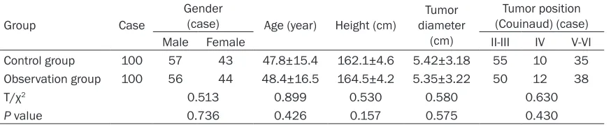
[image:2.612.90.527.86.177.2]length and blood flow occlusion of two groups Table 1. Comparison of general information of patients in two groups
Group Case
Gender
(case) Age (year) Height (cm) diameter Tumor (cm)
Tumor position (Couinaud) (case) Male Female II-III IV V-VI
Control group 100 57 43 47.8±15.4 162.1±4.6 5.42±3.18 55 10 35
Observation group 100 56 44 48.4±16.5 164.5±4.2 5.35±3.22 50 12 38
T/χ2 0.513 0.899 0.530 0.580 0.630
were compared; the postoperative short-term
efficacy indicators: indwelling time of drainage
tube, time to start eating and postoperative
hospital stays were analyzed and compared; the postoperative long-term efficacy index
(metastasis rate, relapse rate, mortality) and complications (perihepatic effusions, ascites, infection, bile leakage, etc.) were compared. Venous blood was extracted at different time points of before operation, 24 h and 72 h after
operation respectively. FACS flow cytometry
(BD Co. USA) was applied to detect CD3+ T lym-phocytes and their subsets (CD4+ and CD8+);
Thermo thermoelectric FC microplate reader
was adopted to detect the levels of serum
inter-leukin-6 (IL-6) and tumor necrosis factor (TNF-α) via enzyme-linked immunosorbent assay
(ELISA).
Follow-up visit
All patients were followed up after surgery once per month by means of outpatient appointment and telephone follow-up for one year to observe
the long-term efficacy index of them.
Statistical processing
SPSS17.0 software was used to process the experimental data, measurement data was expressed as _x ± s, and T test was adopted to compare the data between groups. Enumerat- ion data was represented as percentage, and
The operation time of the observation group was longer than that of the control group; total bleeding volume and blood occlusion rate were less than those of the control group; incision length was shorter than that of the control group. According to the statistical analysis, vari-ous intraoperative differences of patients in
the two groups were statistically significant,
see Table 2.
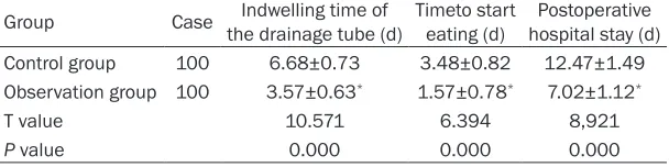
Comparison of the short-term efficacy indexes between the two groups of patients
Compared with the control group, the indwell-ing time of the drainage tube, time to start eat-ing and postoperative hospital stays of patients
in the observation group were significantly less
than those in the control group. According to the statistical analysis, the differences were
statistically significant, see Table 3.
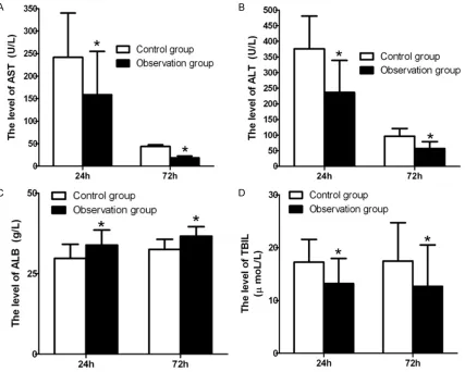
Comparison of liver function indexes between the two groups of patients
Compared with the control group, the indexes of AST (158.68±96.45 U/L vs 241.85±98.32 U/L), ALT (236.67±102.37 U/L vs 376.25±
104.46 U/L) and TBIL (13.22±4.73 μmol/L vs 17.25±4.31 μmol/L) significantly decreased 24
[image:3.612.95.523.85.163.2]h after operation, while the ALB level in the observation group was apparently higher than that in the control group (33.85±4.67 g/L vs
Table 2. Comparison of the various indexes of patients in two groups
Group Case Operation time (min) Total bleeding volume (ml) Incision length (cm) Blood occlusion rate (%)
Control group 100 158.3±24.62 451.5±167.12 25.1±0.14 46
Observation group 100 192.7±22.55* 323.3±158.33* 4.56±0.17* 11*
T/χ2 5.698 2.597 41.233 30.058
P value 0.000 0.012 0.000 0.000
Note: Compared with the control group, *P<0.05.
Table 3. Comparison of short-term efficacy indexes of patients in two
groups
Group Case the drainage tube (d)Indwelling time of Timeto start eating (d) hospital stay (d)Postoperative
Control group 100 6.68±0.73 3.48±0.82 12.47±1.49
Observation group 100 3.57±0.63* 1.57±0.78* 7.02±1.12*
T value 10.571 6.394 8,921
P value 0.000 0.000 0.000
Note: Compared with the control group, *P<0.05.
χ2 test was used to com-pare the data between groups. P<0.05 was con-sidered statistically sig-
nificant.
Results
[image:3.612.91.395.224.300.2]29.72±4.38 g/L). The difference was
statisti-cally significant (P<0.05), see Figure 1.
The levels of AST (18.62±3.95 U/L vs 43.88 ±3.56 U/L), ALT (56.74±22.39 U/L vs 96.52±
24.63 U/L) and TBIL (12.68±7.83 μmol/L vs 17.47±7.23 μmol/L) were significantly lower
than those of the control group, while the level
of ALB in the observation group was
signifi-cantly higher than that in the control group (36.62±2.95 g/L vs 32.48±3.15 g/L) 72 h aft-
rate, relapse rate and mortality both in the ob- servation group and the control group. Com- pared with the observation group, the inci- dence of postoperative complications was
sig-nificantly lower in the control group, reaching statistical significance (P<0.05), see Table 4.
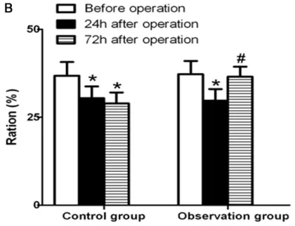
Comparison of the proportion of T lymphocytes between the two groups of patients
[image:4.612.95.522.72.414.2]The ratio of CD3+ T lymphocytes and their sub-sets (CD4+ and CD8+) in two groups significantly
Table 4. Comparison of the long-term efficacy indexes and the inci -dence of complications of patients in two groups
Group Case Long-term efficacy (case) Complications (case) Metastasis Relapse Mortality
Control group 100 4 5 8 30 (30%)
Observation group 100 10 8 7 18 (18%)
χ2 value 1.920 0.740 0.072 3.947
[image:4.612.91.380.496.575.2]P value 0.166 0.390 0.788 0.047
Figure 1. Comparison of postoperative liver function indexes of patients in two groups. A: AST index; B: ALT index; C: ALB index; D: TBIL index. Compared with the control group, *P<0.05.
er operation. The difference
was statistically significant,
see Figure 1.
Comparison of the long-term efficacy and incidence of complications in patients between the two groups
reduced compared to the preoperation 24 h after operation. The difference was statistically
significant. However, the quantity differences
of CD3+ T lymphocytes and their subsets (CD4+ and CD8+) in two groups were not statistical
significant. Compared with the preoperative
condition, there was no statistical difference in the ratio of CD3+ T lymphocytes and their subsets (CD4+ and CD8+) in the observation group 72 h after operation, but the ratio in the control group was still lower (P<0.05). The ratio of CD3+ T lymphocytes and their subsets (CD4+ and CD8+) in the observation group was signi-
ficantly higher than that in the control group (P<0.05), which had statistical significance.
See Figure 2.
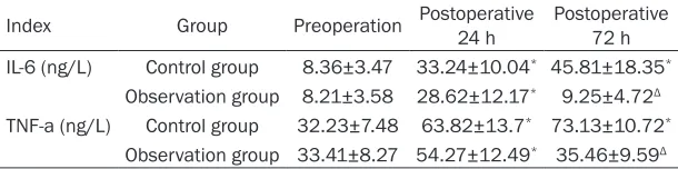
Comparison of the expression levels of IL-6 and TNF-α between the two groups of patients
Compared with the preoperative condition, the postoperative expression levels of IL-6 and
TNF-α in the control group significantly increa-sed with statistical significance (all P=0.000);
those expression levels in observation group 24 h after operation were also obviously incre-
ased with statistical significance (all P=0.000);
however, those expression levels in the obser-vation group 72 h after operation and the pre-operative condition showed no statistical
sig-nificance (P=0.905, P=0.897). Compared with
the control group, those expression levels in
the observation group significantly decreased 72 h after operation (all P=0.000), and the difference was of statistical significance, see Table 5.
Discussion
Hepatectomy is the first choice for the treat -ment of liver cancer. Compared with open hep-atectomy, laparoscopic hepatectomy has
obvi-ous advantages for liver cancer [9, 10]. For
[image:5.612.313.523.73.234.2]tectomy, a set of operation procedures can be completed under the direct vision, bleeding in the liver section can be reduced by the com-bined usage of ultrasonic knife cutting, and it has clear vision, high operational accuracy, as
well as significantly reduced damage of the sur -rounding tissues and organs. The key to laparo-scopic hepatectomy for the treatment of liver cancer lies in the following aspects: whether the liver cancer tissues can be fully resected, whether bile leakage, bleeding and other com-plications can be avoided, and it must ensure that the resected liver cancer tissues can be removed completely and successfully. With the development and progress of laparoscopic technology, the scope of indications of laparo-scopic hepatectomy is further expanded, which can be used to treat liver surface tumors, api-cal tumors and multiple liver cancer, with less and less surgical contraindications [11-13]. The total bleeding volume and blood occlusion rate of the observation group were less than those of the control group; incision length was shorter than that of the control group. All of these sug-gested that laparoscopic hepatectomy was
sig-nificantly less traumatic than open laparotomy,
its safety can be assured, and it had a good prospect, which was consistent with the previ-ous studies [14, 15]. Laparoscopic hepatecto-my, with the ultrasound knife as an intraopera-tive instrument, has the characteristics of the exact location of cutting and less damage on normal tissues. In addition, the surgery be-
comes more difficult due to its small incision
and surgeons must keep good vision and avoid blood occlusion at the same time. Therefore,
normative refined operation is needed, leading
to prolonged operation time, which is, in this study, one of the reasons why the observation group spends more operative time than the control group.
scopic hepatectomy of the observation group
was significantly better than that of the
con-trol group. This was probably because that laparoscopic hepatectomy kept the patients’ abdominal cavity a relative closed state, which effectively reduced the exposure and water evaporation of the internal organs, alleviating the stimulation of the gastrointestinal tract, and the less intraoperative bleeding volume
reduced the postoperative infusion quantity.
With respect to the postoperative recovery of liver function, the results of this study showed that the indexes of AST, ALT, TBIL of patients in
the observation group were significantly lower
than those in the control group at 24 h and 72 h after operation, while the ALB level was apparently higher than that in the control group, with statistical differences. It demonstrated that the patients with laparoscopic hepatecto-my had a better recovery of liver function than those with open hepatectomy, which may result from lighter degree of hepatic section trauma and liver crush injury of laparoscopic surgery.
The long-term efficacy of laparoscopic
hepa-tectomy for liver cancer is always attracting the concern and attention of scholars. In the
early stage, the inaccurate identification of liver
tumor boundary combined with intra-abdomi-nal hypertension, ineffective lymph node dis-section and tumor spread, resulted in limiting the extensive application of laparoscopic hepa-tectomy. This study indicated that the compli-cations of laparoscopic hepatectomy for liver cancer were relatively few; it reached
statisti-cally significance, compared with open hepa -tectomy. By one year of postoperative
follow-up, we found there was no significant difference
in tumor metastasis rate, relapse rate and mor-tality rate in the two groups of patients, which further illustrated that the long-term clinical
[image:6.612.90.395.98.174.2]efficacy of laparoscopic hepatectomy for the Table 5. Comparison of expression levels of cytokines of patientsin two
groups
Index Group Preoperation Postoperative 24 h Postoperative 72 h IL-6 (ng/L) Control group 8.36±3.47 33.24±10.04* 45.81±18.35* Observation group 8.21±3.58 28.62±12.17* 9.25±4.72Δ
TNF-a (ng/L) Control group 32.23±7.48 63.82±13.7* 73.13±10.72* Observation group 33.41±8.27 54.27±12.49* 35.46±9.59Δ
Note: Compared with the preoperative, *P<0.05; compared with the control group, ΔP<0.05.
As for postoperative sh-
ort-term efficacy, through
the study we can see that the indwelling time of drainage tube, time to start eating, postopera-tive hospital stays of the observation group were shorter than those of the control group, which also showed that the
-treatment of liver cancer was the same as that of open laparotomy.
Previous studies have suggested that laparo-scopic hepatectomy cannot completely remove hepatoma carcinoma cells and still remained in the treatment phase of reducing tumor load [16, 17]. At present, it is believed that the tumor metastasis or relapse depends entirely on the cellular immune function. If the patients have normal cellular immune function after hepatec-tomy, it had a certain role in inhibiting or killing tumor cells; on the contrary, tumor cells free of
immune surveillance can be quickly metasta
-sized or relapsed [18]. T lymphocytes were a
multifunctional cell population that plays an important role in humoral immunity and cellular immunity. CD3+ T lymphocytes can help T lym-phocyte antigen receptor to identify the major histocompatibility complex on antigen present-ing cells. CD4+ and CD8+ are two important cell subsets of CD3+ T lymphocytes, which can
reflect the immunoregulation ability of living
organisms. This study aims to detect the chang-es of cellular immune function after hepatec-tomy by measuring the ratio of CD3+ T lympho-cytes and their subsets (CD4+ and CD8+). The results show that the ratio in the observation group is restored to the preoperative level 72 hours after operation, while that in the control group is still lower than the preoperative level, indicating that laparoscopic surgery has little effect on the postoperative cellular immune
function and has a quick recovery. This is prob -ably because that laparoscopic hepatectomy has fewer traumas, small operation incision, and relatively complete skin and mucosal bar-rier, which can exhibit exogenous pathogens into living organisms, and has lighter stress reactions [19]. Although living organisms are subjected to a certain degree of immunosup-pression, laparoscopic hepatectomy can pro-mote the rapid recovery of cellular immune function [20, 21].
IL-6 and TNF-α are cytokines produced by mac
-rophages and monocytes. In the acute inflam -matory phage of trauma, IL-6 can regulate pro-liferation and differentiation of T lymphocytes,
and induce cytotoxic T lymphocytes. And TNF-α, an effector of cell-mediated and tissue dam
-aged immune inflammatory response, can
en-hance the proliferation of T lymphocytes to the
antigens. It can be seen that IL-6 and TNF-α
can complete the immune response and inflam
-mation-mediated response in acute inflamma -tory phage of trauma. In this study, the results showed that the expression levels of IL-6 and
TNF-α significantly increased at 24 h after liver
resection. However, They in the observation group were basically back to the preoperative level at 72 h after operation, while in the con-trol group, the levels still remained at a high level, suggesting laparoscopic surgery had few impacts on the living organism’s immune
func-tion damage and inflammafunc-tion response, fur
-ther reflecting the minimal invasion of laparo -scopic hepatectomy.
In summary, laparoscopic hepatectomy for liver
cancer has definite clinical efficacy and the
advantages such as little trauma, high safety, low incidence of complications, small impact on cellular immune function and rapid postop-erative recovery. However, there are still some limitations in this study, such as small sample
size, single-center research and so on. The result still needs to be further confirmed by
broad scholars’ continuous exploration and
practice through a large sample size, and multi
-center randomized trial. It is believed that
with the constant improvement of technology, laparoscopic hepatectomy will have a broader application prospect.
Disclosure of conflict of interest
None.
Address correspondence to: Zirong Wen, Depart- ment of Liver Diseases, Hospital of Infectious
Disease, No. 9 Fushun Road, Shibei District,
Qing-dao 266000, Shandong, China. Tel: +86-0532-81-
636120; Fax: +86-0532-81636688; E-mail: ziron [email protected]
References
[1] Shiganova AM, Vyzhigina MA, Buniatian KA, In -viiaeva EV, Vinnitskii LI, Balaian OV and Go-lovkin AS. [The role of immune monitoring in major liver resections from the position of the operative trauma and anaesthesia protection
level]. Anesteziol Reanimatol 2013; 30-34.
[2] Jerin A, Pozar-Lukanovic N, Sojar V, Stanisav
-ljevic D, Paver-Erzen V and Osredkar J. Balance of pro- and anti-inflammatory cytokines in liver
[3] Sarpel U, Hefti MM, Wisnievsky JP, Roayaie S,
Schwartz ME and Labow DM. Outcome for pa -tients treated with laparoscopic versus open resection of hepatocellular carcinoma: case-matched analysis. Ann Surg Oncol 2009; 16: 1572-1577.
[4] Nguyen KT, Marsh JW, Tsung A, Steel JJ, Gamb
-lin TC and Geller DA. Comparative benefits of
laparoscopic vs open hepatic resection: a criti-cal appraisal. Arch Surg 2011; 146: 348-356. [5] Zhang X, Yan L, Li B, Wen T, Wang W, Xu M, Wei
Y and Yang J. Comparison of laparoscopic
ra-diofrequency ablation versus open resection in
the treatment of symptomatic-enlarging he-patic hemangiomas: a prospective study. Surg Endosc 2016; 30: 756-763.
[6] Kuntz C, Kienle P, Schmeding M, Benner A, Autschbach F and Schwalbach P. Comparison of laparoscopic versus conventional technique
in colonic and liver resection in a tumor-bear-ing small animal model. Surg Endosc 2002; 16: 1175-1181.
[7] Burpee SE, Kurian M, Murakame Y, Benevides
S and Gagner M. The metabolic and immune response to laparoscopic versus open liver re-section. Surg Endosc 2002; 16: 899-904. [8] Gutt CN, Kim ZG, Schmandra T, Paolucci V and
Lorenz M. Carbon dioxide pneumoperitoneum
is associated with increased liver metastases in a rat model. Surgery 2000; 127: 566-570. [9] Coelho FF, Kruger JA, Fonseca GM, Araujo RL,
Jeismann VB, Perini MV, Lupinacci RM, Cecco-nello I and Herman P. Laparoscopic liver resec-tion: experience based guidelines. World J Gastrointest Surg 2016; 8: 5-26.
[10] Benkabbou A, Souadka A, Serji B, Hachim H, El Malki HO, Mohsine R, Ifrine L and Belkouchi A. Laparoscopic liver resection: initial experience in a North-African single center. Tunis Med 2015; 93: 523-526.
[11] Reddy SK, Tsung A and Geller DA. Laparoscop -ic liver resection. World J Surg 2011; 35: 1478-1486.
[12] Wakabayashi G, Cherqui D, Geller DA, Buell JF, Kaneko H, Han HS, Asbun H, O’Rourke N, Ta
-nabe M, Koffron AJ, Tsung A, Soubrane O,
Machado MA, Gayet B, Troisi RI, Pessaux P, Van
Dam RM, Scatton O, Abu Hilal M, Belli G, Kwon
CH, Edwin B, Choi GH, Aldrighetti LA, Cai X,
Cleary S, Chen KH, Schon MR, Sugioka A, Tang
CN, Herman P, Pekolj J, Chen XP, Dagher I, Jar-nagin W, Yamamoto M, Strong R, Jagannath P,
Lo CM, Clavien PA, Kokudo N, Barkun J and
Strasberg SM. Recommendations for laparo-scopic liver resection: a report from the second international consensus conference held in Morioka. Ann Surg 2015; 261: 619-629.
[13] Xiang L, Xiao L, Li J, Chen J, Fan Y and Zheng S.
Safety and feasibility of laparoscopic hepatec-tomy for hepatocellular carcinoma in the pos-terosuperior liver segments. World J Surg 2015; 39: 1202-1209.
[14] Lee W, Park JH, Kim JY, Kwag SJ, Park T, Jeong SH, Ju YT, Jung EJ, Lee YJ, Hong SC, Choi SK
and Jeong CY. Comparison of perioperative and oncologic outcomes between open and laparoscopic liver resection for intrahepatic cholangiocarcinoma. Surg Endosc 2016; 30: 4835-4840.
[15] Untereiner X, Cagnet A, Memeo R, De Blasi V,
Tzedakis S, Piardi T, Severac F, Mutter D, Kian -manesh R, Marescaux J, Sommacale D and Pessaux P. Short-term and middle-term evalua-tion of laparoscopic hepatectomies compared with open hepatectomies: a propensity score matching analysis. World J Gastrointest Surg 2016; 8: 643-650.
[16] Pulitano C and Aldrighetti L. The current role of laparoscopic liver resection for the treatment of liver tumors. Nat Clin Pract Gastroenterol Hepatol 2008; 5: 648-654.
[17] Bryant R, Laurent A, Tayar C and Cherqui D.
Laparoscopic liver resection-understanding its role in current practice: the Henri Mondor hos-pital experience. Ann Surg 2009; 250: 103-111.
[18] Cariani E, Pilli M, Zerbini A, Rota C, Olivani A, Pelosi G, Schianchi C, Soliani P, Campanini N,
Silini EM, Trenti T, Ferrari C and Missale G. Im -munological and molecular correlates of dis-ease relapse after liver resection for hepato-cellular carcinoma. PLoS One 2012; 7: e32493.
[19] Novitsky YW, Litwin DE and Callery MP. The net immunologic advantage of laparoscopic sur-gery. Surg Endosc 2004; 18: 1411-1419. [20] Chopra SS, Haacke N, Meisel C, Unterwalder
N, Fikatas P and Schmidt SC. Postoperative im -munosuppression after open and laparoscopic liver resection: assessment of cellular immune function and monocytic HLA-DR expression. JSLS 2013; 17: 615-621.
[21] Gutt CN, Kim ZG, Schemmer P, Krahenbuhl L
and Schmedt CG. Impact of laparoscopic and
conventional surgery on Kupffer cells,