R E S E A R C H
Open Access
Molecular evolution of Azagny virus, a newfound
hantavirus harbored by the West African pygmy
shrew (Crocidura obscurior) in Côte d
’
Ivoire
Hae Ji Kang
1, Blaise Kadjo
2, Sylvain Dubey
3, François Jacquet
4and Richard Yanagihara
1*Abstract
Background:Tanganya virus (TGNV), the only shrew-associated hantavirus reported to date from sub-Saharan Africa, is harbored by the Therese’s shrew (Crocidura theresae), and is phylogenetically distinct from Thottapalayam virus (TPMV) in the Asian house shrew (Suncus murinus) and Imjin virus (MJNV) in the Ussuri white-toothed shrew (Crocidura lasiura). The existence of myriad soricid-borne hantaviruses in Eurasia and North America would predict the presence of additional hantaviruses in sub-Saharan Africa, where multiple shrew lineages have evolved and diversified.
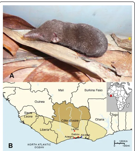
Methods:Lung tissues, collected in RNAlater®, from 39 Buettikofer’s shrews (Crocidura buettikoferi), 5 Jouvenet’s shrews (Crocidura jouvenetae), 9 West African pygmy shrews (Crocidura obscurior) and 21 African giant shrews (Crocidura olivieri) captured in Côte d’Ivoire during 2009, were systematically examined for hantavirus RNA by RT-PCR.
Results:A genetically distinct hantavirus, designated Azagny virus (AZGV), was detected in the West African pygmy shrew. Phylogenetic analysis of the S, M and L segments, using maximum-likelihood and Bayesian methods, under the GTR+I+Γmodel of evolution, showed that AZGV shared a common ancestry with TGNV and was more closely related to hantaviruses harbored by soricine shrews than to TPMV and MJNV. That is, AZGV in the West African pygmy shrew, like TGNV in the Therese’s shrew, did not form a monophyletic group with TPMV and MJNV, which were deeply divergent and basal to other rodent- and soricomorph-borne hantaviruses. Ancestral distributions of each hantavirus lineage, reconstructed using Mesquite 2.74, suggested that the common ancestor of all
hantaviruses was most likely of Eurasian, not African, origin.
Conclusions:Genome-wide analysis of many more hantaviruses from sub-Saharan Africa are required to better understand how the biogeographic origin and radiation of African shrews might have contributed to, or have resulted from, the evolution of hantaviruses.
Background
Sub-Saharan Africa has long been considered the“ birth-place” of many medically important vector-borne and zoonotic viruses. Among myriad examples are human immunodeficiency virus type 1, which resulted from multiple cross-species transmissions among nonhuman primates to become well established in humans [1,2]; Ebola virus, a filovirus harbored by fruit bats [3,4], which is among the deadliest viruses on earth; and
Lassa virus, a rodent-borne arenavirus [5], which causes an estimated 100,000-500,000 human infections each year in West African countries. In addition, the recent report of Lujo virus as the cause of fatal hemorrhagic fever in South Africa [6], where severe arenavirus-asso-ciated human disease had not been previously recog-nized, serves as a startling reminder that other pathogenic zoonotic viruses remain undiscovered.
While rodents (Order Rodentia) have long been known to serve as reservoirs of hantaviruses (Family Bunyaviridae, Genus Hantavirus), recent studies indicate a far richer genetic diversity among hantaviruses har-bored by shrews (Order Soricomorpha, Family * Correspondence: yanagiha@pbrc.hawaii.edu
1
John A. Burns School of Medicine, University of Hawaii at Manoa, Honolulu, HI 96813, USA
Full list of author information is available at the end of the article
Soricidae) and moles (Family Talpidae) of multiple spe-cies spanning across four continents, including Thotta-palayam virus (TPMV) in the Asian house shrew (Suncus murinus) [7,8], Imjin virus (MJNV) in the Ussuri white-toothed shrew (Crocidura lasiura) [9], Cao Bang virus (CBNV) in the Chinese mole shrew ( Anouro-sorex squamipes) [10], Seewis virus (SWSV) in the Eura-sian common shrew (Sorex araneus) [11,12], Ash River virus (ARRV) in the masked shrew (Sorex cinereus) and Jemez Spring virus (JMSV) in the dusky shrew (Sorex monticolus) [13], Kenkeme virus (KKMV) in the flat-skulled shrew (Sorex roboratus) [14], Camp Ripley virus (RPLV) in the northern short-tailed shrew (Blarina bre-vicauda) [15], Asama virus (ASAV) in the Japanese shrew mole (Urotrichus talpoides) [16], Oxbow virus (OXBV) in the American shrew mole (Neurotrichus gibbsii) [17], Nova virus (NVAV) in the European com-mon mole (Talpa europaea) [18] and Rockport virus (RKPV) in the eastern mole (Scalopus aquaticus) [19]. Until recently, conspicuous in their absence have been reports of hantaviruses in Africa. Sangassou virus (SANV) in the African wood mouse (Hylomyscus simus) [20] and Tanganya virus (TGNV) in the Therese’s shrew (Crocidura theresae) [21] are the only known examples to date, but additional hantaviruses likely exist in sub-Saharan Africa, where unique rodent and soricid lineages have evolved and diversified.
In this study, lung tissues from crocidurine shrews captured in Côte d’Ivoire were systematically analyzed for hantavirus RNA by reverse transcription polymerase chain reaction (RT-PCR). Undaunted by the vast genetic diversity of hantaviruses harbored by shrews, we resorted to a brute-force strategy of designing oligonu-cleotide primers based on regions of high sequence con-servation. After exhaustive hit-and-miss attempts, a genetically distinct hantavirus, designated Azagny virus (AZGV), was detected in the West African pygmy shrew (Crocidura obscurior). Sequence and phylogenetic ana-lyses of the S, M and L segments indicated that AZGV shared a common ancestry with TGNV and was evolu-tionarily distant from TPMV and MJNV, two crocidur-ine shrew-borne hantaviruses in Asia.
Methods
Trapping and specimen collection
Shrews were live caught, using pitfall traps, as well as Sherman traps (8 × 9 × 23 cm) (H.B. Sherman, Talla-hassee, FL) and Longworth traps (14 × 6.5 × 8.5 cm) (Penlon Ltd., Oxford, UK). Pitfall traplines were set with drift fences using 1-m plastic sheeting or fine mesh cloth erected between short poles and running across plastic buckets that were 20-cm deep and buried flush with the soil surface. Up to 20 buckets were placed along 100- to 150-m long drift fences at 5- to 10-m
intervals. Sherman and Longworth traps were set at intervals of 5 m during daylight hours of each day. Trapping was conducted over two to 10 consecutive days, at Azagny National Park, Lamto Natural Reserve and Dabou (Figure 1), during March, July, August, November and December 2009. Traplines and pitfalls were checked every morning and coordinates of each trapped animal were recorded by the global positioning system (GPS). Species, gender, weight, reproductive maturity and GPS coordinates of each captured shew were recorded. Lung tissues were collected in RNAla-ter® RNA Stabilization Reagent (Qiagen, Inc., Chats-worth, CA) and processed for RNA extraction, within 4 weeks of collection, for RT-PCR analysis. All experimen-tal procedures on animals followed internationally recognized guidelines and were approved by the Ivorian National Park Office and the Muséum National d’ His-toire Naturelle.
RNA extraction and cDNA synthesis
Total RNA was extracted, using the PureLink Micro-to-Midi total RNA purification kit (Invitrogen, San Diego, CA), from shrew tissues, and cDNA was prepared using
the SuperScript III First-Strand Synthesis System (Invi-trogen) and random hexamers and/or an oligonucleotide primer (OSM55: 5’-TAGTAGTAGACTCC-3’) designed from the conserved 5’-end of the S, M and L segments of hantaviruses.
RT-PCR and DNA sequencing
PCR was performed as described previously [18], with each 20-μL reaction containing 250 μM dNTP, 2 mM MgCl2, 1 U of AmpliTaq polymerase (Roche, Basel,
Swit-zerland) and 0.25μM of oligonucleotide primers, with trial-and-error testing of primers and modified cycling conditions. Initial denaturation at 94°C for 5 min was fol-lowed by two cycles each of denaturation at 94°C for 40 sec, two-degree step-down annealing from 48°C to 38°C for 40 sec, and elongation at 72°C for 1 min, then 32 cycles of denaturation at 94°C for 40 sec, annealing at 42° C for 40 sec, and elongation at 72°C for 1 min, in a Gen-eAmp PCR 9700 thermal cycler (Perkin-Elmer, Waltham, MA). Amplicons were purified using the QIAQuick Gel Extraction Kit (Qiagen, Hilden, Germany), and DNA sequencing was performed using an ABI Prism 377XL Genetic Analyzer (Applied Biosystems, Foster City, CA).
Protein analysis and secondary structure prediction
For secondary structure prediction of the nucleocapsid protein and envelope glycoprotein, amino acid sequences were submitted to NPS@ structure server [22]. Glycosylation sites and transmembrane helices were predicted using NetNlyc 1.0 and Predictprotein [23] and TMHMM version 2.0 [24], respectively.
Phylogenetic analysis
Partial S-, M- and L-segment sequences of AZGV were aligned and compared with publicly available hantavirus sequences, using ClustalW (DNASTAR, Inc., Madison, WI) [25] and transAlign [26]. Representative members of theNairovirus(Dugbe virus, NC_004157) and Phlebo-virus(Rift Valley fever virus, NC_014395) genera were included as outgroups. Phylogenetic trees were gener-ated by maximum likelihood and Bayesian methods, implemented in PAUP* (Phylogenetic Analysis Using Parsimony, 4.0b10) [27], RAxML Blackbox webserver [28] and MrBayes 3.1 [29], under the best-fit GTR+I+Γ model of evolution established using jModeltest 0.1.1 [30]. By employing two complementary and not redun-dant ML methods, additional analytical rigor was achieved. Topologies were evaluated by bootstrap analy-sis of 1,000 iterations, and posterior node probabilities were based on 2 million generations and estimated sam-ple sizes over 100 (imsam-plemented in MrBayes). Since tree topologies for each genomic segment were very similar using RAxML, PAUP* and MrBayes, trees generated by MrBayes were displayed.
Biogeographic analyses
Ancestral distributions of each lineage were recon-structed using a maximum-likelihood approach using Mesquite 2.74 [31]. The current geographic distribution of hantavirus species was coded as 0 for Eurasia, 1 for Africa, and 2 for America. Similarly, we tested for an ancestral Soricomorpha (0) vs. Rodentia (1) origin of hantaviruses. The model of character evolution was a simple stochastic model (Mk1) [32], which assumed an equal and symmetrical rate of change between any two states [33,34], and the character state frequencies were estimated from the transition probabilities. In this likeli-hood-based approach, the probability that a character changes along a branch of the tree is consequently a function of the branch length, with changes being less likely along shorter branches than longer ones.
To have branch lengths representing absolute time of divergence, a calibrated Bayesian tree was built with BEAST 1.4 [35] using a Yule tree prior (adequate to study interspecific diversification). Preliminary analyses were performed with an uncorrelated lognormal relaxed clock to test if a strict molecular clock can be rejected for our dataset (ucld.stdev parameter with a frequency histogram abutting 0). Because in our simulations the
‘’ucld.stdev’’ had a frequency histogram not abutting 0, we chose this molecular clock for the analyses [36]. The analyses were performed with two independent chains and 10 million generations and chains were sampled every 1,000 generations with a burn-in of 1 million gen-erations. Convergences were determined with Tracer v1.4 [37].
mtDNA sequence analysis
The entire 1,140-nucleotide region of the cytochromeb
gene of mtDNA was amplified by PCR, as previously described [17,18], to confirm the taxonomic identity of the hantavirus-infected shrew and to examine its phylo-genetic relationship to other shrew hosts.
Results and Discussion
RT-PCR and sequence analysis
In analyzing lung tissues from 39 Buettikofer’s shrews (Crocidura buettikoferi), 5 Jouvenet’s shrews (Crocidura jouvenetae), 9 West African pygmy shrews (Crocidura obscurior) and 21 African giant shrews (Crocidura oli-vieri) by RT-PCR using multiple primer pairs, novel hantavirus RNAs were detected in a West African pygmy shrew, captured in Azagny National Park (lati-tude, 5°14.5’N; longitude, 4°48.1’W), Côte d’Ivoire, on December 15, 2009 (Figure 1). The identity of the West African pygmy shrew (GenBank JF276229), in which AZGV was detected, was confirmed by phylogenetic analysis of the 1,140-nucleotide mtDNA cytochromeb
Although hantavirus RNAs were not found in tissues of the other three African crocidurine shrew species tested, we believe it is simply because suitable oligonu-cleotide primers were not designed. This conjecture is based on our past experience of repeated failed attempts before the successful detection of soricomorph-borne hantaviruses [17-19]. Thus, continuing efforts are ongoing to retest these specimens, and future expedi-tions are being planned to collect tissues suitable for virus isolation attempts.
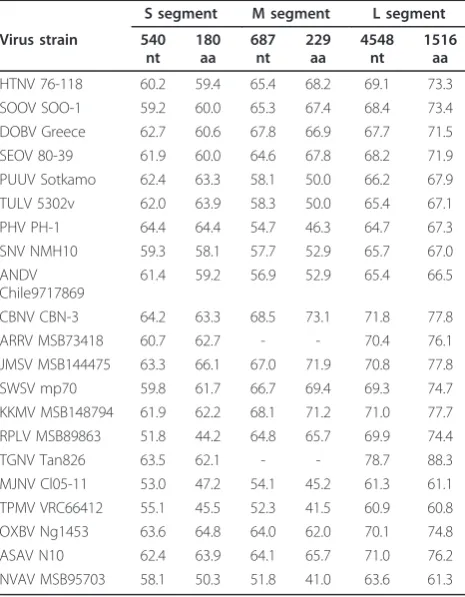
Despite intensive efforts to obtain the full genome of AZGV, the inability to effectively design primers for this novel hantavirus and the limited amount of available RNA were obstacles too great to overcome. Neverthe-less, pairwise alignment and comparison of partial S-, M- and L-genomic segment sequences of AZGV with representative rodent- and soricid-borne hantaviruses clearly demonstrated that AZGV was genetically distinct (Table 1). AZGV was most closely related to TGNV
from neighboring Guinea and was highly divergent from TPMV and MJNV, two crocidurine shrew-borne hanta-viruses from Asia.
In a 540-nucleotide region of the AZGV S segment (GenBank JF276226), the hypothetical nonstructural protein (NSs) open-reading frame, found in arvicolid, neotomine and sigmodontine rodent-borne hantaviruses, was absent. Analysis of a 687-nucleotide region of the AZGV M-genomic segment (GenBank JF276227), which spanned the Gn-Gc juncture, showed the highly con-served WAASA amino acid motif cleavage site at posi-tions 167 to 171. Potential N-linked glycosylation sites were found at amino acid positions 46, 118 and 142 in the Gn glycoprotein and 179 and 216 in the Gc gylco-protein. Analysis of the nearly full-length 4,548-nucleo-tide AZGV L-genomic segment (GenBank JF276228), encoding an incomplete RNA-dependent RNA polymer-ase of 1,516 amino acids, showed conservation of the five major motifs, designated A, B, C, D and E [38].
Extensive analysis of the available AZGV genome, using multiple recombination-detection methods, including GENECONV, Bootscan, Chimaera, 3SEQ, RDP, SiScan, MaxChi and HyPhy Single Recombinant Breakpoint [39], failed to disclose any evidence of genetic recombination.
Phylogenetic analysis and co-phylogeny mapping
High bootstrap support for a shared ancestry between AZGV and TGNV, as evidenced by bootstrap values of >70% and posterior node probabilities of >0.70, was found in phylogenetic trees, based on available S- and L-segment sequences, using the maximum-likelihood (ML) and Bayesian methods (Figure 2). That is, AZGV in the West African pygmy shrew, like TGNV in the Therese’s shrew [21], did not form a monophyletic group with TPMV in the Asian house shrew [8] and MJNV in the Ussuri white-toothed shrew [9], which were deeply divergent and basal to other rodent- and soricomorph-borne hantaviruses, with longer branch lengths suggesting greater evolutionary change and pos-sibly older age.
Phylogenetic trees, reconstructed for co-phylogeny mapping using TreeMap 2.0b[40], exhibited high boot-strap support for each genomic segment (at both the nucleotide and amino acid levels), with segregation of hantaviruses, according to the Subfamily of their sorico-morph reservoir hosts. That is, congruent topologies were found in the host-virus phylogenetic relationships, except for previously reported hantaviruses hosted by two species of shrew moles [16,17].
Biogeographic analyses
Because the choice of basal taxa can strongly influence the biogeographic reconstruction of the target taxa in
Table 1 Sequence similarities (%) of the partial S, M and L segments of AZGV strain KBM15 and representative hantaviruses harbored by rodents and soricomorphs
S segment M segment L segment
Virus strain 540
HTNV 76-118 60.2 59.4 65.4 68.2 69.1 73.3 SOOV SOO-1 59.2 60.0 65.3 67.4 68.4 73.4 DOBV Greece 62.7 60.6 67.8 66.9 67.7 71.5 SEOV 80-39 61.9 60.0 64.6 67.8 68.2 71.9 PUUV Sotkamo 62.4 63.3 58.1 50.0 66.2 67.9 TULV 5302v 62.0 63.9 58.3 50.0 65.4 67.1 PHV PH-1 64.4 64.4 54.7 46.3 64.7 67.3 SNV NMH10 59.3 58.1 57.7 52.9 65.7 67.0 ANDV
Chile9717869
61.4 59.2 56.9 52.9 65.4 66.5
CBNV CBN-3 64.2 63.3 68.5 73.1 71.8 77.8 ARRV MSB73418 60.7 62.7 - - 70.4 76.1 JMSV MSB144475 63.3 66.1 67.0 71.9 70.8 77.8 SWSV mp70 59.8 61.7 66.7 69.4 69.3 74.7 KKMV MSB148794 61.9 62.2 68.1 71.2 71.0 77.7 RPLV MSB89863 51.8 44.2 64.8 65.7 69.9 74.4 TGNV Tan826 63.5 62.1 - - 78.7 88.3 MJNV Cl05-11 53.0 47.2 54.1 45.2 61.3 61.1 TPMV VRC66412 55.1 45.5 52.3 41.5 60.9 60.8 OXBV Ng1453 63.6 64.8 64.0 62.0 70.1 74.8 ASAV N10 62.4 63.9 64.1 65.7 71.0 76.2 NVAV MSB95703 58.1 50.3 51.8 41.0 63.6 61.3
such analyses, the outgroups (Dugbe virus and Rift Val-ley fever virus) were not included in the analyses. As shown in Figure 3, the common ancestor of all hanta-viruses likely emerged in Eurasia rather than Africa (ML support, 87%). Moreover, the analyses suggested that the ancestral hosts of hantaviruses may have been members of the Soricomorpha, rather than Rodentia (ML support, 56%). However, genomic analyses of many more sorico-morph-borne hantaviruses, and studies in search of han-taviruses in other potential reservoir hosts, especially insectivorous bats (Order Chiroptera), are necessary to validate these tentative conclusions.
Biogeographic reconstruction, based on fossil records and assuming equally probable exchanges between con-tinents in both directions, suggests that the Family Sori-cidae originated in Eurasia [33]. And based on karyologic, paleontologic and molecular genetic data, a Palaearctic-Oriental origin has been proposed for the genus Crocidura, dating to the Upper Miocene (6.8 mil-lion years before present) [34]. Moreover, in-depth ana-lysis of 3,560-base pairs of mitochondrial and nuclear DNA of crocidurine shrews from across Eurasia and
Africa, reveals that shrews belonging to the genus Croci-dura segregate into an Afrotropical clade, an Asian clade and an Old World clade, which includes Afrotro-pical, East Palaearctic-Oriental and West Palaearctic species [34]. Crocidura obscurior groups into the Old World clade, whileCrocidura theresae, as well as Croci-dura buettikoferi, CrociCroci-dura jouvenetaeand Crocidura olivieri, group in the Afrotropical clade, and Crocidura lasiurain the Asian clade.
Apart from Côte d’Ivoire, the West African pygmy shrew is found in subtropical or tropical moist lowland forests in Ghana, Guinea, Liberia, Sierra Leone and pos-sibly Nigeria. A similar West African distribution is noted forCrocidura buettikoferiandCrocidura jouvene-tae, while Crocidura olivieriis far more widespread, from west to east across sub-Saharan Africa [41]. Although previous ecological studies had indicated that
Therese’s shrew or to analyze the genetic diversity and phylogeography of TGNV.
In this regard, future studies are warranted to confirm that the West African pygmy shrew is the true reservoir host of AZGV. Also, whole genome sequence analysis of many more hantaviruses from sub-Saharan Africa are required to gain a better understanding about how the biogeographic origin and radiation of African shrews might have contributed to, or have resulted from, the evolution and cross-species transmission of hantaviruses. Intensified efforts are underway in search of hanta-viruses in other shrew species, including Myosorexand
Sylvisorex shrews, which are not found elsewhere and are unique to Africa. Meanwhile, the disproportionately high number of hantaviruses hitherto discovered in sori-comorphs over a relatively brief period of searching, and the far greater genetic diversity of shrew- and mole-borne hantaviruses, compared to those harbored by rodents, provide additional support for the emerging concept that soricomorphs may have served as the early reservoirs of ancestral hantaviruses.
Acknowledgements
We thank the director of the Ivorian National Park Office and Dr.
Souleymane Konate, director of the Lamto Natural Reserve, for permission to conduct studies in protected sites. Also, we thank Professor Christiane Denys for supporting the field work. Financial support for this research was provided in part by U.S. Public Health Service grant R01AI075057 from the National Institute of Allergy and Infectious Diseases, and grants P20RR018727
and G12RR003061 from the National Center for Research Resources, National Institutes of Health.
Author details
1
John A. Burns School of Medicine, University of Hawaii at Manoa, Honolulu, HI 96813, USA.2Department of Biology, Université de Cocody, Abidjan, Côte
d’Ivoire.3School of Biological Sciences, University of Sydney, New South Wales 2006, Australia.4Department Systématics and Evolution, Muséum
National d’Histoire Naturelle, Paris, France.
Authors’contributions
HJK performed the RNA extraction, RT-PCR, and genetic and phylogenetic analyses. BK and FJ conducted the field expeditions and provided the background data on wild-caught shrews. SD conducted the biogeographic analysis and data interpretation. RY conceived the research design, arranged the collaboration and provided scientific oversight. All authors read and approved the final manuscript.
Competing interests
The authors declare that they have no competing interests.
Received: 22 June 2011 Accepted: 28 July 2011 Published: 28 July 2011
References
1. Van Heuverswyn F, Peeters M:The origins of HIV and implications for the global epidemic.Curr Infect Dis Rep2007,9:338-346.
2. Sharp PM, Hahn BH:The evolution of HIV-1 and the origin of AIDS.Philos Trans R Soc Lond B Biol Sci2010,365:2487-2494.
3. Leroy EM, Kumulungui B, Pourrut X, Rouquet P, Hassanin A, Yaba P, Délicat A, Paweska JT, Gonzalez JP, Swanepoel R:Fruit bats as reservoirs of Ebola virus.Nature2005,438:575-576.
4. Biek R, Walsh PD, Leroy EM, Real LA:Recent common ancestry of Ebola Zaire virus found in a bat reservoir.PLoS Pathog2006,2:e90. 5. Frame JD, Baldwin JM Jr, Gocke DJ, Troup JM:Lassa fever, a new virus
disease of man from West Africa. I. Clinical description and pathological findings.Am J Trop Med Hyg1970,19:670-676.
6. Briese T, Paweska JT, McMullan LK, Hutchison SK, Street C, Palacios G, Khristova ML, Weyer J, Swanepoel R, Egholm M, Nichol ST, Lipkin WI:
Genetic detection and characterization of Lujo virus, a new hemorrhagic fever-associated arenavirus from Southern Africa.PLoS Pathog2009,4: e1000455.
7. Carey DE, Reuben R, Panicker KN, Shope RE, Myers RM:Thottapalayam virus: a presumptive arbovirus isolated from a shrew in India.Indian J Med Res1971,59:1758-1760.
8. Yadav PD, Vincent MJ, Nichol ST:Thottapalayam virus is genetically distant to the rodent-borne hantaviruses, consistent with its isolation from the Asian house shrew (Suncus murinus).Virol J2007,4:80. 9. Song JW, Kang HJ, Gu SH, Moon SS, Bennett SN, Song KJ, Baek LJ, Kim HC,
O’Guinn ML, Chong ST, Klein TA, Yanagihara R:Characterization of Imjin virus, a newly isolated hantavirus from the Ussuri white-toothed shrew (Crocidura lasiura).J Virol2009,83:6184-6191.
10. Song JW, Kang HJ, Song KJ, Truong TT, Bennett SN, Arai S, Truong NU, Yanagihara R:Newfound hantavirus in Chinese mole shrew, Vietnam.
Emerg Infect Dis2007,13:1784-1787.
11. Song JW, Gu SH, Bennett SN, Arai S, Puorger M, Hilbe M, Yanagihara R:
Seewis virus, a genetically distinct hantavirus in the Eurasian common shrew (Sorex araneus).Virol J2007,4:114.
12. Kang HJ, Arai S, Hope AG, Song JW, Cook JA, Yanagihara R:Genetic diversity and phylogeography of Seewis virus in the Eurasian common shrew in Finland and Hungary.Virol J2009,6:208.
13. Arai S, Bennett SN, Sumibcay L, Cook JA, Song JW, Hope A, Parmenter C, Nerurkar VR, Yates TL, Yanagihara R:Phylogenetically distinct hantaviruses in the masked shrew (Sorex cinereus) and dusky shrew (Sorex monticolus) in the United States.Am J Trop Med Hyg 2008,78:348-351.
14. Kang HJ, Arai S, Hope AG, Cook JA, Yanagihara R:Novel hantavirus in the flat-skulled shrew (Sorex roboratus).Vector Borne Zoonotic Dis2010,
10:593-597.
15. Arai S, Song JW, Sumibcay L, Bennett SN, Nerurkar VR, Parmenter C, Cook JA, Yates TL, Yanagihara R:Hantavirus in northern short-tailed shrew, United States.Emerg Infect Dis2007,13:1420-1423. Figure 3 Phylogenetic distribution and reconstruction.
16. Arai S, Ohdachi SD, Asakawa M, Kang HJ, Mocz G, Arikawa J, Okabe N, Yanagihara R:Molecular phylogeny of a newfound hantavirus in the Japanese shrew mole (Urotrichus talpoides).Proc Natl Acad Sci USA2008,
105:16296-16301.
17. Kang HJ, Bennett SN, Dizney L, Sumibcay L, Arai S, Ruedas LA, Song JW, Yanagihara R:Host switch during evolution of a genetically distinct hantavirus in the American shrew mole (Neurotrichus gibbsii).Virology 2009,388:8-14.
18. Kang HJ, Bennett SN, Sumibcay L, Arai S, Hope AG, Mocz G, Song JW, Cook JA, Yanagihara R:Evolutionary insights from a genetically divergent hantavirus harbored by the European common mole (Talpa europaea).
PLoS One2009,4:e6149.
19. Kang HJ, Bennett SN, Hope AG, Cook JA, Yanagihara R:Shared ancestry between a mole-borne hantavirus and hantaviruses harbored by cricetid rodents.J Virol2011,85:7496-7503.
20. Klempa B, Fichet-Calvet E, Lecompte E, Auste B, Aniskin V, Meisel H, Denys C, Koivogui L, ter Meulen J, Krüger DH:Hantavirus in African wood mouse, Guinea.Emerg Infect Dis2006,12:838-840.
21. Klempa B, Fichet-Calvet E, Lecompte E, Auste B, Aniskin V, Meisel H, Barrière P, Koivogui L, ter Meulen J, Krüger DH:Novel hantavirus sequences in shrew, Guinea.Emerg Infect Dis2007,13:520-522. 22. Combet C, Blanchet C, Geourjon C, Deléage G:NPS@: Network Protein
Sequence Analysis.Trends Biochem Sci2000,25:147-150.
23. Gavel Y, von Heijne G:Sequence differences between glycosylated and non-glycosylated Asn-X-Thr/Ser acceptor sites: implications for protein engineering.Protein Eng1990,3:433-442.
24. Krogh A, Larsson B, von Heijne G, Sonnhammer EL:Predicting transmembrane protein topology with a hidden Markov model: application to complete genomes.J Mol Biol2001,305:567-580. 25. Thompson JD, Higgins DG, Gibson TJ:CLUSTAL W: improving the
sensitivity of progressive multiple sequence alignment through sequence weighting, position-specific gap penalties and weight matrix choice.Nucleic Acids Res1994,22:4673-4680.
26. Bininda-Emonds OR:transAlign: using amino acids to facilitate the multiple alignment of protein-coding DNA sequences.BMC Bioinformatics 2005,6:156.
27. Swofford D:PAUP*: Phylogenetic Analysis Using Parsimony (*and Other Methods).Sunderland, Massachusetts: Sinauer Associates; 2003.
28. Stamatakis A, Hoover P, Rougemont J:A rapid bootstrap algorithm for the RAxML web servers.Syst Biol2008,57:758-771.
29. Ronquist F, Huelsenbeck JP:MrBayes 3: Bayesian phylogenetic inference under mixed models.Bioinformatics2003,19:1572-1574.
30. Posada D:jModelTest: Phylogenetic Model Averaging.Mol Biol Evol2008,
25:1253-1256.
31. Maddison WP, Maddison DR:Mesquite: a modular system for evolutionary analysis.2010 [http://mesquiteproject.org], Version 2.74. 32. Lewis PO:A likelihood approach to estimating phylogeny from discrete
morphological character data.Syst Biol2001,50:913-925. 33. Dubey S, Salamin N, Ohdachi SD, Barrière P, Vogel P:Molecular
phylogenetics of shrews (Mammalia: Soricidae) reveals timing of transcontinental colonizations.Mol Phylogenet Evol2007,44:126-137. 34. Dubey S, Salamin N, Ruedi M, Barrière P, Colyn M, Vogel P:Biogeographic
origin and radiation of the Old World crocidurine shrews (Mammalia: Soricidae) inferred from mitochondrial and nuclear genes.Mol Phylogenet Evol2008,48:953-963.
35. Drummond AJ, Rambaut A:BEAST v1.4.2006 [http://beast.bio.ed.ac.uk/]. 36. Drummond AJ, Ho SYW, Rawlence N, Rambaut A:A Rough Guide to
BEAST 1.4.2007 [http://beast.bio.ed.ac.uk/].
37. Rambaut A, Drummond AJ:Tracer v1.4.2007 [http://beast.bio.ed.ac.uk/ Tracer].
38. Kukkonen SK, Vaheri A, Plyusnin A:L protein, the RNA-dependent RNA polymerase of hantaviruses.Arch Virol2005,150:533-556.
39. Pond SL, Frost SDW, Muse SV:HyPhy: hypothesis testing using phylogenies.Bioinformatics2005,21:676-679.
40. Charleston MA, Page RDM:TreeMap 2.0b. Macintosh program for co-phylogenetic analysis, 2.0b ed.1998.
41. Dubey S, Antonin M, Denys C, Vogel P:Use of phylogeny to resolve the taxonomy of the widespread and highly polymorphic African giant shrews (Crocidura olivierigroup, Crocidurinae, Mammalia).Zoology2007,
110:48-57.
42. Kadjo B:Peuplement en Rongeurs dans quatre milieux de savane préforestière de Lamto, Côte d’Ivoire.Mémoire de DEA1992, 71. 43. Kadjo B, Dosso H, Vuattoux R:Influence de la végétation sur le
peuplement des rongeurs dans la région centre de Côte d’Ivoire.
Agronomie Africaine1996,8:169-180.
doi:10.1186/1743-422X-8-373
Cite this article as:Kanget al.:Molecular evolution of Azagny virus, a
newfound hantavirus harbored by the West African pygmy shrew
(Crocidura obscurior) in Côte d’Ivoire.Virology Journal20118:373.
Submit your next manuscript to BioMed Central and take full advantage of:
• Convenient online submission
• Thorough peer review
• No space constraints or color figure charges
• Immediate publication on acceptance
• Inclusion in PubMed, CAS, Scopus and Google Scholar
• Research which is freely available for redistribution