CLINICAL ANDDIAGNOSTICLABORATORYIMMUNOLOGY,
1071-412X/99/$04.00⫹0 Nov. 1999, p. 938–945 Vol. 6, No. 6
Copyright © 1999, American Society for Microbiology. All Rights Reserved.
Hemoglobin Toxicity in Experimental Bacterial Peritonitis Is
Due to Production of Reactive Oxygen Species
YEONG-MIN YOO,1,2KI-MO KIM,1SUNG-SOO KIM,3JEONG-A HAN,1HO-ZOO LEA,2
ANDYOUNG-MYEONG KIM1*
Departments of Molecular and Cellular Biochemistry1and Pharmacology,3College of Medicine, and Department of Biology, College of Natural Science,2Kangwon National University,
Chunchon, Kangwon-do, Korea
Received 9 December 1998/Returned for modification 31 March 1999/Accepted 12 July 1999
Hemoglobin (Hb) is a toxic molecule responsible for the extreme lethality associated with experimental
Escherichia coliperitonitis, but the mechanism has yet to be elucidated. Hb, but not globin, showed toxic effects
in a liveE. colimodel but not in a model using killedE. coli. Methemoglobin, hematin, and the well-known
Fenton reagents iron and iron-EDTA demonstrated the same lethal effect inE. coliperitonitis as Hb, while the
addition of the Fenton inhibitors desferrioxamine (DF) and diethylenetriamine pentaacetate removed most of the cytotoxic activity of iron. Administration of a combined dose of superoxide dismutase and catalase
mini-mized the action of Hb and iron-EDTA, suggesting that both O2˙ⴚand H2O2are involved in the toxic action of
Hb in this rat model. The combination of the antioxidative enzymes and DF further suppressed iron-mediated
lethality. An electron spin resonance technique with the spin-trapping reagent 5,5-dimethyl-1-pyroline-N-oxide
(DMPO) showed O2˙ⴚgeneration in the peritoneal fluid of rats injected withE. colialone orE. coliplus
iron-DF, and ˙OH generation was detected in the peritoneal fluid of the rats injected with iron-EDTA. Hb did not show any spin adduct of oxygen radicals, suggesting that Hb produces non-spin-trapping radical ferryl ion,
which decayed the spin adduct of DMPO. In the presence of Hb or iron-EDTA, O2ⴚ-generating activity and
viability of phagocytes decreased, whereas lipid peroxidation of peritoneal phagocytes increased. Generation of oxygen radicals and lipid peroxidation did not differ in the live and dead bacterial models. Bacterial numbers in the peritoneal cavity and blood were markedly increased in the live bacterial model with Hb and iron-EDTA. The Fenton inhibitor iron-DF prevented the loss of phagocyte function, lipid peroxidation, and bacterial proliferation. These results led us to conclude that the lethal toxicity of Hb in bacterial peritonitis is associated with a Fenton-type reaction, the products of which decrease phagocyte viability, through the induction of lipid peroxidation, allowing bacterial proliferation and resulting in mortality.
Phagocytes (macrophages and neutrophils) are primary host defense systems against peritoneal infection. They induce an important biochemical pathway, a respiratory burst, which re-sults in generation of O2˙⫺and H2O2, followed by the forma-tion of more-toxic secondary oxidants such as hydroxyl radical (˙OH) and HOCl. These oxidizing species are responsible for pathological events (29) such as destruction of biomolecules, killing of infected microorganisms, and autokilling of phago-cytes. The hydroxyl radical is generated by transition metals, particularly iron, via the Fenton reaction in an O2˙⫺ -generat-ing environment. This radical is thought to be a major spe-cies for oxygen toxicity in biological systems. Yamazaki and Piette (31) have stoichiometrically measured the formation of ˙OH and confirmed the formation of not only ˙OH but also ferryl ion from the Fenton reaction by using a spin-trapping technique.
Hemoglobin (Hb) may also act as a Fenton reagent in bio-logical systems (22, 24). This possibility has been supported by many lines of evidences that show that Hb causes the oxidation of several organic molecules by producing ˙OH and/or ferryl ion [Fe(IV)AO] in the presence of H2O2 (9, 23). Free Hb causes injury to the central nervous system through
peroxida-tion of membrane lipid (26). We confirmed that neutrophils were inactivated by the oxidizing species, ferryl ion, produced from Hb in O2˙⫺-generating systems (17).
Hb has been known as a potent toxic molecule responsible for the high lethality associated with experimentalEscherichia coliperitonitis since the early 1960s, when Davis and Yull (3, 4) discovered a synergistic effect on mortality between bacteria and erythrocytes in the peritoneums of rats. This synergistic action is a perplexing problem caused by the combination of blood and bacteria during surgery. Recently, Kim et al. re-ported that erythrocytes scavenge both O2˙⫺and nitric oxide and then inhibit the formation of the bactericidal molecule peroxynitrite (14). Although the lethal toxicity of purified Hb has been thought to be associated with various biochemical events in the peritoneal cavity (1, 7), the lethal effect of Hb in the animal model is not fully understood.
We have therefore examined the possibilities that Hb pro-duces strong oxidizing species by reacting with O2˙⫺and H2O2 generated by recruited neutrophils in the peritoneal cavity and that these oxidizing species cause deleterious effects on the host defense system. By comparing Hb toxicity with the effects of well-known Fenton reagents and inhibitors on the mor-tality of rats, reactive oxygen production, and the pathobio-chemical changes in this animal model, we demonstrated that a Fenton-type reaction producing reactive oxygen spe-cies may be involved in the lethality of Hb in experimental E. coliperitonitis.
* Corresponding author. Mailing address: Department of Molecular and Cellular Biochemistry, College of Medicine, Kangwon National University, Chunchon, Kangwon-do, Korea. Phone: 82-361-250-8831. Fax: 82-361-242-7571. E-mail: ymkim@cc.kangwon.ac.kr.
938
on August 17, 2020 by guest
http://cvi.asm.org/
MATERIALS AND METHODS
Materials.Catalase (CAT) (from bovine liver), superoxide dismutase (SOD) (from bovine erythrocytes), phorbal myristate acetate, cytochromec(from horse heart), phosphate-buffered saline (PBS) (pH 7.4), 5,5-dimethyl-1-pyroline-N -oxide (DMPO), globin, and hemin were obtained from Sigma. DMPO was used after redistillation. Desferrioxamine B (deferoxamine) (DF) was from CIBA Pharmaceutical Co. Hematin solutions were prepared by dissolving hemin in a small volume of weak alkaline solution and diluting with PBS within 1 h of experiments. Hb was prepared from bovine erythrocytes by a one-step procedure with a DEAE-Sephadex A-50 column (30). Methemoglobin (MetHb) was pre-pared by oxidizing Hb with a slight excess of potassium ferricyanide and sepa-rated from the added components through a Sephadex G-25 column. Hb and MetHb were concentrated with a Centricon apparatus (Amicon). The Hb con-centration was calculated as a heme concon-centration by spectrophotometry.
Preparation of bacterial solution.E. coli(ATCC 25922) was incubated on a blood agar plate at 37°C for 17 h, and a single colony was cultured in tryptic soy broth for an additional 18 h to produce a suspension of approximately 109 cells/ml. The bacteria were harvested by centrifugation, washed three times with saline, and resuspended in saline. The cell concentration was measured by spectrophotometry at 550 nm. The bacterial solution (absorbance was⬃1.0 at 550 nm) was diluted in PBS by 106- and 107-fold, inoculated on agar plates, and incubated at 37°C for 18 h (17). The number of viable bacteria was measured by counting developed colonies. Killed E. coliwas prepared by treatment with formalin (5).
Animal mortality.Male Sprague-Dawley rats (180 to 200 g) were given stan-dard rat pellets and water ad libitum. Experimental peritonitis was induced in rats by intraperitoneal (i.p.) injection ofE. coli(4⫻109CFU/kg of body weight) with or without iron complexes (12.4 mol of iron/kg of body weight). The injection mixtures were prepared immediately before injection, and the injection volume was 5 ml/kg of body weight. Mortality of rats was determined at 24 h after administration. The effects of SOD and CAT were examined by two i.p. injec-tions of 2,600 and 11,000 U/kg of body weight, respectively, at 0 and 8 h following injection of bacteria.
Measurement of O2˙ⴚproduction in the peritoneal fluid.The rats injected i.p.
with viableE. coliwith or without an iron complex were anesthetized with an intramuscular injection of Ketamine (44 mg/kg) at various time points. The rats were i.p. injected with 1 ml of saline and then physically shaken for 1 min to ensure adequate mixing of peritoneal contents. A midline laparotomy was per-formed, and the fluid was obtained from all regions of the peritoneal cavity. O2˙⫺ generation in an ex vivo system was measured by SOD (100 U/ml)-inhibitable cytochromecreduction, monitored at 550 nm (⌬ε550⫽21 mM⫺1 cm⫺1for reduced oxidized cytochromec), in PBS (1 ml) containing cytochrome c(100M), peritoneal fluid (250l), and CAT (100 U).
Isolation, quantitation, and viability of peritoneal phagocytes.All peritoneal fluid was obtained from the rats injected i.p. with viableE. coliwith or without an iron complex. Peritoneal phagocytes were prepared by centrifugation at 300⫻
gfor 8 min and washed twice with ice-cold PBS. The cell pellets were resus-pended in PBS. Total phagocytes (macrophages and neutrophils) were counted by light microscopy with trypan blue staining. For counting the macrophage population, a cell suspension was plated on 24-well plates and incubated for 2 h at 37°C in a CO2incubator. The plates were washed three times with PBS, and
the adhering cells were counted by light microscopy. Cell viability was deter-mined by trypan blue dye exclusion.
Bacterial counts in peritoneal fluid and blood.Blood was collected by heart puncture and collected in a Vacutainer (Becton Dickinson; 7-ml capacity) con-taining 10.5 mg of EDTA. Peritoneal fluid and blood samples were diluted with ice-cold sterile water. The number of viable bacteria was measured as described above.
Lipid peroxidation.Peritoneal exudate cells were prepared by centrifugation at 500⫻gfor 10 min and suspended in a small volume of PBS. Lipid peroxi-dation was assayed by measuring thiobarbituric acid (TBA)-reactive substance (TBARS) at 532 nm (2). A sample (0.25 ml) was mixed with 0.5 ml of trichlo-roacetic acid-TBA-HCl (15%, 0.375%, and 0.25 N, respectively) containing 0.05% butylated hydroxytoluene. The mixture was heated in a boiling water bath for 20 min. After being cooled with tap water, the mixture was centrifuged at 2,200⫻gand 4°C for 20 min. The absorbance of the supernatant was measured at 532 nm. TBARS as a lipid peroxide level was calculated by using an extinction coefficient of 1.56⫻105M⫺1cm⫺1.
ESR spectroscopy of DMPO spin adducts.Peritoneal fluid was collected from the rats 4 h after the i.p. injection ofE. coliwith or without iron complexes. The fluid (460l) was mixed with 40l of 1 M DMPO and incubated at 37°C for 8 min. The mixture was immediately transferred into an electron spin resonance (ESR) flat cell, and ESR spectra were recorded with a Varian E-109 spectrom-eter. Spectrometer settings were as follows: microwave power, 20 mW; modula-tion frequency, 100 kHz; modulamodula-tion amplitude, 1.0 G; time constant, 0.25 s; receiver gain, 1.25⫻104. For decay of DMPO-OH by ferryl ion, DMPO-OH was formed by incubation of DMPO (10 mM), 100M Fe2⫹-EDTA, and 200M H2O2for 1 min and then addition of CAT (100 U/ml) to remove excessive H2O2. The reaction mixture was incubated with or without ferryl ion, which is generated by mixing Hb (0.4 mM) with H2O2(0.4 mM) for 4 min and then incubating with 100 U of CAT/ml for 1 min. After 8 min, ESR spectra were recorded with a Varian E-109 spectrometer with a receiver gain of 2⫻102.
Statistical analysis.Data were analyzed by the chi-square test for animal mortality and by the Studentttest. Statistical significance was established at aP
value of⬍0.05.
RESULTS
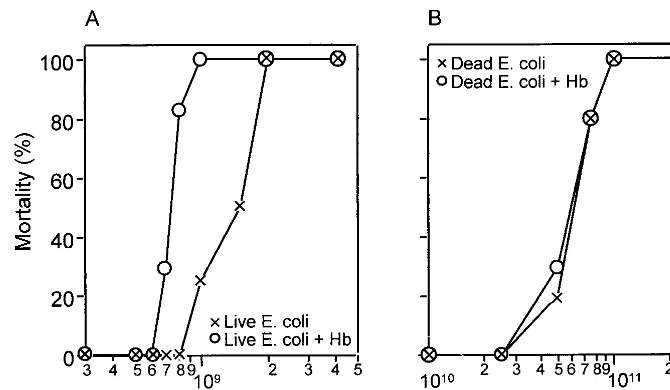
As shown by Simmons’ group (5, 6, 10, 11, 19, 22), purified Hb administered simultaneously with E. coliinto the perito-neal cavity results in a potentiated lethal effect (Fig. 1). Viable E. coli at 8 ⫻ 108bacteria/200 g of body weight, which is a nonlethal dose, resulted in more than 80% mortality when Hb was administered within 24 h of injection (Fig. 1A). However, formalin-killedE. coli, showing a similar dose-response mor-tality curve at a concentration about 100 times higher than that for live bacteria, did not show a synergistic effect on mortality at 5⫻1010bacteria/200 g of body weight when concomitantly injected with Hb (Fig. 1B), indicating that the viability of E. FIG. 1. Effect of Hb on mortality in experimentalE. coliperitonitis. Six or seven animals were studied in each group. Mortality was measured at 24 h after i.p. injection of liveE. coli(A) or formalin-killedE. coli(B) with or without Hb (12.4 mmol/kg of body weight [BW]). The total volume of the injection was 5 ml/kg of body weight.
VOL. 6, 1999 HEMOGLOBIN TOXICITY IN BACTERIAL PERITONITIS 939
on August 17, 2020 by guest
http://cvi.asm.org/
coliis an important factor in the toxic effect of Hb in experi-mental peritonitis.
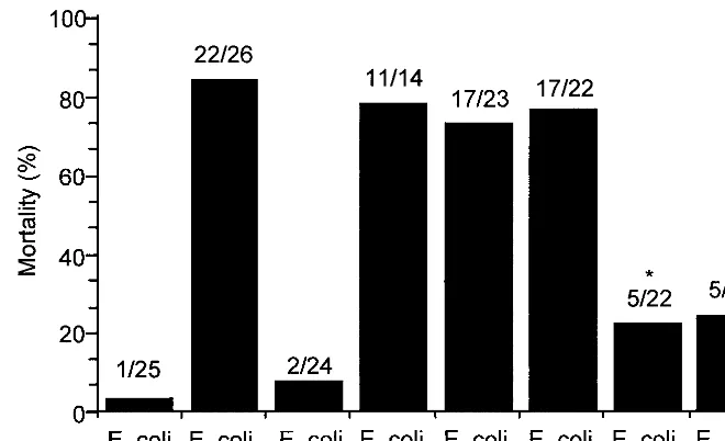
It is well known that iron from a variety of sources, including Hb, can enter into Fenton-type chemistry (25) and that neu-trophils infiltrating the infectious site produce O2˙⫺(13). It is therefore possible that Hb as a Fenton reagent produces a highly toxic oxidant in the peritoneal cavity during bacterial peritonitis. To test the hypothesis that a Fenton-type reaction is responsible for the lethal effect of Hb in bacterial peritonitis, we investigated the effects of globin, hematin, and several iron(II) chelates on mortality in the rat peritonitis model (Fig. 2). Hematin, iron itself, and iron-EDTA showed effects on mortality similar to those of Hb, whereas globin did not show a toxic effect. The initial valence state of iron, either ferrous or ferric, did not change the mortality (data not shown). Unlike EDTA, DF and diethylenetriamine pentaacetate (DTPA) sig-nificantly reduced the toxic effect of iron itself in experimental peritonitis. These data suggest that a Fenton-type reaction may be involved in the toxic effect of Hb in this animal model.
To test whether iron complexes play a role as a Fenton reagent in the rat peritonitis model, the effects of SOD and CAT on mortality and on generation of O2˙⫺and H2O2in the peritoneal cavity (since these oxygen metabolites are important reagents in Fenton chemistry) were investigated. A mixture of SOD and CAT was injected twice i.p. withE. coliand Hb or iron-EDTA into rats, simultaneously and 8 h after injection of bacteria. The toxic effect of the iron complexes on mortality was examined at 24 h after treatment. Administration of an SOD-CAT mixture reduced the toxic action of Hb (P⬍0.01) and iron-EDTA (P⬍ 0.01), though not completely (Fig. 3). Furthermore, the combination of the antioxidative enzymes and DF strongly suppressed iron-mediated lethality. The sults suggest that phagocytes (resident macrophages and re-cruited neutrophils) produced activated oxygen species (O2˙⫺ and H2O2), which react with Hb and iron-EDTA to generate more-toxic species such as ferryl ion and ˙OH.
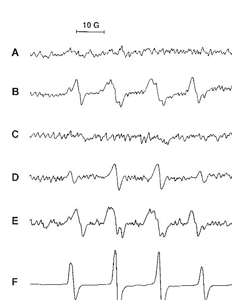
To establish definitively the generation of reactive oxygen species in our experimental model, we used ESR spectroscopy
to detect spin adducts of DMPO with oxygen radicals which were generated in the peritoneal fluid collected from rats at 4 h after injection with bacteria alone or bacteria plus iron che-lates. As shown in Fig. 4A, DMPO spin adducts were not found in the peritoneal fluid of rats which received saline as a control. The mixtures of DMPO spin adducts of O2˙⫺(DMPO-OOH) and ˙OH (DMPO-OH), but mostly DMPO-OH, were detect-ed directly in the peritoneal fluid of rats injectdetect-ed with bacteria alone (Fig. 4B). The spectrum obtained for the system of bacteria plus iron-DF was not different from that obtained for rats injected with bacteria alone (Fig. 4E). Under the same experimental conditions, the spectrum of DMPO-OH disap-peared with the addition of SOD (data not shown), indicat-ing that DMPO-OH was generated by decay of DMPO-OOH, as previously reported (20). However, the spectrum of only DMPO-OH was identified in the peritoneal fluid of rats in-jected with bacteria plus iron-EDTA (Fig. 4D), but no spin adduct was found in rats injected with bacteria plus Hb (Fig. 4C). Since DMPO spin adducts can be rapidly decayed by the reaction product of Hb and H2O2, the disappearance of the DMPO adduct of ˙OH was examined. An ESR spectrum of DMPO-OH was detected in the mixture of iron-EDTA and H2O2(Fig. 4F), and this spectrum disappeared with the addi-tion of ferryl ion generated from the reacaddi-tion of Hb with H2O2 (Fig. 4G).
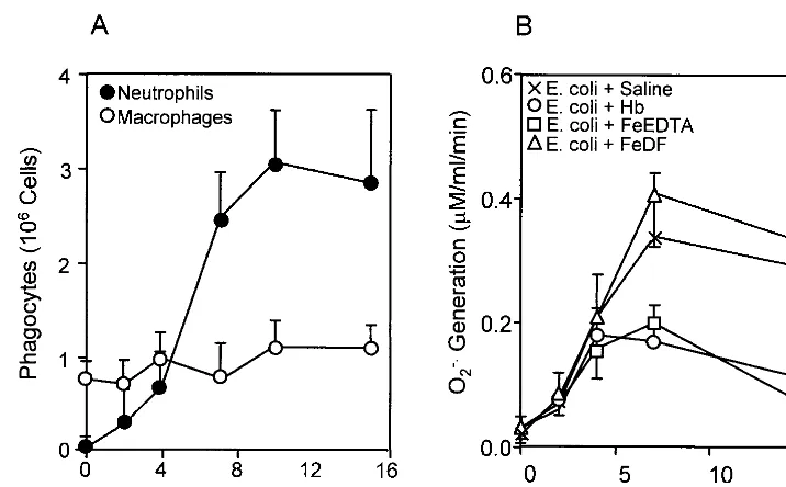
Our previous data showed that neutrophils are inactivated by toxic oxidants generated from Fenton-type reagents, includ-ing Hb in an O2˙⫺-generating system (17). To test if peritoneal phagocytes are inactivated by iron complexes in experimental bacterial peritonitis, we examined the phagocyte population and its O2˙⫺-generating activity in the peritoneal cavity (Fig. 5). The numbers of peritoneal macrophages were nearly un-changed, while the neutrophil population was significantly in-creased after 4 h of bacterial injection (Fig. 5A). The O2˙⫺ -generating activity of the peritoneal phagocytes isolated from rats injected withE. colialone orE. coliplus iron-DF increased after 2 h and reached maximum levels after 7 h (P⬍0.01) (Fig. 5B). Under the same conditions, however, treatment with iron-FIG. 2. Effects of Hb, globin, hematin, and iron chelates on mortality in experimentalE. coliperitonitis. Rats were injected i.p. withE. coli(4⫻109cells/kg of body weight) and iron complex or globin (12.4 mmol/kg of body weight), and mortality was determined at 24 h after injection. The injection mixtures were prepared immediately before injection. The total volume injected was 5 ml/kg of body weight. The molar ratio of iron(II) to chelator was 1:2. The ratio of dead rats to treated rats is indicated above each bar. Statistically significant mortality reductions (asterisks) with chelators, DF, and DTPA versus iron ion itself (P⬍0.001) were determined by the chi-square test. MetHb and iron(III) ion had the same adjuvant effects as Hb and Fe(II) ion, respectively.
940 YOO ET AL. CLIN. DIAGN. LAB. IMMUNOL.
on August 17, 2020 by guest
http://cvi.asm.org/
EDTA or Hb attenuated the O2˙⫺-generating activity of peri-toneal phagocytes.
Toxic oxidants produced from a Fenton-type reaction cause oxidative injury through lipid peroxidation (28), resulting in cell death and inactivation of neutrophil function (17). To address this concern, lipid peroxidation and phagocyte viability were measured in the peritoneal phagocytes of rats injected withE. coliand iron complexes. Figure 6A shows that injection ofE. colialone into the peritoneal cavity did not significantly increase lipid peroxidation in peritoneal phagocytes, but its injection with Hb or iron-EDTA markedly increased lipid per-oxidation in peritoneal phagocytes (P⬍0.02 at 15 h). Iron-DF had no effect on lipid peroxidation. Phagocyte viability in the peritoneal cavity decreased (P ⬍ 0.01 at 15 h) following injection with Hb or iron-EDTA but did not decrease sig-nificantly (P ⬎ 0.1 at 15 h) when bacteria were injected alone or with iron-DF (Fig. 6B). Furthermore, we examined the effects of live and deadE. colion reactive oxygen gen-eration and lipid peroxidation in the peritoneal cavity. The formation of DMPO spin adducts and lipid peroxidation in the peritoneal cavity were not significantly different between these peritonitis models with or without Hb (Table 1). These results suggest that bacterial proliferation through phago-cyte inactivation is a critical factor for Hb toxicity inE. coli peritonitis.
From work performed by Dunn et al. (5) and O’Brien (21), one might conclude that an important role for Hb or any iron source in experimental peritonitis is to cause uncontrolled bac-terial proliferation in the peritoneal cavity. Although forE. coli alone the number of cells in the peritoneal cavity began to decrease after an initial weak increase, the number ofE. coli cells injected with Fenton-type reagents, Hb, or iron-EDTA rapidly increased in the peritoneal cavity, by about 103-fold within 15 h (P⬍0.01) (Fig. 7A). The number of viable bacteria in blood was lower than that in the peritoneal cavity but its time dependence was identical to that in the peritoneal cavity (Fig. 7B). When Fe-DF (ratio of 1 to 2) was injected, the numbers of viable bacteria in the peritonal cavity and blood
FIG. 3. Effect of both SOD and CAT on the adjuvant effect of Hb or iron-EDTA in experimentalE. coliperitonitis. Experimental conditions were as described in the legend to Fig. 2, except that SOD and CAT were injected i.p. twice, at 2,600 and 11,000 U/kg, respectively, at 0 and 8 h after i.p. injection ofE. coliand adjuvant. The ratio of dead rats to treated rats is indicated above each bar. Statistically significant mortality reductions versus each control were determined by the chi-square test (ⴱ,P⬍0.01;ⴱⴱ,P⬍0.05).
FIG. 4. ESR spectrum of DMPO spin adducts of oxygen radical generated in peritoneal fluid. DMPO spin adducts in peritoneal fluids collected from rats injected with saline (A),E. coli(B),E. coliplus Hb (C),E. coliplus iron-EDTA (D), orE. coliplus iron-DF (E) were measured. In vitro formation of DMPO-OH was detected in the reaction mixture of Fe2⫹-EDTA and H
2O2(F) in the presence of ferryl ion (G), which is formed by incubation of Hb and H2O2, by ESR spectroscopy. Spectrometer settings are described in Materials and Meth-ods. The spectra were from one of three experiments, which showed similar results.
VOL. 6, 1999 HEMOGLOBIN TOXICITY IN BACTERIAL PERITONITIS 941
on August 17, 2020 by guest
http://cvi.asm.org/
were similar to those in animals injected with bacteria alone until 10 h and thereafter were a little higher (P⬍0.05 at 15 h), probably due to the suppression of phagocyte-dependent bac-terial killing by the excessive amount of DF (27).
DISCUSSION
As reported previously (6, 18), Hb increased lethality in experimental bacterial peritonitis. The most prominent char-acteristic reported so far regarding the toxic effect of Hb in experimental peritonitis seems to be the significantly increased rate of bacterial proliferation in vivo (7, 11). Hb did not in-crease the lethality in a peritonitis model of killed E. coli,
supporting a role for Hb in increasing bacterial growth. We previously reported that Hb diminished the microbicidal activ-ity of neutrophils in an O2˙⫺-generating system (17), and we postulated that a Fenton reaction producing toxic radicals played an important role in neutrophil inactivation. We here report that Hb and the Fenton reagent iron-EDTA, but not the Fenton inhibitor iron-DF, increased lethal toxicity by decreas-ing phagocyte viability and increasdecreas-ing bacterial growth in ex-perimental bacterial peritonitis.
Peritoneal phagocytes such as resident macrophages and infiltrating neutrophils are the first line of defense against bacterial peritonitis. They produce O2˙⫺and H2O2, which are FIG. 5. Quantitation of peritoneal phagocytes and their O2˙⫺-generating activity in experimentalE. coliperitonitis. Numbers of total peritoneal phagocytes were determined by light spectroscopy with the trypan blue staining method. Macrophages were counted from adhered cells on culture plates. Subpopulations of neutrophils and macrophages (A) were calculated by the subtraction method. O2˙⫺production (B) was measured by SOD-inhibitable cytochromecreduction in peritoneal phagocytes obtained from the rats injected with bacteria in the absence of iron complex and in the presence of Hb, iron-EDTA, or iron-DF. Each point represents the mean⫾standard deviation for three individual rats.
FIG. 6. Effects of Hb and iron chelates on lipid peroxidation (A) and viability (B) of peritoneal phagocytes duringE. coliperitonitis. Peritoneal phagocytes were prepared by centrifugation immediately after collection of the peritoneal fluids and washed with PBS. Data represent the means⫾standard deviations for three individual rats, with results obtained in duplicate for each experiment.
942 YOO ET AL. CLIN. DIAGN. LAB. IMMUNOL.
on August 17, 2020 by guest
http://cvi.asm.org/
implicated in bacterial killing. O2˙⫺ supplied by phagocytes interacts with oxidized iron to generate the reduced form of iron. The reduced forms of iron ion and its chelate complexes are well-known Fenton reagents that react with H2O2to pro-duce strong oxidizing species such as ˙OH and Fe(IV)AO, which oxidize several cellular components (17, 24).
Many investigators (9, 23, 25, 26, 30) have reported that Hb produces oxidizing species, ˙OH and/or Fe(IV)AO, in the presence of H2O2through a Fenton-type reaction. It has been suggested that these oxidizing species are responsible for oxy-gen toxicity, including neutrophil inactivation (17) and protein degradation (16), in various biological systems. Although iron(II)-DF and iron(II)-DTPA produce ˙OH in the presence of H2O2, DF and DTPA, probably because of the slow oxida-tion-reduction cycle of these iron chelates, are known inhibi-tors of the Fenton reaction. Here, we will first focus our dis-cussion on the possibility that the lethal effects of Hb and iron-EDTA in our peritonitis model are associated with the production of oxidizing species through a Fenton-type reaction (Fig. 2). Dead bacteria or lipopolysaccharide can also stimulate phagocytes and produce reactive oxygen species (12). The oxy-gen radicals inactivate the O2˙⫺-generating activity of neutro-phils through lipid peroxidation in the presence of a Fenton reagent in vitro (17). Although Hb also increases peroxidation
through oxygen radical generation in deadE. coliperitonitis to a level similar to that in liveE. coliperitonitis (Table 1), Hb did not enhance the lethal effect (Fig. 1B). These results suggest that bacterial proliferation through decreased phagocyte func-tion in the peritoneal cavity is one critical factor for Hb-medi-ated adjuvant mortality in bacterial peritonitis.
SOD and CAT are powerful scavengers of O2˙⫺and H2O2. Therefore, they are effective in inhibiting a Fenton reaction (18) and neutralizing neutrophil-mediated tissue damage (8). In our peritonitis model, administration of SOD and CAT reduced the lethal effect of Hb and iron-EDTA (Fig. 3), and the Fenton inhibitors DF and DTPA also suppressed the lethal effect of iron (Fig. 2). Furthermore, the combination of SOD, CAT, and DF strongly inhibited iron-mediated mortality in our peritonitis model. These results suggest that O2˙⫺and H2O2 are involved in the production of oxidizing species from the Fenton reaction of Hb and cause the toxic effect of Hb in our peritonitis model.
Phagocytes, i.e., macrophages and neutrophils, are known to produce O2˙⫺and H2O2, which normally are involved in the killing of infectious bacteria, and such was the case whenE. colialone was injected. These oxygen species can interact with Fenton reagents such as iron-EDTA and Hb and produce stronger oxidants such as ˙OH and ferryl ion, which are in-volved in tissue injury through lipid peroxidation (17). ESR spectroscopy showed direct evidence that peritoneal phago-cytes produce reactive oxygen species during bacterial perito-nitis, followed by the formation of more-autotoxic secondary oxidants such as ˙OH or ferryl ion in the presence of Fenton reagents. Neither O2˙⫺nor ˙OH was detected in the perito-neal cavities of the rats injected with bacteria plus Hb. A major reactive species produced by the reaction of Hb with H2O2is thought to be ferryl ion (17), which is not directly trapped by DMPO (31). The ferryl species rapidly decayed DMPO spin adducts to other products that were not detected by ESR spectroscopy (15). This evidence may explain why no ESR spectrum was detected when Hb was injected into the perito-neal cavity. These results indicate that the large number of neutrophils which migrated into the peritoneal cavity 4 h after bacterial injection appear to be the major source of O2˙⫺
FIG. 7. Effects of Hb and iron chelates on bacterial proliferation in the peritoneal cavity (A) and blood (B) duringE. coliperitonitis. Experimental conditions were the same as described in the legend to Fig. 5. Each point represents the mean⫾standard deviation of CFU from two individual rats, with results obtained in triplicate, at each indicated time point after i.p. inoculation withE. coli.
TABLE 1. Effects of live and deadE. colion reactive oxygen generation and lipid peroxidation in bacterial peritonitis
E. coli Injection DMPO spinadductsa (mean⫾SD)
Lipid peroxidationb (mean⫾SD)
Live Saline 100.0⫾10.2 35.8⫾5.3
Hb 5.6⫾2.1 76.5⫾6.3
Dead Saline 84.2⫾11.5 28.8⫾5.3
Hb 4.2⫾1.7 62.5⫾7.4
aDMPO spin adducts were measured 4 h after peritoneal injection ofE. coli and Hb. Data are relative intensities of reactive oxygen spin adducts for three animals.
bLipid peroxidation was measured 12 h after injection. Data are nanomoles of TBARS per milligram of protein.
VOL. 6, 1999 HEMOGLOBIN TOXICITY IN BACTERIAL PERITONITIS 943
on August 17, 2020 by guest
http://cvi.asm.org/
and H2O2, participating in lipid peroxidation, inactivation of phagocytic function, and bacterial proliferation via a Fenton reaction.
˙OH and ferryl ion are highly reactive species and react with a variety of biological molecules such as protein, membrane lipid, and nucleic acids. Peroxidation of membrane lipid is a major cellular injury caused by these oxidizing species. This markedly decreased the O2˙⫺ production in the peritoneal cavity in the presence of Hb or iron-EDTA (Fig. 5), probably because of the decreased phagocyte viability through Fenton-type reactions (Fig. 6B). Inactivation of phagocyte function allowed increased bacterial proliferation (Fig. 7). The protec-tive effect of DF on O2˙⫺production suggests that the Fenton-type reaction causes inactivation of the peritoneal phagocytes. The results are in accordance with in vitro neutrophil inacti-vation by Fenton-type iron complexes in the O2˙⫺-generating system (17).
Hb can interact with NO, synthesized fromL-arginine by any of the three isotypes of NO synthase and oxidized to nitrate, which is a nontoxic stable product. Hb is known to be a bio-logical scavenger of NO. NO interacts with O2˙⫺ and then produces highly toxic peroxynitite, which reacts against bacte-ria and host cells. Recent studies have reported that erythro-cytes inhibit the formation of peroxynitrite and allow bacteria to proliferate in the peritoneal cavity, resulting in increased mortality (14). Purified Hb is very much different from eryth-rocytes, because erythrocytes contain many different types of antioxidative enzymes and antioxidants. The toxic mechanism of purified Hb may be different from that of Hb in erythro-cytes. Like Hb, the Fenton inhibitor Fe-DTPA can interact with NO and neutralize the biological and pathological func-tions of NO (13). However, Fe-DTPA did not show a syner-gistic effect on mortality with bacteria in our animal model. The production of reactive oxygen species, but not NO, in the peritoneal cavity was detected by ESR 4 h after bacterial in-jection, because NO is produced in vivo by the induction of inducibe NO synthase 8 to 10 h after injection of lipopolysac-charide or bacteria. These results suggest that inactivation of phagocyte function may be due to a reactive oxygen-dependent Fenton reaction.
The products formed by the Fenton reaction, ferryl ion and ˙OH, may indiscriminately destroy biomolecules to kill not only host cells but also microorganisms in the infectious site. In our peritonitis model, however, apparently the host cells are destroyed and the numbers of viable bacteria in the peritoneal cavity and blood increase (Fig. 6). This finding could be ex-plained by assuming that the phagocytes are more susceptible to the oxidizing species than the bacteria because of differences in the cell wall structures or proximity of the oxidant forma-tion, resulting in bacterial proliferation. These phenomena are enhanced by the addition of Hb or EDTA but not iron-DF, supporting the hypothesis that Hb in this rat model pro-duces oxidizing species which decrease phagocyte viability in the peritoneal cavity to allow bacterial proliferation, resulting in lethality.
ACKNOWLEDGMENT
This work was supported by Korea Science and Engineering Foun-dation grant 981-0714-100-2 and Korea Research FounFoun-dation grant 1998-021-F00050.
REFERENCES
1.Bornside, G. H., P. J. Bouis, Jr., and I. Cohn, Jr.1970. Enhancement of
Escherichia coliinfection and endotoxic activity by hemoglobin and ferric
ammonium citrate. Surgery68:350–355.
2.Buege, J. A., and S. D. Aust.1978. Microsomal lipid peroxidation. Methods Enzymol.52:302–310.
3.Davis, J. H., and A. B. Yull.1962. A possible toxic factor in abdominal injury. J. Trauma2:291–300.
4.Davis, J. H., and A. B. Yull.1964. A toxic factor in abdominal injury. II. The role of the red cell component. J. Trauma4:84–90.
5.Dunn, D. L., R. A. Barke, J. T. Lee, Jr., R. M. Condie, E. W. Humphrey, and R. L. Simmons.1983. Mechanisms of the adjuvant effect of hemoglobin in experimental peritonitis. VII. Hemoglobin does not inhibit clearance of Escherichia coli from the peritoneal cavity. Surgery94:487–493.
6.Dunn, D. L., R. D. Nelson, R. M. Condie, and R. L. Simmons.1983. Mech-anisms of the adjuvant effect of hemoglobin in experimental peritonitis. VI. Effects of stroma-free hemoglobin and red blood cell stroma on mortality and neutrophil function. Surgery93:653–659.
7.Filler, R. M., and H. K. Sleeman.1967. Pathogenesis of peritonitis. I. The effect of Escherichia coli and hemoglobin on peritoneal absorption. Surgery
61:385–392.
8.Grisham, M. B., and D. N. Granger.1988. Neutrophil-mediated mucosal injury. Dig. Dis. Sci.33(Suppl.):6S–15S.
9.Gutteridge, J. M. C.1986. Iron promoters of the Fenton reaction and lipid peroxidation can be released from haemoglobin by peroxides. FEBS Lett.
201:291–295.
10. Hau, T., R. D. Nelson, V. D. Fiegel, R. Levenson, and R. L. Simmons.1977. Mechanisms of the adjuvant action of hemoglobin in experimental peritoni-tis. 2. Influence of hemoglobin on human leukocyte chemotaxis in vitro. J. Surg. Res.22:174–180.
11. Hau, T., R. Hoffman, and R. L. Simmons.1978. Mechanisms of the adjuvant effect of hemoglobin in experimental peritonitis. I. In vivo inhibition of peritoneal lekocytosis. Surgery83:223–229.
12. Jersmann, H. P., D. A. Rathjen, and A. Ferrante.1998. Enhancement of lipopolysaccharide-induced neutrophil oxygen radical production by tumor necrosis factor alpha. Infect. Immun.66:1744–1747.
13. Kazmierski, W. M., G. Wolberg, J. G. Wilson, S. R. Smith, D. S. Williams, H. H. Thorp, and L. Molina.1996. Iron chelates bind nitric oxide and decrease mortality in an experimental model of septic shock. Proc. Natl. Acad. Sci. USA93:9138–9141.
14. Kim, Y. M., S. J. Hong, T. R. Billiar, and R. L. Simmons.1996. Counter-protective effect of erythrocytes in experimental bacterial peritonitis is due to scavenging of nitric oxide and reactive oxygen intermediates. Infect. Immun.
64:3074–3080.
15. Kim, Y. M., S. H. Jeong, I. Yamazaki, L. H. Piette, S. Han, and S. J. Hong.
1995. Decay studies of DMPO-spin adducts of free radicals produced by reactions of metmyoglobin and methemoglobin with hydrogen peroxide. Free Radic. Res.22:11–21.
16. Kim, Y. M., S. S. Kim, G. Kang, Y. M. Yoo, K. M. Kim, M. E. Lee, J. A. Han, and S. J. Hong.1998. Comparative studies of protein modification me-diated by Fenton-like reaction of iron, hematin, and hemoglobin: gener-ation of different reactive oxidizing species. J. Biochem. Mol. Biol.31:161– 169.
17. Kim, Y. M., I. Yamazaki, and L. H. Piette.1994. The effect of hemoglobin, hematin, and iron on neutrophil inactivation in superoxide generating sys-tem. Arch. Biochem. Biophys.309:308–314.
18. Kuppusamy, P., and J. L. Zweier.1989. Characterization of free radical generation by xanthine oxidase. Evidence for hydroxyl radical generation. J. Biol. Chem.264:9880–9884.
19. Lee, J. T., Jr., D. H. Ahrenholz, R. D. Nelson, and R. L. Simmons.1979. Mechanisms of the adjuvant effect of hemoglobin in experimental peritonitis. V. The significance of the coordinated iron component. Surgery86:41– 48.
20. Lioyd, R. V., and R. P. Mason.1990. Evidence against transition metal-independent hydroxyl radical generation by xanthine oxidase. J. Biol. Chem.
265:16733–16736.
21. O’Brien, A. D.1982. Innate resistance of mice toSalmonella typhiinfection. Infect. Immun.38:948–952.
22. Pruett, T. L., O. D. Rotstein, V. D. Fiegel, J. J. Sorenson, R. D. Nelson, and R. L. Simmons.1984. Mechanism of the adjuvant effect of hemoglobin in experimental peritonitis. VIII. A leukotoxin is produced by Escherichia coli metabolism in hemoglobin. Surgery96:375–383.
23. Puppo, A. and B. Halliwell. 1988. Formation of hydroxyl radicals from hydrogen peroxide in the presence of iron. Is haemoglobin a biological Fenton reagent? Biochem. J.249:185–190.
24. Rush, J. D., and W. H. Koppenol.1986. Oxidizing intermediates in the reaction of ferrous EDTA with hydrogen peroxide. Reactions with organic molecules and ferrocytochrome c. J. Biol. Chem.261:6730–6733. 25. Sadrzadeh, S. M. H., E. Graf, S. S. Panter, P. E. Hallaway, and J. W. Eaton.
1984. Hemoglobin. A biologic Fenton reagent. J. Biol. Chem.259:14354– 14356.
26. Sadrzadeh, S. M. H., and J. W. Eaton.1988. Hemoglobin-mediated oxidant damage to the central nervous system requires endogenous ascorbate. J. Clin. Investig.82:1510–1515.
27. Sane, A., S. Manzi, J. Perfect, A. J. Herzberg, and J. O. Moore.1989.
944 YOO ET AL. CLIN. DIAGN. LAB. IMMUNOL.
on August 17, 2020 by guest
http://cvi.asm.org/
Desferoxamine treatment as a risk factor for zygomycete infection. J. Infect. Dis.159:151–152.
28. van-Bebber, I. P., W. K. Boekholz, R. J. Goris, P. H. Schillings, H. P. Dinges, S. Bahrami, H. Redl, and G. Schlag.1989. Neutrophil fuction and lipid peroxidation in a rat model of multiple organ failure. J. Surg. Res.47:471– 475.
29. Weiss, S. J., and A. F. LoBuglio.1982. Biology of disease.
Phagocyte-gener-ated oxygen metabolites and cellular injury. Lab. Investig.47:5–18. 30. Winterbourn, C. C.1985. Reaction with superoxide with hemoglobin, p.
137–141.InR. A. Greenwald (ed.), CRC handbook of methods for oxygen radical research. CRC Press Inc., Boca Raton, Fla.
31. Yamazaki, I., and L. H. Piette.1990. ESR spin-trapping studies on the reaction of Fe2⫹ions with H
2O2-reactive species in oxygen toxicity in biol-ogy. J. Biol. Chem.265:13589–13594.
VOL. 6, 1999 HEMOGLOBIN TOXICITY IN BACTERIAL PERITONITIS 945