EVOLUTION OF ACID PHOSPHATASE-1 IN THE GENUS DROSOPHILA AS ESTIMATED
BY
SUBUNITHYBRIDIZATION
I:
METHODOLOGYROSS J. MACINTYRE
Section of Genetics, Development, and Physiology, Cornell University, Ithaca, New York
Received August 14, 1970
M A N Y evolutionary changes in macromolecules have been detected in recent years. The most precise information has come from amino acid sequence comparisons of homologous proteins (see NOLAN and MARGOLIASH (1968) for a review). With the exception of hemoglobin where X-ray diffraction studies on its three-dimensional structure are available ( PERUTZ and LEHMANN 1968), it is
usually impossible to implicate a functional importance to the differences be-
tween two homologous proteins. Indeed, there is a lively dispute concerning the relative importance of natural selection in evolution at the molecular level (KING
and JUKES 1969; ARNHEIM and
TAYLOR
1969; RICHMOND 1970). Other inter- specific comparisons such as electrophoretic mobilities or immunological charac- teristics of homologous proteins, in the absence of evidence to the contrary (DUKE and GLASSMAN 1968; HUBBY and THROCKMORTON 1968; ARNHEIM, PRAGER andWILSON
1969), may in fact measure properties of the macromolecule that can vary widely without significantly affecting its performance in vivo. Experimental proof that an homologous protein from species X is as or less efficient than the protein from species Y within the cells of an individual from speciesY
will prob- ably not be obtained in the near future.It is feasible, however, to investigate a property of a protein that directly affects its biological activity. I n multimeric enzymes in which the subunits are inactive, the bonding between the subunits is of considerable importance. Not only must the regions of the subunits involved in this interaction be highly specific for sub- unit recognition inside the cell, but also the number of kinds of intersubunit bonds may be instrumental in maintaining a subunit-multimer equilibrium (GUIDOTTI, KONIGSBERG and CRAIG 1963). This equilibrium may be important in the regulation of the enzyme’s activity. Again, the only specific information available is on hemoglobin, and it is evident that intersubunit bonding involves a considerable number of amino acids (PERUTZ et al. 1968). Since intersubunit bonding is such a complex property of the macromolecule, it is presumably sub- ject to the action of natural selection.
There have been a number of qualitative studies on the abilities of homologous subunits from different species to form active heterospecific enzymes (HUBBY
484 R. J. MACINTYRE
combinations. This alone would indicate that the integrity of the regions involved in subunit attachment is selectively important. Can differences in these regions be detected? W e have approached this question by measuring the relative activi- ties of the heterospecific and homospecific enzymes formed when subunits of a n enzyme from two species are allowed to reassociate together.
For this investigation, the gene-enzyme system acid phosphatase-1 in species of the genus Drosophila was chosen. This system has several advantages. First,
available evidence indicated it is a simple dimer (MACINTYRE 1966), and methods for reversibly dissociating the enzyme into its constituent subunits have been described (MACINTYRE and DEAN 1967). Secondly, as I will show in the next paper of this series, there is considerable interspecific variation in the electro- phoretic mobility of this enzyme. The separability of homospecific and hetero- specific enzymes in gels is essential for the measurement of their activities since a quantitation system for measuring the activities of acid phosphatases separated in acrylamide gels has been developed (MACINTYRE 1971). Thirdly, the evolu- tionary relationships of extant species in the genus Drosophila have been thoroughly studied from many points of view (PATTERSON and STONE 1952;
THROCKMORTON 1962). Even though the genus Drosophila is a small taxonomic group, there is evidence that substantial amounts of gene substitution have taken place during its evolution (LAIRD and MCCARTHY 1968). Several excellent studies on protein evolution have already been made on species within this genus
(HUBBY and NARISE 1967; DUKE and GLASSMAN 1968; HUBBY and THROCK-
MORTON 1968).
The rationale behind the experiments is straightforward. Dissociated subunits from the acid phosphatases from species X and species Y are allowed to join and form the two homospecific enzymes XX and YY, and the heterospecific enzyme XY. The relative activities of each can be measured, and the results compared to a n expected distribution based on the assumed amount of X and Y in the observed results using the expansion of (X
+
Y) z. It is the purpose of this paper to examineand justify several basic assumptions underlying the experimental approach. These assumptions are: (1) the methods used will not favor the formation or quantitation of one of the three enzymes in the reassociated mixture; (2) the methods used do not affect the functional integrity of the dissociated subunits; and ( 3 ) acid phosphatase-1 is a dimeric enzyme, or at least is separated into half- size molecules by the dissociation treatment.
Homologous acid phosphatase-l enzymes from three species will be used in the experiments designed to test the assumptions outlined above. The three include the sibling species D. melanogaster and
D.
simulans and the more distantly related D. uirilis.MATERIALS A N D METHODS
Enzyme purification: Acid phosphatase-1 from the three species was partially purified by the
ACID PHOSPHATASE-I EVOLUTION
485
against 0.005 M acetate buffer p H 5.0, before layering on the column. After adsorption of the enzyme preparation, 2 column volumes of 0.05 M acetate buffer p H 5.0 were applied. Then 10-ml steps of increasing molarity of acetate buffer were run through the column with enough pressure to maintain a flow rate of 1-2 ml/min. 5-ml fractions were collected. Peak fractions as detected by the enzyme assay described below were pooled and concentrated by making the pooled frac- tions 70% saturated with ammonium sulfate. The suspension was centrifuged (27,000 X g for 10 min) and the precipitate redissolved in 0.05 M NaCl.
Enzyme actiuity assays: Both test-tube and gel assays utilized the same substrate, alpha
naphthyl acid phosphate. The hydrolysis releases a-naphthol which couples to the diazonium salt, Fast Red TR. The dye complex is water insoluble. The test-tube reaction involves coupling the a-naphthol with Fast Red TR after a certain period of time. The presence of 4% sodium dodecyl sulfate with the added Fast Red TR solubilizes the dye complex. The gel assay employs the staining mixture described in MACINTYRE (1966) with the omission of magnesium and manganese ions. The bands are cut out of the gel and the dye eluted in measured volumes of glacial acetic acid. The amount of the dye complex in both assays is determined from spectrophotometric read- ings at 340 nm. Complete details and comparisons of both assays can be found in MACINTYRE
(1971).
Electrophoretic procedures: These are described in more detail in MACINTYRE (1971). The
apparatus for acrylamide gel electrophoresis described by RAYMOND (1964) for 3 mm thick gels was used and a continuous buffer system of 0.1 M Tris-borate pH 9.2 was employed. Specific modi- fications included a 5% gel, a prerun of 2% h r without circulation followed by drainage and re- plac-ment of the buffer in the lower compartment, insertion of extracts in 10% dextrose, expo- sure to a 450-volt potential for the run, and soaking of the gel overnight in cold acetate buffer p H 5.0 before staining.
Checks were always made as described by MACINTYRE (1971) to insure that the enzymes were still catalyzing the reaction at maximum rates when the staining of the gel was terminated.
Dissociation and reassociation procedures: Dissociation of acid phosphatase-I was accom-
plished by raising the p H of the extract to between 10 and 11 with dilute NaOH. The description of this procedure and the experimental verification of reversible dissociation can be found in MACINTYRE and DEAN (1967). Reassociation was accomplished by dialysis of alkali-inactivated extracts against 0.4 M Tris-maleate buffers of various p H values. Specific details f o r each experi-
ment will be given in the RESULTS section. Reassociation of subunits subjected to electrophoresis
was accomplished by soaking the gel overnight in a 0.4 M Tris-maleate buffer p H 6.0. The gel
was then rinsed in 0.05 M acetate buffer pH 5.0, and stained for acid phosphatase activity.
Methods of estimating molecular weights: 5-20% sucrose gradients were prepared according
486 R. J. MACINTYRE
of acid phosphate-I subunits are described above. Methods for staining the standards can be found in KOEN and SHAW (1968). Hemoglobin was visible in the unstained gel.
Interspecific hybrids bewteen D. melanogaster and D . simulans were obtained from reciprocal crosses observing the precautions discussed in MACINTYRE (1966). Single flies were crushed in 0.08 ml of 0.05 M NaCl, 10% dextrose, and the extract was inserted into acrylamide gel pockets.
Electrophoresis, staining, and quantitation of the acid phosphatase zones are described above.
RESULTS
The experimental results will be grouped into three sections, each concerned with one of the three assumptions outlined above. Before the interspecific subunit hybridization experiments can be described, certain preliminary information about the enzymes and the methods of dissociation and reassociation will be pre- sented. This information is relevant to the first assumption and will be presented under that section.
Assumption no. 1 : The methods used do not fauor the formation or quantitation of one of the three enzymes in the reassociated mixture.
A ) Preliminary information
1) pH optima of the species’ enzymes: If the two homospecific enzymes
XX
and
W
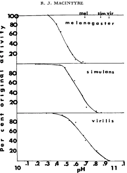
had different pH optima, the measurements of acid phosphatase activity of the three enzymes after electrophoresis and staining would be biased in favor of the enzyme whose pH optimum was closer to the pH of the staining solution. Figure 1 shows that for the three species used in these preliminary experiments, differences in the pH optima of the homologous acid phosphatase-1 enzymes do not exist. Therefore, by incubating the gel in stain in pH 5.0-5.1, there should be no accentuation of the activity of one of the three enzymes. This assumes, however, that the heterospecific enzyme XY will have the same pH optimum as the two homospecific acid phosphatases.2) Effect of dissociation on acid phosphatase-1 subunits: The pH at which 100% inactivation occurs was determined for each of the three homologous enzymes. The inactivation curves are shown in Figure 2. The experiments have been repeated several times, and the slight differences between the inactivation curves are always obtained. It is thought that dissociation results from the nega- tive intermolecular repulsive forces which build up as the increase in OH- con- centration titrates the positively charged polar amino acids. The increase in the net negative charge on the subunit surfaces finally breaks the weak, noncovalent intersubunit bonds (SCHACHMAN 1963). Separated subunits, however, can be further affected by greater increases in pH. The data in Table 1 show that if the pH is raised too high during the dissociation treatment the activity regained after reassociation can be drastically reduced. This may be due to either alterations in the structures of all the subunits or a reduction in the population of subunits that can still reassociate. For this reason, when making a n interspecific test it is
ACID P H O S P H A T A S E - I E V O L U T I O N
.20
.I 0
48
7\ O
\
.O
\
e
.
O..
e-.
..
!
v i r i l i s
\
\
o \
-
acetate\ t ris-maleate
\ buffers
\
o \
0,
*
.
-,.
I \
I
a
\ . I
I 0 .
a.
I I
I
I
. O 0 .
1
'
' " * ' ' . _ _ \ .20.10
h.
simulans
C
FIGURE I.-pH optima of acid phosphatase-I from D. melanogaster, D . simulans, and D.
uirilis. Acetate buffers and Tris-maleate buffers were made at the indicated pH. The acetate
488
._
>
a,
.I
c
R. J. MACINTYRE
P
2.
c
v i r i l i s
C
a m
;
4 120
.1
O9 11
.1
2
3
A
.5 .6 J'a
P"
10
FIGURE 2.-Inactivation curves of acid phosphatase-I from D. melanogaster, D. simulans, and
D. uirilis. 0.1 N NaOH was added to 5.0 ml of a partially purified enzyme preparation in 0.05 M
NaCl at 25°C. After mixing, the p H was measured with a combination microelectrode. At the pH's indicated on the graph, 0.2 ml aliquots were removed and immediately assayed for acid phosphatase activity (see MATERIALS AND METHODS). Activities were corrected for the volume of
NaOH added. The pH at which 100% inactivation occurs for each species' enzyme is indicated on the top line.
11 .OO ( t o completely dissociate the more resistant D. uirilis acid phosphatase). Far fewer D. melanogaster subunits could effectively reassociate, however. This would bias the results strongly toward the formation or measurement of D. uirilis
homospecific enzyme.
ACID PHOSPHATASE-1 EVOLUTION
TABLE 1
489
Effect of the final p H of the dissociation treatment on the percent of
the initial activity regained after reassociation
Percent initial activity
regained after reassociation Final pH of dissociation'
D. melanogaster
10.79 11.03 11.16 11.44
11.00 11.15 11.37 11.55
D. virilis
44.1 16.2
9.0 6.4
65.3 35.9 14.0 8.1
* Aliquots of partially purified acid phosphatase-I preparations from D. virilis and D. melano-
gaster were taken to the indicated p H s by addition of 0.1 N NaOH. The aliquots were then
assayed to confirm complete inactivation, and dialyzed for 72 hr against 01.4 M Tris-maleate buffer,
pH 6.0 at 4°C. They were then assayed for acid phosphatase activity as indicated in MATERIALS .4ND METHODS.
conditions favoring one or the other of the three enzymes in the mixture, they should be evident from the comparison of the two sets of data.
4)
Length of time of reassociation: I t is important to allow sufficient time for all undamaged subunits to reassociate in the interspecific tests. Other experiments have indicated that the initial rate of increase in enzyme activity is directly dependent upon the concentration of subunits and the temperature of the Tris- maleate buff er. The final levels of reassociation attained are independent of sub-unit concentration if dialysis is carried out long enough (MACINTYRE, unpub- lished). Figure 4 shows that at 4"C, reassociation is virtually complete after 4 8 hr.
In the trial experiments involving reassociation of subunits from
D.
melanogaster,D.
simulans, and D. virilis, the pH-inactivated enzymes were dialyzed for 72 h rat 4°C. This length of time should have assured complete reassociation.
B)
Results from interspecific tests1) Initial tests: Other information relevant to the first assumption depends upon the results of experiments in which dissociated subunits reassociate and form heterospecific enzymes. Three painvise tests were made;
D.
melanogaster XD.
virilis,D.
simulansx
D. uirilis, and D. melanogaster XD.
simulans. In each490
0.9
I
R. J. MACINTYRE
E'
0
P In
0
0
FIGURE 3
v i r i l i s .15
.10
.05
' 0
4.9 5.2 5 5 5.8 A1
6A
6J 7.3 7.6 7.9 8.2P"
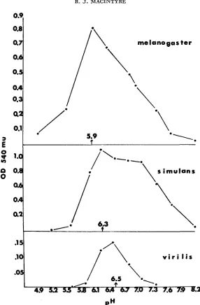
-pH optima for reassociation of subunits of acid phosphatase-I from D. melano-
, .-
gaster, D. simulans, and D. uirilis. 2.0 ml aliquots of pH-inactivated acid phosphatase-1 from
each of the three species ( D . melanogaster, pH 10.7; D. simulans, pH 10.8; D. uirilis, pH 11.0) were dialyzed against 0.4 Tris-maleate buffers at the indicated p H s for 72 hr at 4°C. The aliquots were then assayed for acid phosphatase activity. Apparent pH optima for each species are indi- cated on the abscissae.
ACID P H O S P H A T A S E - I E V O L U T I O N
49
127 36
18 45 54 63 7 2
9
HOURS
FIGURE 4.-Time course of reactivation of pH-inactivated extracts of acid phosphatase-1 from
D. melunoguster. 2.0 ml aliquots of a pH-inactivated (10.7) preparation of acid phosphatase-1
from D. melanogaster were dialyzed at 4°C against 2 liters of 0.4 M Tris-maleate buffer, p H 6.0.
At the times indicated, an aliquot was removed and assayed for acid phosphatase activity as indi- cated in MATERIALS AND METHODS. The activity of the 72-hr aliquot was 30% of the activity of the
initial undissociated preparation.
subjected to electrophoresis, and the acid phosphatase activities of the separated enzymes were estimated as described in MATERIALS AND METHODS.
Figure
5
is an electropherogram of the acid phosphatases from the three species and mixtures (with homospecific and heterospecific enzymes) from each of the three pairwise tests. The heterospecific enzyme forms in each case, strongly sug- gesting that the three enzymes are homologous. Quantitative results from the three pairwise tests, which were repeated, are in Tables 2-4. Table5
summarizes the data including the observed and expected (homospecific:heterospecific) en- zyme activity ratios and the differences between them. In Table 2, there is a n example of how the expected ratio is calculated. Note that the method assumes the enzyme is a dimer. The sign of the difference between the expected and observed ratios indicates which type of enzyme activity, i.e., homospecific orheterospecific, is disproportionally represented in the mixture. A minus sign means the heterospecific enzyme activity is in excess of expected levels. A plus
TABLE 2 Results of experiments inuoluing reussociation of acid phosphatase-I subunits of D. melanogaster and D. virilis Percent of total acid phosphatase in each zone' Experi- Percent Ratio* pH of ment reactivation melanogaster uirilis (homospecific: Expected reassociation number Subunits after dialysis homospecific heterospecific homospecific heteroswific) ratio** Difference melanogaster 31 5.9 1 uirilis 34 9 70 21 0.43 1
.oo
-0.57 mixture 52 (7-11) (67-76) (17-22) (0.32-0.49) (1 .oO-1.04) (-0.51--0.68) w melanogaster 33 mixture 29 (11-13) (68-74) (15-19) (0.35-0.47) (1 .OO) (-0.53- -0.65)E
melanogaster 22 mixture 38 (9-12) (64-69) (21-25) (0.45-0.56) (1.00-1.04) (-0.44- -0.57)2
melumgusier 224
n
5.9 2 uirilis 35 12 70 18 0.42 1
.oo
-0.582
6.5 1 uirilis 49 10 67 23 0.50 1.oo
-0.50 6.5 2 uirilis 47 9 71 20 0.41 1.oo
-0.59 mixture 30 (8-10) (70-71) (19-22) (0.41-0.43) (1 .OO) (-0.59--0.61) * Ranges in parentheses. Number of determinations: experiment 1 (pH 5.9)-8, (pH 6.5)-7; experiment 2 (pH 5.9)-5, (pH 6.5)-4. ** The expected ratio is calculated as follows: p (melunogus~er subunits) = mean proportion of melanogaster homospecific enzyme+
'/z mean For example, in experiment 1 at pH 5.9, p = P2+
q2 proportion of heterospecific enzyme. q (uirilis subunits) = 1-p. Expected ratio =2DU ,,
ACID P H O S P H A T A S E - 1 EVOLUTION 493
FIGURE 5.--Electropherogram of acid phosphntase-1 from D . virilis ( v i r ) , D . sirrrulans (sim), and D. melanoRastrr (mel) (slot 1 ) ; the reassociated subunits from D. simulans and D . uirilis
(slot 2); from D. mrlanogaster and D. uirilis (slot 3 ) ; and D. melanogaster and D . simulans (slot 4 ) . In each mixture, the heterospecific enzyme is in the middle of each pattern.
pected. An observed ratio of 1.5 with a n expected ratio of 1.0 means that the
activity of each homospecific enzyme is about 1
%
times that of the heterospecific enzyme. On the other hand, an observed ratio of 0.5 with a n expected ratio of1.0 means that the heterospecific enzyme activity is twice those of the homo- specific enzymes. I t should be mentioned here that in the tests involving the more distantly related species
D.
virilis xD.
melanogaster andD.
virilis XD.
simulans,the heterospecific subunit combination apparently predominates. The opposite appears to be true in the test between the sibling species
D.
melanogaster andD.
simulans. A possible reason for this will be discussed below.There are two observations from the results of the interspecific tests pertinent
to the first assumption about the methods. The first is the similarity of the results from the two experiments (Table 5 ) . This means that slight differences in enzyme preparations, final pH of dissociation, electrophoretic conditions, etc. will not significantly affect the final results. It is also evident that the results will not be significantly affected if the pH a t which reassociation takes place is closer to one species' pH optimum. As long as the pH of reassociation is a t one species'
TABLE 3 Results of experiments involuing reassociation of acid phosphatase-1 subunits of D. simulans and D. virilis Experi- pH of ment reassociation number Subunits
simulans mixture
6.3
1
uirilis simulans mixture simulans mixture
6.3 2 uirilis 6.5 1 virilis Percent of total acid phosphatase in each zone' Percent Ratio'
E$$?
Difference reactivation simuh uirilis (homcspedfic: after dialysis homospecific heterospecific homospecific heterospecific)53 57
23 63 14 0.60 1.00 58 (19-28) (59-67) (11-18) (0.49-0.69) (1 .O%I .08) (-0.31- -0.51) 4 -0.40 w 27
8
62 21 56 23 0.79 1 .00 -0.218
41 (19-22) (54-60) (21-24) (0.67-0.85) (1 .OO) (-0.15- -0.33)5
55 52
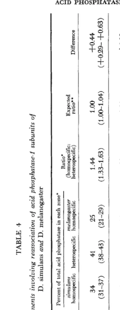
TABLE 4 Kesulls of experirnenls involving reussociation of acid phusphatase-I subunits of D. simulans and D. melanogaster Percent of total acid phosphatase in each zone*
*
Experi- Percent Ratio' pH,of, ment reactivation simulans melunogaster (homospecific: Expected vi redssoclation mimh Subunits after dialysis homospecific heterospecific homospecific he teruspecific) ra ti@ * Difference simulans 53E
5.9 1 melanogaster 47 34 41 25 1 .M 1.oo
3-0.44 mixture 60 (31-37) (38-43) (21-29) (1.33-1.63) (1.00-1.04) (f0.29- +0.63)9
H+
mixture 34 (33-35) (4143) (23-24) (1.33-1.44) (1 (+0.33- f0.44)
;I;
csimulans 58 0 mixture 61 (37-45) (39-41) (15-24) (1 .%1.56) ( 1 .OO-1.17 ) (+0.27- +0.56) simulans 62 mixture 41 (33-35) (41-46) (19-26) (1 .I 7-1.56) (1.W (+0.17- +0.56)
--
simulans 28 v, 5.9 2 melanogaster 36 35 4.2 23 1.38 1.oo
3-0.38Y
____
6.3 1 melanogusler 4.1. 41 40 19 1.50 1.08 3-0.426"
Z496 R. J . M A C I N T Y R E TABLE 5
Summary of results from acid phosphatase-1 subunit reassociation experiments
involving D. melanogaster, D. simulans, and D. virilis
Interspecific test
Observed (homospecific:
Reassociated hetemspecific) Difference from at pH enzyme ratio' expected ratio
melanogaster
X
uirilis
( 1 ) .43 - .57
(2) ,442 - .58 5.9
( 1 ) .50 - .50 6.5
( 2 ) .41 - .59
simulans
( 1 ) .60 - .40
(2) .79 - .21 6.3
X
( 1 ) .79 - .21
( 1 ) 1.44
+
.44uirilis 6.5
(2) .75 - .25
melanogaster 5.9
( 2 ) 1.38
+
.38( 1 ) 1.50
+
.42X
simulans 6.3
(2) 1.33
+
.33* Results of two experiments for each interspecific test.
provided a n opportunity to obtain more supporting evidence for the first assump- tion. Specifically, the apparent differences in the reassociation observed in the preliminary interspecific tests are assumed to be due entirely to the properties of the subunits. Biases in the results could occur if other molecules in the partially purified preparations had some effect on the reassociation process. If such mole- cules do exist, another purification step might change the results of the inter- specific tests. This assumes, of course, that the additional purification step would separate acid phosphatase-1 and the other hypothetical macromolecule. Acid phosphatase-l from D. melanogaster and D. uirilis was eluted from phospho- cellulose columns and concentrated as described in MATERIALS AND METHODS. The enzyme from D. uirilis came off the column when the molarity of the acetate buffer used for elution was between 0.30-0.40. The enzyme from D. melanogaster eluted in 0.55-0.65 M. acetate buffer. After concentration of the peak fractions.
enzyme preparations were then dissociated, mixed, and reassociated as described above. Measurements of the activities of the three enzymes which formed in the mixture of subunits are in Table 6, experiment 1. A comparison of this ratio with the ratios from the D. melanogaster X D. uirilis tests (reported in Table 5) indi- cates an increase in purification does not significantly affect the reassociation results.
ACID PHOSPHATASE- 1 E V O L U T I O N 49 7
2
8 3
8
P
3
-
5
-J
Y
Y
.U
Y
Q -E:
F4
:
2?
UE
B
E
s
PI
.s
k
2
Y
Y
(0
4
z t
E
M
g
.U-J
4
'X
.s
2
0
;
.$
.g
I
s
._ U-e
E
e
D %
y1
U
Y Y
ix
r; m c
8
3
am
. ^
2
8
c
5
.E
5
.n
00
-
e5
E.E
g
.e
2
'f
Y
2
4
B
z
2
3
3
g
8J
5
cU
LI
g
g
c.e
Fr;
498 R . J. M A C I N T Y R E
trations, it was possible to purify the heterospecific enzyme from the melano- gaster X virilis test. Dissociation and reassociation of this enzyme would be a repeat of the original test, but one in which the initial numbers of subunits from each species should be equal. In the preliminary tests, acid phosphatase activities of the two enzyme preparations were equalized, with the assumption that the numbers of the two kinds of subunits after dissociation would be approximately the same in the mixture. Figure 6 shows that when a reassociated mixture of sub- units from
D.
melanogaster andD.
uirilis is eluted from a phosphocellulose column, three peaks of acid phosphatase activity are obtained. If the most active fractions under each peak are pooled, concentrated, and subjected to electro- phoresis, the enzymes in each peak can be identified. Figure 7 shows the electro- pherogram which was obtained. Peak 1 contains theD.
virilis homospecific enzyme, and peak 3 theD.
melamgaster homospecific enzyme. The pooled frac- tions from peak 2 contain only the heterospecific enzyme.The pH-inactivation curve of this enzyme fell between the curves for
D.
melanogaster and
D.
virilis(MACINTYRE,
unpublished). Two preparations of this.2 0
.16
.1 2
.08
.04
?
E
0 e
Y)
n
0
c
3
f!
1
' 0 2!=I
1
I \
.
.
\*1
\ *\1
k.
.
./
.'.
4 ( 2 20 28 36 4 4 5 2 6 0
~
68
t u b e no.
FIGURE 6.-Phosphocellulose chromatography profile of acid phosphatase-I enzymes from re- associated subunits of D . melanogaster and D. uirilis. The column was equilibrated as described in MATERIALS AND METHODS. 20 m l steps of acetate buffer (pH 5.0) of increasing molarity were
used for elution. From 0.30 M to 0.40 M, steps were 0.005 M ; and from 0.40 M to 0.70 M, steps
ACID PHOSPHATASE- 1 EVOLUTION 499
FIGURE ‘I.-Electropherogram of acid phosphatases in peak fractions from the elution profile
in Figure 6. Slot 1 contains the three enzymes in the reassociated mixture of D. mrlunopnsfer and
D. virilis subunits before adsorption to the column. Slot 2 contains pooled and concentrated frac-
tions from peak 1. Slot 3-pooled and concentrated fractions from peak 2. Slot &-pooled and concentrated fractions from peak 3.
enzyme were completely dissociated a t high pH, and dialyzed back against 0.4 M Tris-maleate buffer pH 6.0. T h e preparations were then subjected to electro- phoresis. Both the homospecific enzymes and the heterospecific enzyme were regenerated, and the measurements of their activities are included in Table 6,
experiments 2 and 3. Again, virtually identical differences between observed and expected ratios in this and the earlier tests are obtained. It appears, then, that small differences in the numbers of subunits from the two species will not sig- nificantly affect the results of the interspecific tests.
Assumption no. 2: The methods used do not affect the functional integrity of
the dissociated subunits: The effects on the acid phosphatase-1 enzymes prior to
and during the dissociation and reassociation treatments could conceivably alter their structure resulting in distorted (homospecific:heterospecific) enzyme ratios
in vitro. This can be checked by examining the ratios of the three enzymes in
interspecific hybrid flies. It is possible to obtain interspecific hybrids only between the sibling species
D.
melanogaster andD.
simulans. Another comparison be- tween enzyme ratios obtained in vivo and in vitro is available, however. This test is between subunits from the two electrophoretic variants of the enzyme inD.
UI 0 0 TABLE 7 Acid phosphatase activities of enzymes from D. melanogaster x D. simulans interspecific hybrids and D. melanogaster Acph-lA/Acph-IB heterozygotes Also included are the results from reassociated mixtures of subunits from purified and dissociated AA and BB forms of the enzyme from D. melanogaster Percent acid phosphatase activity in each zone' Ratio' mehogaster sirnulans (homospecific: Expected Test homospecific heterospecific homospecific heterospecific) ratio** Difference 0 D. simulans 9 x 21 44 35 1.27 1.04. 30.23
4
5
D. melanogaster 9 X 22 43 35 1.33 1.00 +0.33
G
3
ACID PHOSPHATASE-I EVOLUTION 501 which are controlled by the Acph-lA and A ~ p h - 1 ~ alleles, respectively
(MAC-
INTYRE 1966). The electrophoretic patterns of both reciprocal interspecific hy- brids between
D.
melanogaster (Acph-ln homozygotes) andD.
simulans andAcph-IA/Acph-ln heterozygotes of
D.
melanogaster were assayed. Data are pre-sented in Table 7. Results from the interspecific hybrids can be compared with the ratios reported in Tables 4 and
5.
The data from the adult Acph-lA/Acph-IB heterozygotes are compared with the patterns in a reassociated mixture of subunits from partially purified and dissociated AA and
BB
enzymes fromD.
melanogaster. The similarities betweenin uiuo and in uitro results in both comparisons make it quite probable that the
subunits combine in the same proportions or the enzymes which form have the same activities both in the in uitro tests and inside living cells. Presumably, there- fore, the purification procedures and experimental methods do not significantly alter the functional integrity of the subunits of the enzyme.
Assumption no. 3: Acid phosphatase-1 is a dimeric enzyme. Measurements of the molecular weight of the enzyme were made to see if its size is compatible with those of other dimeric enzymes. The acid phosphatases from
D.
melanogaster andD.
uirilis were each centrifuged in sucrose gradients with known standards. Theresults from the various assays of the collected fractions are depicted in Figure 8.
The calculations, following MARTIN and AMES (1961), from the data in A, B, & C
of Figure 8 indicate acid phosphatase-I of each species has a molecular weight of
100,000. The single peak of acid phosphatase activity from the gradient contain- ing a mixture of
D.
uirilis andD.
melanogaster enzymes also indicates the homologous acid phosphatases probably have identical molecular weights.The ability to detect dissociated subunits in an acrylamide gel following electro- phoresis (Figure 9) made it possible to estimate their molecular weight. ZWANN
(1967) found that the retardation of the migration of globular proteins in acryl- amide gels with smaller pore sizes is inversely related to the log of their molecular weights. Acid phosphatase-1, its subunits, and several known standards were subjected to electrophoresis in 6% and 9% gels. The retardation coefficients have been plotted against the log molecular weight of the standards in Figure IO. The three standards and acid phosphatase-1
,
whose molecular weights were deter- mined by other methods, fall on the same straight line. The retardation coefficient of the subunits indicates they have a molecular weight of about 55,000. The dissociation treatment, therefore, is splitting acid phosphatase-1 into subunits about half the weight of the native enzyme. This is compatible with a dimer model for active acid phosphatase-I.DISCUSSION
502 R. J. MACINTYRE
;
0
n n 0:
0
l
a
I E ;
0 'E
a -
I '
sa
34a
I E d
" - I .
2'
I47 U l E NO
FIGURE 8.-Sucrose density gradient profiles of acid phosphatase-1 from D. melanogaster and
D. uirilis. Gradient A contained acid phosphatase-I from D. melanogaster and purified horse
hemoglobin (M.W 67,000). Gradients B and C contained acid phosphatase-I from D. melano-
gaster and D. uirilis, respectively, with yeast alcohol dehydrogenase (M.W. 152,000). Gradient
ACID P H O S P H A T A S E - I E V O L U T I O N 5 03
FIGURE 9.-Electropherogram of acid phosphatase-1 from D. melanogaskr and D. uirilis and dissociated subunits of each. Electrophoretic procedures can he found in MATERIALS A N D METHODS.
The gel buffer, however, was 0.1 M Tris-borate at pH 9.8. The first pocket contains a mixture of acid phosphatase-I from D. melanogaster and D. virilis. pH-inactivated enzyme was inserted into the 2nd (D. melanogaster and D. virilis), 3rd (D. virilis), and 4th (D. melanogaster) pockets in the gel. Following electrophoresis but before staining, the gel was incubated overnight in 0.4 M Tris-maleate buffer, pH 6.0, to allow the subunits to reassociate.
ing the regions of subunit attachment or the active site itself.
The experiments and the data reported in this paper were designed to test the validity of that interpretation. Before an experiment involving the reassociation of acid phosphatase-1 subunits from two different species is conducted, however, the pH optima, the inactivation curves at high pH, and the p H optima for reassociation should be determined for the enzymes from each species.
If
this is done, and the proper adjustments are made during the dissociation, reassociation, and gel assay steps in the experiment, there appears to be no reason to suspect bias in the results.504 R. J. MACINTYRE
o I standards
-
I unknownsY Y Y
0
v
z
0
-
I-
a
CI
I
a c
Y
U
L o g . M o l . w t .
FIGURE 10.-Retardation coefficients and log of the molecular weights horse hemoglobin (M.W. 67,000), E. coli alkaline phosphatase (M.W. 80,000), yeast alcohol dehydrogenase (M.W. 152,000), acid phosphatase-I and dissociated subunits from D. melanogaster. Retardation coeffi- cients are distance (mm) traveled in 9% gel/distance traveled in 6% gel per 1400 volt hours.
The estimates of the molecular weights of the enzyme and its subunits are compatible with a dimeric structure. Many enzymes of similar size are thought, with varying degrees of certainty, to be composed of only two subunits (KLOTZ
and DARNALL 1969). Other evidence pointing to a dimeric structure is available. First, an electrophoretic pattern of three bands is not only found in Acph-lA/ A ~ p h - 1 ~ heterozygotes and interspecific hybrids but also is obtained after re- association of subunits from different species and after dissociation and reassoci- ation of the purified heterospecific enzyme from the
D.
melanogaster XD.
uirilis test. Such an electrophoretic pattern is considered characteristic of dimeric enzymes (SHAW 1964; MARKERT and WHITT 1968). Secondly, the subunits pro- duced after pH inactivation migrate to a single position in acrylamide gels (Figure 11 ).
Yet they can apparently reassociate and form active enzymes. This observation almost certainly rules out a heteromultimeric structure for acid phosphatase-1, for the subunit controlled by a hypothetical second structural gene would have to have the same net charge and size as the subunit made at the Acph-l locus. This conclusion is supported by complementation tests between fifteen ethyl methanesulfonate induced “zero” mutants of acid phosphatase-I.
ACID PHOSPHATASE-I EVOLUTION
5
05B
A
M E L A N O G A S T E R H O M O S P E C I F I C
E N Z Y M E
m e I
a n
0
:
t e
r
C
V l R l L l S
H O M O S P E C I F I c E N Z Y M E
I
U
b n
U
I
t
H E T E R O S P E C I F I c E N Z Y M E
FIGURE 11 .-Hypothetical scheme of intersubunit bonds between the homospecific enzymes
(A = D. melanogaster, B = D. uirilis) and the heterospecific enzyme (C) formed in the reasso-
ciation of D. melanogaster and D. virilis subunits. ( f ) and (-) are positively and negatively charged side chains of amino acids. - - - indicates weaker hydrogen or hydrophobic bonds between subunits.
is itself composed of more than one polypeptide chain, the calculation of the expected amounts of enzyme which should form during reassociation is still valid. This calculation which follows a binomial expansion assumes only that the treat- ment splits the enzyme into two identical molecules one-half the size of the native enzyme. The estimate of the molecular weight of the pH-induced subunit of 55,000 makes it quite likely that the native enzyme has been split into halves at high pH.
506 R. J. MACINTYRE
the substrate turnover numbers of the various homospecific and heterospecific enzymes may not be the same. It has been impossible to distinguish between these alternative explanations of the results, which themselves are not necessarily mutually exclusive. So far, direct measurements of protein in the zones of acid phosphatase activity have not been possible. The amounts of protein are appar- ently too small to measure without resorting to radioisotope or immunological techniques. In order to resolve this very serious problem, we are presently trying to directly determine the specific activities of the homospecific and heterospecific enzymes from the three species. Even if we can eventually determine the amount of protein in each zone, however, the results could be misleading since the enzyme has been only partially purified. A contaminating protein migrating to the posi- tion of one of the acid phosphatases could lead to a wrong interpretation. Thus we hope to supplement the specific activity determinations with other more in- direct experiments. In lieu of completely pure enzyme preparations, however, the issue may never be satisfactorily resolved. Nevertheless, the fact that the observed (homospecific:heterospecific) enzyme activity ratios do not follow the predictions of the binomial expansion suggests that amino acid substitutions affecting either the regions of subunit contact or the conformation of the active site of acid phosphatase-1 have occurred during the evolution of the genus Dro- sophila. Unfortunately, it is impossible to ascertain how many or what kind of amino acid changes these are. It seems unlikely, however, that the amino acid substitutions underlying these differences would be selectively neutral, since they apparently affect such important properties of the enzyme, namely its activity and/or its quaternary structure.
The observation that there may be a preferential formation or an increase in the activity of the heterospecific enzyme in the tests between
D.
uirilis and bothD.
melunoguster andD.
simuluns is at first glance rather surprising. The former species is in a different subgenus thanD.
melunoguster andD.
simulans. One might expect that evolutionary changes in the long time interval separating these species from a common ancestor would have resulted in a decrease in the amount of heterospecific enzyme forming in a mixture of the two kinds of subunits or in its activity. An apparent increase in the expected amount of heterospecific acid phosphatase-I is seen frequently in other interspecific tests (MACINTYRE. manu- script in preparation).ACID PHOSPHATASE-I EVOLUTION 507 of subunit reassociation between
D.
melanogaster andD.
uirilis for example, stronger bonds, i.e., ionic rather than hydrophobic, might form in the dimeriza- tion of unlike subunits. Figure 11 is a diagrammatic representation of the possible although highly hypothetical interactions. The heterospecific enzyme in vivo would be damaging to individuals from either species since it would indiscrimi- nately shift the equilibrium in favor of the active enzyme. At any rate, if the subunit-enzyme equilibrium does exist and is physiologically important, natural selection may not invariably act in such way as to reduce the affinities of subunits of distantly related species.SUMMARY
A
method for detecting what should be selectively important evolutionary changes in a homomultimeric enzyme, acid phosphatase-I, in species of the genus Drosophila is proposed. By measuring activity ratios of homospecific and hetero- specific enzymes which form after reaggregation of dissociated subunits, differ- ences between amino acid sequences which affect regions of subunit attachment or the conformation of the active site can be detected. Three assumptions about the methodology were examined. First, preliminary information about the homo- logous enzymes from three species,D.
melanogaster,D.
simulans, andD.
uirilis and results from the interspecific subunit reassociation tests indicate the methods used do not bias the results in favor of one or another of the three enzymes which form when subunits from two species reassociate. Secondly, comparisons were made between the results of in vitro subunit reassociation experiments and enzyme patterns obtained from both interspecific hybrids and heterozygotes for alleles specifying electrophoretic variants. The similarities betweenin
vitro andin vivo results make it quite probable that the methods used do not impair the properties of the subunits which are important in reassociation. Thirdly, molecu- lar weight estimates of acid phosphatase-I and its subunits are compatible with the assumption that the active enzyme is a dimer. Alternative explanations of the results and a hypothetical model of subunit attachment are considered.
LITERATURE CITED
ARNHEIM, N., E. M. PRAGER and A. C. WILSON, 1969
ARNHEIM, N. and C. E. TAYLOR, 1969
DUKE, E. J. and E. GLASSMAN, 1968
GUIDOTTI, C., W. KONIGSBERG and L. C. CRAIG, 1963 globin. Proc. Natl. Acad. Sci. U.S. 5 0 : 774-781. HUBBY, J. L. and S. NARISE, 1967
species differences in in vitro hybrid enzyme formation. Genetics 57: 291-300.
HUBBY, J. L. and L. THROCKMORTON, 1968 sibling species. Am. Naturalist 102 : 193-205. KING, J. L. and T. H. JLJKES, 1969
Immunological prediction of sequence
Non-Darwinian evolution: Consequences for neutral al-
Evolution of xanthine dehydrogenase in Drosophila. Ge-
On the dissociation of normal adult hemo- differences among proteins. Biol. Chem. 244: 2085-2094.
lelic \ariation. Nature 223: 900-903.
netics 5 8 : 101-112.
Protein differences in Drosophila. 111: Allelic differences and
Protein differences in Drosophila. IV: A study of
508 R . J. MACINTYRE
KLOTZ, I. M. and D. W. DARNALL, 1969
KOEN, A. and C. W. SHAW, 1968
Subunit structured proteins: A table. Science 166:
Starch-gel electrophoresis of enzymes. pp. 325-364. In: Chro-
matographic and Electrophoretic Techniques, vol. 2. Edited by IVOR SMITH. Interscience
Publ., New York.
Magnitude of interspecific nucleotide sequence diver- gence in Drosophila. Genetics 6 0 : 303-322.
The genetics of an acid phosphatase in Drosophila melanogaster and
D . simulans. Genetics 53: 461-474. -, 1968 A simple and reliable method for quan-
titating the activity of acid phosphatase isozymes after electrophoresis in polyacrylamide gels. Isozyme Bull. 1 : 27. A method for measuring activities of acid phos- phatases separated by acrylamide gel electrophoresis. Biochem. Genet. 5 : 45-56.
Subunits of acid phosphatase-I in Drosophila nzelano-
gaster: Reversible dissociation in uitro. Nature 214: 274-275.
Molecular varieties of isozymes. Experientia 24: 977- 1088.
A method for determining the sedimentation behavior
Comparative aspects of primary structures of proteins.
Evolution in the Genus Drosophila. MacMillan, New
Molecular pathology of human haemoglobin. Nature
Three-dimensional Fourier synthesis of horse oxyhaemoglobin at 2.8 A resolution: The atomic model. Nature 219: 131- 139.
126-1 28.
LAIRD, C. D. and B. J. MCCARTHY, 1968
MACINTYRE, R. J., 1966
-, 1971
MACINTYRE, R. J. and M. DEAN, 1967
MARKERT, C. L. and G. S. WHITT, 1968
MARTIN, R. L. and B. N. AMES, 1961
of enzymes: Application to protein mixtures. J. Biol. Chem. '236: 1372-1379. NOLAN, C. and E. MARGOLIASH, 1968
Ann. Rev. Biochem. 31: 727-790. PA'ITERSON, J. T. and W. S. STONE, 1952
PERUTZ, M. F. and H. LEHMANN, 1968
PERUTZ, M. F., H. MUIRHEAD, J. M. Cox, and L. C. G. GOAMAN, 1968 York.
219: 902-909.
RAYMOND, S., 1964 RICHMOND, R., 1970
SCHACHMAN, H., 1963
Acrylamide gel electrophoresis. Ann. N.Y. Acad. Sci. 121: 350-361.. Non-Darwinian evolution: A Critique. Nature 225: 1025-1028.
Considerations on the tertiary structure of proteins. Cold Spring Harbor Symp. Quant. 2.8: 409-430.
SHAW, C. R., 1964
Symp. Biol. 1 7 : 117-130. THROCKMORTON, L., 1962
Publ. 6 2 0 5 : 207-344. ZWANN, J., 1967
The use of genetic variation in the analysis of isozyme structure. Brookhaven
The problem of phylogeny in the genus Drosophila. Univ. Texas