_____________________________________________________________________________________________________
*Corresponding author: E-mail: [email protected];
32(6): 33-39, 2020; Article no.JAMMR.56213
ISSN: 2456-8899
(Past name: British Journal of Medicine and Medical Research, Past ISSN: 2231-0614, NLM ID: 101570965)
Effect of Antiasthma Medication on Blood
Eosinophil Count and Serum Immunoglobulin E
Levels: A Randomized Controlled Study
Wasim A. Wani
1, Sheeraz A. Dar
1*, Khalid M. Kawosa
1, Mudasir Nazir
1,
Ikhlas Ahmad
1and Asif Ahmed
21Department of Pediatrics and Neonatology, Sher-I-Kashmir Institute of Medical Sciences Hospital,
Srinagar, Jammu and Kashmir, India. 2
Department of Pediatrics, SKIMS MC Bemina, Srinagar, Jammu and Kashmir, India.
Authors’ contributions
This work was carried out in collaboration among all authors. Author SAD conceptualized and designed the study, drafted the initial manuscript, coordinated and supervised data collection and approved the final manuscript as submitted. Authors WAW and IA carried out the initial analyses, reviewed and revised the manuscript and approved the final manuscript as submitted. Authors KMK, MN and SAD designed the data collection instruments, and coordinated and supervised data collection, critically reviewed the manuscript and approved the final manuscript as submitted. All authors read and approved the final manuscript.
Article Information
DOI: 10.9734/JAMMR/2020/v32i630430 Editor(s): (1) Dr. Babatunde Olanrewaju Motayo, Federal Medical Center, Nigeria and University of Ibadan, Nigeria. Reviewers: (1) Agnes Hamzaoui, University of Tunis El Manar, Tunisia. (2) Ajit K. Gangawane, Parul University, India. Complete Peer review History:http://www.sdiarticle4.com/review-history/56213
Received 21 February 2020 Accepted 27 April 2020 Published 05 May 2020
ABSTRACT
Background: Asthma is a chronic inflammatory condition of lung airways resulting in episodic airflow obstruction.
Aims: The main objective of this study is to find the effect of antiasthma medication on serum IgE levels and blood eosinophil count.
Study Design: This randomized controlled trial study was performed in children aged 6-15 years of age, with clinically stable and moderate persistent asthma.
Results: The findings of this study indicate both group (Budesonide/formoterol group and budesonide group) patients experienced a significant decrease in serum IgE levels and blood
eosinophil counts over the study period. However, the difference in two groups was not statistically significant.
Conclusions: Inhaled steroids are effective in controlling systemic inflammation in asthma as evidenced by a decrease in IgE levels and eosinophil counts. However addition of LABA doesn’t have any additive effect.
Keywords: Asthma; budesonide; eosinophil; IgE levels; inhaled steroids; LABA.
1. INTRODUCTION
Bronchial asthma (BA) is a chronic inflammatory disease with a marked heterogeneity in etiology, pathophysiology and clinical aspects, leading to the identification of different phenotypes (early onset atopic/allergic, eosinophilic, exercise-induced, obesity and paucigranulocitic [1]. Subsequently, taking into account the molecular mechanisms underlying the pathophysiology of bronchial inflammation, the concept of endotypes has been introduced [2]. BA is often sustained by allergic sensitization, which leads to bronchial hyper-responsiveness and acute bronchoconstriction in response to specific and non-specific triggers. For many years, from a pathogenic perspective, the focus of research has been on the role of T cells in the initiation and perpetuation of inflammation [3,4]. In particular, T helper 2 (Th2) cells have been identified as the cells involved in controlling immunoglobulin E (IgE) production because of their ability to produce interleukin (IL)-4 and IL-13, and influence the functioning of eosinophils through the actions of IL-5 [5].
The biological role of IgE is complex and related to its ability to influence the functioning of several immune and structural cells involved in the pathogenesis of chronic allergic inflammation [6]. The biological pathways that IgE uses to influence cell activity rely on interactions with specific receptors. Two classes of receptors have been identified: high-affinity (FcεRI) receptors and low-affinity CD23 (or FcεRII) receptors. FcεRI receptors are not only expressed by mast cells and basophils but also by dendritic cells (DCs), airway smooth muscle cells (ASMCs), epithelial cells, endothelial cells, and eosinophils [7,8,9,10]. DCs can provide all of the co-stimulatory signals required for activation of T cells, which play a key role in the pathogenesis of BA. DCs are dedicated antigen-presenting cells and are key in the induction of Th2 cell activation in the primary immune response to allergens [11,12]. The ability to present antigens is amplified by IgE bound to FCεRI receptors expressed on the surface of DCs [6]. It has been
shown that IgE captures the allergens, facilitating their presentation to memory Th2 lymphocytes [13].FcεRI–IgE-dependent allergen presentation by DCs may critically lower the atopic individual’s threshold to mount allergen-specific T cell responses. In fact, the targeting of allergens to FcεRI via IgE leads to a 1000-fold increase in the activation of T cells in addition to the production of chemokine ligand 28 (CCL28), a chemokine that selectively attracts Th2 lymphocytes [14]. Furthermore, the activation of allergen-specific Th2 cells is associated with an amplification of allergen-specific IgE production in a vicious cycle of the pathogenic mechanisms of allergic asthma [6]. IgE antibodies are also able to negatively modulate the innate function of plasmocytoid DCs. In these cells, the activation of FcεRI receptors blocks, or at least reduces, the intracellular signals involved in type I interferon (IFN) production [15]. The reduction in IFN production correlates with the defect in anti-viral response in allergic asthma patients [16]. Moreover, viral respiratory infections can be the initial cause of asthma, and can result in exacerbations and worsen severity. Research to understand the relationship between IgE and virus infection susceptibility was spurred, in part, by the results of the clinical trial performed using omalizumab in children with asthma [6]. The study found that anti-IgE therapy was able to reduce the exacerbations typically occurring in the spring but mainly in the fall when the children go back to school. It is known that the majority of exacerbations are induced by viral respiratory infections [17].
circulates through the blood and arrives at the bone marrow where it stimulates eosinophil progenitors, which migrate towards the bronchial walls under the effect of chemokines such as eotaxins (CCL11) [21]. However, other cytokines are also able to directly influence eosinophils. Granulocyte–macrophage colony stimulating factor (GM-CSF) and IL-3 activate and enhance eosinophil functions, such as cytotoxic killing, superoxide production, leukotriene production, and Ig-induced degranulation [22].
In our study, we tried to find the effect of antiasthma medication on serum IgE levels and blood eosinophil count.
2. MATERIALS AND METHODS
2.1 Study Design
This randomized controlled trial study was performed in Department of Paediatrics, Sher-I-Kashmir Institute of Medical Sciences, Srinagar, between March 2016 and February 2018 in children aged 6-15 years of age. Children with clinically stable and moderate persistent asthma and with forced expiratory volume in one second
(FEV1) of more than 60% after the
bronchodilator had been withheld for 6 hours were included in the study. Children with history of life-threatening asthma and adverse reactions to the medications used in the study were excluded from the study.
2.2 Data Collection
Children with signs and symptoms of asthma and who satisfied the inclusion criteria were selected. History of day time symptoms (use of short acting beta agonists/week), nighttime
awakening/month, number of acute
exacerbations, was obtained from the patients. The children were divided into 2 groups. Both groups received fixed dose of inhalational steroids (budesonide 400micrograms/day). While as only one group received long-acting beta agonists (formoterol 12 micrograms/day). The Children were free to use short acting beta agonists (albuterol).
The children were assessed at 3, 6 and 9 months for asthma control and for eosinophil count and IgE levels.
2.3 Statistical Analysis
All the continuous variables of the study have been shown in terms of descriptive statistics. To analyse the data, we have applied standard statistical tests like unpaired t- test, chi square test, mcnemar test. The results obtained have been discussed on 5% level of significance i.e p< 0.05 considered significant. Moreover, the appropriate statistical charts have been used to represent the data. The statistical software SPSS V-20 has been used for the statistics.
3. RESULTS AND DISCUSSION
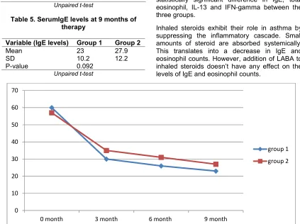
Comparison of serumIgE Levels in the two groups: (Tables 2,3,4,5 and Fig. 1).
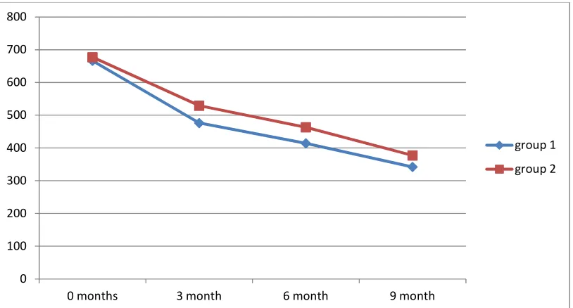
Comparison of eosinophil count in the two groups: (Tables 6,7,8,9 and Fig. 2).
Asthma is characterized by airway inflammation and hyper responsiveness. Besides local inflammation, asthma is also found to have systemic inflammation [23]. Eosinophils and IgE are considered to play a significant role in allergic immune responses. The reduction of IgE levels and eosinophils counts seems to be an attractive target in the management of allergic diseases like asthma, allergic rhinitis etc. Inhaled steroids inhibit the number of circulating eosinophils in asthma [24]. In the present study, we determined to examine whether the addition of LABA to inhaled steroids causes greater fall in IgE levels and eosinophil counts.
Sixty children who met the inclusion criteria were included. They were randomly divided into two treatment groups. Efficacy variables studied were blood eosinophil count and serum IgE levels in each group at each visit. The mean (±SD) age of the patients in our study was 9.3(±3.1) years in group 1 and 8.9(±2.6) years in group 2. The age distribution in the two groups was statistically insignificant (>0.05).
Table 1. Comparison of age in the two study groups
Variable(age) Group 1 (steroid+LABA) Group 2(steroid)
Mean 9.3 8.9
SD 3.1 2.6
95% CI 8.2-10.4 7.9-9.9
Table 2. Serum IgE levels at the start of therapy
Variable (IgE levels) Group 1 Group 2
Mean 59.7 57.8
SD 24 28
P-value 0.78
Unpaired t-test
Table 3. Serum IgE levels at 3 months of therapy
Variable (IgE levels) Group 1 Group 2
Mean 30.2 35.7
SD 17.6 18.3
P-value 0.2
Unpaired t-test
Table 4. Serum IgE levels at 6 months of therapy
Variable (IgE levels) Group 1 Group 2
Mean 26 31.6
SD 13 14
P-value 0.12
Unpaired t-test
Table 5. SerumIgE levels at 9 months of therapy
Variable (IgE levels) Group 1 Group 2
Mean 23 27.9
SD 10.2 12.2
P-value 0.092
Unpaired t-test
We studied and compared serum IgE between two groups at the start, 3 months, 6months and 9months of therapy. Both groups experienced a statistically significant decrease inserum IgEover the study period. There was a greater decrease in IgE levels in budesonide/formoterol group when compared to budesonide alone group. However, the difference in the two groups was not statistically significant.
The second parameter we studied and compared was blood eosinophil count between two groups at the start, 3 months, 6months and 9months of therapy. There was a decrease in eosinophil count in both budesonide/formoterol group and budesonide alone group throughout study period. However, the difference in two groups was not statistically significant.
Our results were consistent with the study done by Kamaran O et al. [25]. They randomized the patients into 3 groups. Group 1 used inhaled budesonide, group 2 used inhaled budesonide/ formoterol and group 3 used inhaled budesonide and oral montelukast. They didn’t find any statistically significant difference in IgE, total eosinophil, IL-13 and IFN-gamma between the three groups.
Inhaled steroids exhibit their role in asthma by suppressing the inflammatory cascade. Small amounts of steroid are absorbed systemically. This translates into a decrease in IgE and eosinophil counts. However, addition of LABA to inhaled steroids doesn’t have any effect on the levels of IgE and eosinophil counts.
Fig. 1. Comparison of a decrease in IgE levels in the two groups 0
10 20 30 40 50 60 70
0 month 3 month 6 month 9 month
group 1
Fig. 2. Comparison of decrease in eosinophil count between the two groups
Table 6. Blood eosinophil count at the start of therapy
Variable (eosinophils) Group 1 Group 2
Mean (MEDIAN) 666 (695) 677 (630)
SD 184 206
P-value 0.82
Unpaired t-test
Table 7. Blood eosinophil count at 3 months of therapy
Variable (eosinophils) Group 1 Group 2
Mean (MEDIAN) 476 (431) 529 (450)
SD 172 206
P-value 0.29
Unpaired t-test
Table 8. Blood eosinophil count at 6 months of therapy
Variable(eosinophils) Group 1 Group 2
Mean (MEDIAN) 414 (390) 463 (400)
SD 134 196
P-value 0.26
Unpaired t-test
Table 9. Blood eosinophil count at 9 months of therapy
Variable(eosinophils) Group 1 Group 2
Mean (MEDIAN) 342(330) 377(325)
SD 104 143
P-value 0.285
Unpaired t-test
0 100 200 300 400 500 600 700 800
0 months 3 month 6 month 9 month
group 1
4. CONCLUSION
Inhaled steroids are effective in controlling systemic inflammation in asthma as evidenced by a decrease in IgE levels and eosinophil counts. However addition of LABA doesn’t have any additive effect.
CONSENT
As per international standard or university standard written parents’ consent has been collected and preserved by the author(s).
ETHICAL APPROVAL
It is not applicable.
COMPETING INTERESTS
Authors have declared that no competing interests exist.
REFERENCES
1. Asthma WSE. Defining of the persistent adult phenotypes. Lancet. 2006;368:804– 13.
2. Lotvall J, Akdis CA, Bacharier LB, Bjermer L, Casale TB, Custovic A, et al. Asthma
endotypes: A new approach to
classification of disease entities within the asthma syndrome. J Allergy ClinImmunol. 2011;127:355–6.
3. Ricci M, Rossi O, Bertoni M, Matucci A. The importance of Th2-like cells in the
pathogenesis of airway allergic
inflammation. ClinExp Allergy. 1993;23: 360–9.
4. Woodfolk JA. T-cell responses to allergens. J Allergy ClinImmunol. 2007; 119:280–94.
5. Romagnani S. Immunologic influences on allergy and the TH1/TH2 balance. J Allergy ClinImmunol. 2004;113:395–400.
6. Matucci A, Vultaggio A, Maggi E, Kasujee I. Is IgE or eosinophils the key player in allergic asthma pathogenesis? Are we asking the right question?. Respiratory Research. 2018;19(1): 113.
7. Maurer D, Fiebiger S, Ebner C, Reininger B, Fischer GF, Wichlas S, et al. Peripheral blood dendritic cells express Fc epsilon RI as a complex composed of Fc epsilon RI alpha-and Fc epsilon RI gamma-chains and can use this receptor for IgE-mediated
allergen presentation. J Immunol. 1996; 157:607–16.
8. Redhu NS, Gounni AS. The high affinity IgE receptor (FcεRI) expression and function in airway smooth muscle. Pulm Pharmacol Ther. 2013;26:86–94.
9. Sihra BS, Kon OM, Grant JA, Kay AB. Expression of high-affinity IgE receptors (Fc epsilon RI) on peripheral blood basophils, monocytes, and eosinophils in
atopic and nonatopic subjects:
Relationship to total serum IgE concentrations. J Allergy ClinImmunol. 1997;99:699–706.
10. Kraft S, Kinet JP. New developments in FcεRI regulation, function and inhibition. Nat Rev Immunol. 2007;7:365–78.
11. Gaurav R, Agrawal DK. Clinical view on the importance of dendritic cells in asthma. Expert Rev ClinImmunol. 2013;9:899–919. 12. Froidure A, Shen C, Pilette C. Dendritic
cells revisited in human allergic rhinitis and asthma. Allergy. 2016;71:137–48.
13. Schroeder JT, Bieneman AP, Chichester KL, Hamilton RG, Xiao H, Saini S, et al. Decreases in human dendritic cell-dependent T(H)2-like responses after acute in vivo IgE neutralization. J Allergy ClinImmunol. 2010;125:896–901.
14. Khan SH, Grayson MH. Cross-linking IgE augments human conventional dendritic cell production of CC chemokine ligand 28. J Allergy ClinImmunol. 2010;125:265–7. 15. Lynch P, Mazzone SB, Rogers MJ, Arikkatt
JJ, Loh Z, Pritchard AL, et al. The plasmacytoid dendritic cell: At the
cross-roads in asthma. EurRespir J.
2014;43:264–75.
16. Kelly JT, Busse WW. Host immune responses to rhinovirus: mechanisms in asthma. J Allergy ClinImmunol. 2008;122: 671–84.
17. Busse WW, Morgan WJ, Gergen PJ, Mitchell HE, Gern JE, Liu AH, et al. Randomized trial of omalizumab (anti-IgE) for asthma in inner-city children. New Engl J Med. 2011;364:1005–15.
18. Bousquet J, Chanez P, Lacoste JY, Barneon G, Ghavanian N, Enander I, et al. Eosinophilic inflammation in asthma. N Engl J Med. 1990;323:1033–9.
19. Garcia G, Taille C, Laveneziana P, Bourdin A, Chanez P, Humbert M. Anti-interleukin-5 therapy in severe asthma. EurRespir Rev. 2013;22:251–7.
al. Blood eosinophil count and prospective annual asthma disease burden: A UK cohort study. Lancet Respir Med. 2015;3:849–58.
21. Kita H. Eosinophils: Multifaceted biologic properties and roles in health and disease. Immunol Rev. 2011;242:161–77.
22. Nagata K, Hirai H, Tanaka K, Ogawa K, Aso T, Sugamura K, et al. CRTH2, an orphan receptor of T-helper-2-cells, is expressed on basophils and eosinophils
and responds to mast cell- derived factor(s). FEBS Lett. 1999;459:
195–9.
23. Jousilahti P, Salomaa V, Hakala K, Rasi V, Vahtera E, Palosuo T. The association of
sensitive systemic inflammation markers with bronchial asthma. Ann Allergy Asthma Immunol. 2002; 89:381-5.
24. Kerrebijin KF, Von Essen-Zandvliet EMM, Neijens HJ. Effect of long term treatment with an inhaled corticosteroid and beta agonists on bronchial responsiveness in asthmatic children. J Allergy ClinImmunol. 1987;79:653-9.
25. Kamaran O, Arli O, Uzuner N, Islekel H, Babayigit A, Olmez D, et al. The
effectiveness of asthma therapy
alternatives and evaluating the effectivity of asthma therapy by interleukin 13 and interferon gamma levels in children. Allergy Asthma Proc. 2007;28(2):204-9. _________________________________________________________________________________
© 2020 Wani et al.; This is an Open Access article distributed under the terms of the Creative Commons Attribution License (http://creativecommons.org/licenses/by/4.0), which permits unrestricted use, distribution, and reproduction in any medium, provided the original work is properly cited.
Peer-review history: