im Biomedizinischen Centrum München (BMC)
der
LUDWIG-MAXIMILIANS-UNIVERSITÄT MÜNCHEN
Vorstand: Prof. Dr. Peter B. Becker
und der
JUSTUS-LIEBIG-UNIVERSITÄT GIESSEN
Analysis of the multivalent binding
properties of the novel H2A.Z interactor
PWWP2A
Dissertation zum Erwerb des Doktorgrades der Naturwissenschaften
an der Medizinischen Fakultät der Ludwig-Maximilians-Universität München
Ramona Spitzer
aus Günzburg
Betreuerin: Prof. Dr. Sandra B. Hake
Zweitgutachter: Prof. Dr. Andreas Ladurner
Dekan: Prof. Dr. med. dent. Reinhard Hickel
Ich, Ramona Spitzer, erkläre hiermit an Eides statt, dass ich die vorliegende Dissertation mit dem Titel:
“Analysis of the multivalent binding properties of the novel H2A.Z interactor
PWWP2A”
selbständig verfasst, mich außer der angegebenen keiner weiteren Hilfsmittel bedient und alle Erkenntnisse, die aus dem Schrifttum ganz oder annähernd übernommen sind, als solche kenntlich gemacht und nach ihrer Herkunft unter Bezeichnung der Fundstelle einzeln nachgewiesen habe. Ich erkläre des Weiteren, dass die hier vorgelegte Dissertation nicht in gleicher oder in ähnlicher Form bei einer anderen Stelle zur Erlangung eines akademischen Grades eingereicht wurde.
München, den ……14.09.2018……. ………...
First, I would like to thank my supervisor Prof. Dr. Sandra B. Hake for the opportunity to work as a PhD student in her lab. Sandra, you always pushed my thesis with constant input and ideas and it was always fun to discuss my project or to defend my point of view. You gave me the opportunity to improve myself in giving talks, discussing and defending scientific issues and strengthening my personality. On top, you always had a sympathetic ear for me, no matter if it was about scientific questions or personal problems. Thank you so much for letting me be a part of the amazing “HAKE-GIRLS”.
Next, I want to thank Dr. Catherine Regnard (“PWWP”-Team) and Dr. Felix Müller-Planitz for being members of my thesis advisory committee. There was always an “open door” to discuss questions and to get helpful advice for my PhD project.
I am happy for the opportunity to work in the Biomedical Center (BMC) and being part of a great “chromatin-network-community” and the IRTG1064 (thanks Elizabeth for being such a great coordinator). There was no question, method or reagent which could not be answered, learned or “borrowed”. I want to thank all former members of the Hake-Lab and of course our former master student Lucia (sushi & rice-balls-4-ever). It was a pleasure to work together with you and I really enjoyed your company. Furthermore, I want to thank Stephi Link (the crazy cat lady) for being the other 50% of the Hake-Pair (LMU-Crew). It was always fun to work together with you and discuss all the gossip, which was floating around. I think with you, it is not possible to miss anything new or secret in the lab community.
I was also lucky to share the lab with the former and current FMP´s (especially Sabrina, Ashish and Petra), where I got constantly input for “in vitro” questions like purification (Äkta), assemblies and cloning strategies or just having a little chit-chat.
In addition, I am grateful that I could experience collaborative work with outstanding scientists here in Munich and all over the world: Stephanie Link, Moritz Völker-Albert, Axel Imhof, Masahiko Harata, Thomas Burgold, Brian Hendrich, Mariam Sana, Mario Torrado del Rey and Joel Mackay.
In silent remembrance of Karl Bader who always believed in science and technical progress
I
Summary ... 1
Zusammenfassung ... 3
1 Introduction ... 5
1.1 The structure of chromatin ... 5
1.2 Alteration of chromatin structure ... 8
1.2.1 Posttranslational modifications of histones ... 11
1.2.2 Histone variants ... 13
1.2.3 H2A variants ... 15
1.3 The histone variant H2A.Z ... 17
1.4 H2A.Z binding proteins ... 21
1.5 PWWP domain-containing proteins ... 25
1.6 Objectives ... 27
2 Material and Methods ... 28
2.1 Materials ... 28
2.1.1 Technical devices ... 28
2.1.2 Consumables ... 30
2.1.3 Chemicals ... 31
2.1.4 Kits, enzymes and markers ... 33
2.1.5 Antibodies ... 34
2.1.6 Plasmids ... 35
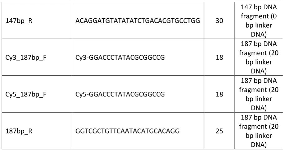
2.1.7 Oligonucleotides ... 36
2.1.8 Bacterial strains and cell lines ... 38
2.1.9 Software ... 38
2.1.10 Buffers and solutions ... 39
2.2 Cell biological methods ... 41
2.2.1 Mammalian cell cultivation ... 41
2.2.2 Transient transfection of mammalian cells ... 42
2.3 Molecular biological methods ... 42
2.3.1 Agarose gel electrophoresis ... 42
2.3.2 Cloning of PWWP2A truncations ... 42
II
2.4.2 Coomassie staining of SDS-PAGE gels ... 44
2.4.3 Immunoblotting ... 44
2.4.4 Mononucleosome preparation of HeLa Kyoto cells ... 45
2.4.5 Purification of MNase digested DNA ... 46
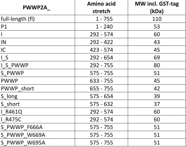
2.4.6 Purification of recombinant GST-tagged PWWP2A and PWWP2A truncations 47 2.4.7 Identification of histone modifications by Mass-Spectrometry (MS) ... 50
2.4.8 Reconstitution of recombinant mononucleosomes ... 52
2.4.9 In vitro binding assays with recombinant PWWP2A constructs ... 58
2.4.10 NMR spectroscopy ... 62
2.5 Bioinformatics ... 62
2.5.1 MS Data analysis... 62
3 Results ... 64
3.1 PWWP2A is a direct nucleosome interaction protein ... 64
3.2 PWWP2A´s internal region divides labor – general nucleosome binding and specific H2A.Z recognition ... 67
3.3 H2A.Z´s C-terminus affects binding to PWWP2A in a HK cell-derived system ... 70
3.4 Point mutations within IC impair binding to H2A and H2A.Z ... 73
3.5 Single histone tail deletions do not impair binding of IN in vitro ... 77
3.6 IN interacts with free DNA and binds nucleosomal linker DNA ... 79
3.7 PWWP2A´s PWWP domain directly interacts with recombinant mononucleosomes .. ... 83
3.8 The PWWP domain interacts with free nucleic acids and nucleosomal linker DNA . 85 3.9 Combination of S and PWWP domain interacts with in vivo-derived H3K36me3-mononucleosomes ... 91
3.10 PWWP2A interacts with γ-H2A.X mononucleosomes ... 102
4 Discussion ... 106
4.1 PWWP2A is a multivalent chromatin binder and specifically recognizes H2A.Z .... 106
4.2 A combination of S and PWWP interacts in a sequence- and methylation state-specific manner with H3K36me3-nucleosomes ... 112
4.3 PWWP2A´s C-terminus interacts with the modified histone variant γ-H2A.X ... 115
4.4 Summary and future perspectives ... 117
ABBREVIATIONS... 122
III
1
Summary
The constraint space of the eukaryotic cell nucleus requires the specific packing of DNA with
basic histone proteins into chromatin, with the nucleosome as its basic subunit. The
nucleosome itself consists of approximately 147 bp of DNA wrapped around a histone
octamer, which in turn is composed of two copies of each core histone H2A, H2B, H3 and H4.
In order to control DNA-related processes like replication or transcription, the packaged DNA
has to be always accessible. One possibility to achieve or prevent access to DNA sequences is
the replacement of canonical histones with histone variants, which can lead to altered
chromatin structure, different posttranslational histone modifications or the interaction with
distinct, specific effector proteins. To date, several human histone H2A variants are known
including controverse H2A.Z. In order to explain the different functions of the essential histone
variant H2A.Z, our lab performed pull-down experiments with GFP-tagged H2A.Z and
discovered a novel H2A.Z-interaction protein – PWWP2A. PWWP2A is a vertebrate-specific
protein, involved in cell cycle control and essential for eye and brain development in frogs.
Interestingly, PWWP2A possesses multiple domains, with the predicted to be disordered
internal region (I) and the eponymous PWWP domain being the most interesting. In my PhD
thesis, I aimed to investigate the multivalent binding properties of PWWP2A´s distinct
domains – especially in regard of H2A.Z – and to determine whether it interacts with further
members of the H2A variant family. First, I showed PWWP2A´s direct interaction with
H2A.Z-containing mononucleosomes and its ability to distinguish between canonical H2A and H2A.Z
in vitro. In order to identify the domains of PWWP2A being able to interact with chromatin
and H2A.Z, pull-down experiments with HeLa Kyoto cell-derived mononucleosomes or
competitive Electrophoretic Mobility Shift Assays (cEMSAs) with in vitro reconstituted
mononucleosomes were performed. I demonstrated that PWWP2A harbors at least two
interaction domains – the internal region (I) and the C-terminal PWWP domain, whereby the
internal region can be further subdivided into an N- (IN) and C-terminal part (IC). Here, IN
mediates general nucleosome binding via DNA interaction and IC confers to H2A.Z-specific
nucleosome recognition. Moreover, I showed that the C-terminal PWWP domain interacts
with free nucleic acids and nucleosomal flanking linker DNA. Furthermore, I unraveled that
the combination of the serine rich stretch S and PWWP is crucial to recognize
H3K36me3-nucleosomes in a methylation state- and site-specific manner. Additionally, I demonstrated
2
Altogether, I gained insights how PWWP2A is able to establish a strong chromatin association
and specific interaction with H2A.Z, where it might act as an adaptor to recruit further
chromatin interactors. Furthermore, the association of PWWP2A with histone mark
H3K36me3 and γ-H2A.X-nucleosomes indicates next to H2A.Z-nucleosome binding additional
interaction modes, which might be crucial for the regulation of cellular mechanisms like DNA
damage response. Hence, future experiments are indispensable to further investigate
PWWP2A´s various functions at distinct chromatin moieties and may help to understand how
3
Zusammenfassung
Der begrenzte Raum des eukaryotischen Zellkerns erfordert die spezifische Verpackung der
DNA mit basischen Histonproteinen in Chromatin, wobei das Nukleosom als grundlegende
Untereinheit dient. Das Nukleosom selbst besteht aus circa 147 bp DNA, die um ein
Histonoktamer gewickelt ist, welches wiederum aus zwei Kopien jedes Kernhistons H2A, H2B,
H3 und H4 besteht. Um DNA-relevante Prozesse wie Replikation oder Transkription zu
steuern, muss die verpackte DNA jederzeit zugänglich sein. Eine Möglichkeit, die den Zugang
zu DNA-Sequenzen ermöglicht oder verhindert, ist der Austausch kanonischer Histone durch
Histon-Varianten, was zu einer veränderten Chromatin-Struktur, verschiedenen
posttranslationalen Histon-Modifikationen oder der Interaktion mit unterschiedlichen,
spezifischen Effektor-Proteinen führen kann. Bis heute sind mehrere humane Histon
H2A-Varianten bekannt, mitunter die kontroverse Variante H2A.Z. Um die verschiedenen
Funktionen der essentiellen Histonvariante H2A.Z zu erklären, führte unser Labor
Pull-Down-Experimente mit GFP-markiertem H2A.Z durch und entdeckte ein neuartiges
H2A.Z-Interaktionsprotein – PWWP2A. PWWP2A ist ein vertebraten-spezifisches Protein, das an der
Zellzykluskontrolle beteiligt ist und für die Augen- und Gehirnentwicklung bei Fröschen
essentiell ist. Interessanterweise besitzt PWWP2A mehrere Domänen, wobei die vermutlich
ungeordnete interne Region (I) und die namensgebende PWWP-Domäne am interessantesten
sind. Das Ziel meiner Dissertation war die Untersuchung, der durch PWWP2A´s verschiedenen
Domänen vermittelten, multivalenten Bindungseigenschaften – insbesondere im Hinblick auf
H2A.Z – und herauszufinden, ob es mit weiteren Mitgliedern der H2A-Variantenfamilie
interagiert. Zuerst zeigte ich PWWP2A´s direkte Interaktion mit H2A.Z-haltigen
Mononukleosomen und seine Fähigkeit, zwischen kanonischem H2A und H2A.Z in vitro zu
unterscheiden. Um die Domänen von PWWP2A zu identifizieren, die in der Lage sind, mit
Chromatin und H2A.Z zu interagieren, wurden Pull-Down-Experimente mit aus HeLa
Kyoto-Zellen isolierten Mononukleosomen oder kompetitive Electrophoretic Mobility Shift Assays
(cEMSAs) mit in vitro rekonstituierten Mononukleosomen durchgeführt. Ich habe gezeigt, dass
PWWP2A mindestens zwei Interaktionsdomänen beherbergt - die interne Region (I) und die
terminale PWWP-Domäne, wobei die interne Region weiter in einen N- (IN) und
C-terminalen Teil (IC) unterteilt werden kann. IN vermittelt dabei eine generelle Bindung an
Nukleosomen, indem es mit DNA interagiert und IC eine spezifische Erkennung von H2A.Z.
4
und nukleosomaler flankierender Linker-DNA interagiert. Des Weiteren konnte ich klären,
dass die Kombination aus der Serin-reichen Region S und PWWP entscheidend ist, um
H3K36me3-Nukleosomen in einer Methylierungszustands- und ortsspezifischen Weise zu
erkennen. Zusätzlich habe ich bewiesen, dass PWWP2A mit einem weiteren Mitglied der
Histon H2A-Variantenfamilie interagiert – γ-H2A.X.
Insgesamt habe ich Einblicke gewonnen, wie PWWP2A in der Lage ist, eine starke Assoziation
mit Chromatin und eine spezifische Interaktion mit H2A.Z auszubilden, wo es als Adapter
fungieren könnte, um weitere Chromatin-Interaktoren zu rekrutieren. Die Assoziation von
PWWP2A mit der Histon-Modifikation H3K36me3 und γ-H2A.X-Nukleosomen zeigt, dass
neben der H2A.Z-Nukleosomenbindung noch zusätzliche Interaktionsmodi vorhanden sind,
die für die Regulation von zellulären Mechanismen wie der DNA-Schadensantwort
entscheidend sein könnten. Zukünftige Experimente zur Untersuchung von PWWP2A´s
verschiedenen Funktionen an unterschiedlichen Chromatin-Einheiten sind daher unerlässlich
und können helfen, dass komplexe Zusammenspiel verschiedener Chromatin-Eigenschaften
5
1
Introduction
1.1
The structure of chromatin
The most important function of DNA is to carry the genetic code, the major sequence
information to generate proteins that make up every organism [1]. The DNA of each single
human cell has a length of almost 2 m [2, 3] (3.2 x 10⁹ nucleotides) but fits easily into the cell nucleus with a diameter around 6 µm, thus being geometrically comparable with packing 40
km of wafer-thin fine wire into a tennis ball [4]. Moreover, since the human genome got
sequenced in 2001 [5, 6] it became obvious, that the genetic code itself cannot explain each
cellular process [7] such as the differentiation of multicellular organisms from a unique DNA
sequence present in each single cell. At this point, chromatin enters the stage. DNA is not
packed as a naked molecule into the eukaryotic nucleus, but is assembled as a nucleoprotein
complex – the so-called chromatin – which is composed of a basic repeating subunit, the
nucleosome [8, 9]. The nucleosome itself consists of a disc-shaped nucleosome core particle
(NCP), composed of two copies of each histone H2A, H2B, H3 and H4 and 145-147 bp of
superhelical, left-handed DNA wrapped 1.75 times around the octamer in a left-handed
manner [3, 10, 11] (Figure 1.1.1).
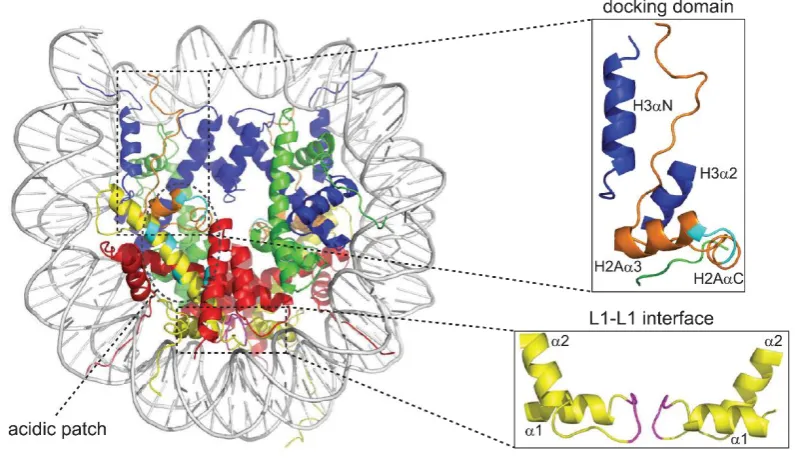
Figure 1.1.1 The nucleosome crystal structure. Nucleosomal crystal structure as described by Luger et
6
The four “core” histones can be functionally and structurally divided into two parts: the
histone tail and the histone fold [2]. The N-terminal core histone tail is largely unstructured
and consists of up to 35 residues, which corresponds to almost 20% of its total amino acids.
The core histone fold domain (HFD) in turn consists of 80-90 amino acid residues and exhibits
an additional, unique secondary structure mediating interactions between histones [13] and
DNA [14]. Further, the different HFDs of all core histones are highly similar within their
structural “α1-L1-α2-L2-α3” motif, where each HFD forms three α-helices (α1- α3) connected
by two loops (L1 and L2) [10]. In solution, histones obligatory form heterodimers, whereby
H2A only pairs with H2B and H3 only pairs with H4 [2]. Two histone H3-H4 pairs form via a
4-helix bundle a (H3-H4)2 tetramer in a twisted W-shaped arrangement. The histone octamer is formed either in presence of DNA or in high-salt conditions (2 M) by the interaction of two
H2A-H2B dimers with one (H3-H4)2 tetramer [13, 15]. Therefore, H2B is connected via a four-helix bundle with H4 and a further interaction profile is formed between the (H3-H4)2 tetramer and H2A´s docking domain [2]. Finally, a 147 bp DNA fragment – its minor groove always facing
the basic surface of histones – interacts base-unspecific with the octamer, thereby forming
the nucleosome [2, 8, 10]. NCPs, connected via linker DNA to the neighbored nucleosome
core, form the primary so-called nucleosomal ´beads-on-a-string´ structure [16-18]. The
secondary chromatin structure is created by further organizing the nucleosomal arrays into a
highly condensed 30 nm fiber by the linker histones H1 and H5 [19, 20] whereas the tertiary
structure is formed by large-scale configurations that build the entire chromosome [17].
Focusing on the 30 nm fiber, extensive biochemical and biophysical studies were carried out
but the existence of a 30 nm fiber in vivo is still under discussion [21]. Nevertheless, there exist
two competing models for its structure: the solenoid model and the zigzag model [17] (Figure
1.1.2). The one-start helix or solenoid model proposes that neighbored nucleosomes are
tightly coiled up and connected via variable linker DNA lengths to a compact structure [20,
22]. In turn, the two-start helix or zigzag model suggests that nucleosomes are arranged with
7
Figure 1.1.2 Two current models of the 30 nm chromatin fiber. (Left) Solenoid model of the 30 nm
fiber modeled without DNA, proposed by [25]. Helical gyres are shown in magenta and blue, respectively. (Right) Zigzag model of the 30 nm fiber with linker DNA criss-crossing between NCPs, proposed by [26]. Helical gyres are shown in orange and blue, respectively. Figure reprinted from [17], with permission from Elsevier.
To understand the specific regulation of gene expression, recent technological advances
fostered the research on certain chromatin states that repress or activate gene activity [27].
Cytologically, chromatin can be partitioned into two major states: heterochromatin and
euchromatin [27, 28]. Heterochromatin mainly consists of repressive, condensed chromatin
regions and harbors repetitive DNA elements [28, 29] whereas euchromatin is active,
gene-rich and less condensed [30]. It is assumed, that the switch between eu- and heterochromatin
is fundamental for the accessibility of DNA thus enabling repressive or active transcriptional
states [27].
Since the main focus of this thesis is set on the work with human cells, I refer to the situation
8
1.2
Alteration of chromatin structure
As mentioned in 1.1, chromatin is partitioned into two major states – euchromatin and
heterochromatin. Heterochromatin can be additionally subdivided into a constitutive and a
facultative form, whereas constitutive heterochromatin describes a permanent,
transcriptionally silenced and facultative heterochromatin just a temporarily silenced state
[31]. However, regarding the lacking explanation for distinct cellular processes and
differentiation events only based on DNA [7], it was obvious that there has to be a connection
between the biological activity and chromatin structure [32]. Already in 1942, Conrad
Waddington pointed out that epigenetics is the causal mechanism that brings the genotype
into phenotype [33], whereas todays widely accepted opinion describes epigenetics as the
study of genomic alterations without changing the DNA sequence [34]. This definition is based
on heritable, cell identity-defining instructions that regulate how, when and whether genetic
information will be transcribed and translated [35]. In general, chromatin structure and thus
DNA packaging dynamics are altered by distinct, but interconnected key players like DNA
methylation, non-coding RNAs (ncRNAs), nuclear architecture, ATP-dependent chromatin
remodelers, post-translational modifications (PTM) of histones or the
incorporation/replacement of histone variants [36].
The chemical modification of DNA by methylating the fifth position of cytosine (5mC) is one
possibility to alter chromatin structure [37, 38]. The covalent modification occurs at 60-80%
of palindromic, symmetrical CpG sites [39-41], is associated with direct or indirect
transcription silencing and has fluctuating levels for different cell types, gene states (active
versus silenced) and genetic locations (gene body versus promoter) [42]. A direct way to
repress gene transcription is either to prevent binding of transcription factors (TFs) to their
recognition motif [43] or the recruitment of methyl-CpG domain-binding proteins fostering
local chromatin condensation [44, 45]. However, recent studies also imply that methylated
cytosines might play a role in gene activation [46]. Moreover, 5mC is linked to biological
functions such as nucleosome positioning [47, 48], splicing [49] or genomic and chromosomal
stability [50, 51]. In general, the modification is established and maintained by three
conserved DNA methyltransferases (DNMT1, DNMT3A and DNMT3B) [52, 53]. DNMT3A and
DNMT3B are fundamental for de novo cytosine methylation [53], whereas DNMT1 is
9
synthesized CpGs during DNA replication [54]. Interestingly, the absence of DNMT1 results in
decreasing 5mC levels [55] pointing towards a specific demethylation of CpG sites [56].
Indeed, members of the Ten Eleven Translocation (TET) dioxygenase family (TET1, TET2 and
TET3) were shown to catalyze the demethylation of DNA [55] by oxidizing 5mC into three
distinct forms: hydroxymethylcytosine (5hmC), formylcytosine (5fC) and
5-carboxylcytosine (5caC) [57-59]. The stable modification 5hmC [60, 61] is associated with
promoters and gene bodies of highly expressed genes [62, 63], whereas 5fC plays a role in
transcription regulation [64] and 5caC reduces the substrate specificity of RNA polymerase II
[65].
Second, the structure of chromatin can be influenced by long noncoding RNAs (lncRNAs)
constituting a heterogenic class of RNAs [66]. To distinguish lncRNAs from small noncoding
RNAs [67] they are defined as “non-protein-coding transcripts larger than 200 nucleotides”
[68]. However, there is no standard classification and lncRNA nomenclature is rather based on
features like transcription origin, molecular function or tissue specificity [69]. Surprisingly,
lncRNAs are evolutionary poorly conserved [70]. Nevertheless, their function (recruitment of
chromatin/histone modifying enzymes, nucleosome positioning modulation) can be
maintained via a high structural conservation [71-73]. One of the most popular functions of
lncRNAs is the inactivation of the X-chromosome in female mammals [74, 75]. There, the
important lncRNA Xist is sufficient to silence X-chromosome linked genes [76-78] by coating
the inactive X [79]. However, in addition to their role in dosage compensation, lncRNAs seem
to be involved in distinct cancer types [80] thus being in focus of recent research.
In addition to DNA methylation and lncRNAs, chromatin structure can be also altered by the
presence of architectural proteins. These proteins guarantee via intra- and inter-nucleosome
interactions the accessibility of eukaryotic DNA by influencing the conformation of the
chromatin fiber (see also Figure 1.1.2) [81]. In the genome, architectural proteins are generally
localized at architectural protein binding sites (ABPSs) [82] that can be subdivided, dependent
on the number of architectural and associated accessory proteins, in low-occupancy or
high-occupancy APBS [83]. Thereby, the specific binding of architectural proteins to different
binding sites leads to fundamental roles in genome organization and function [82] like 3D
genome organization or the mediation of functional chromatin interactions [83]. Amongst
10
architectural protein in vertebrates [84]. In general, CTCF-binding sites are often associated
with different methylation states of DNA (see above) [85], but also DNA
methylation-independent binding sites exist, pointing towards CTCF´s complex functionality like enhancer
blocking, transcriptional pausing, alternative mRNA splicing or as boundary element [84]. To
control biological functions such as gene expression, architectural proteins like CTCF facilitate
the contact between different genome regions by bringing them into close proximity [84].
Moreover, to prevent the interaction of adjacent domains, topologically associating domains
(TADs) are enriched for clusters of CTCF binding sites [86] thereby establishing TAD borders
[87]. In addition to its function as TAD border, CTCF was also identified as boundary element
for lamin-associated domains (LADs), which are linked to the nuclear lamina [84, 88]. Although
it is tempting to assume CTCF being the main contributor of border formation, it is unlikely
that this is one of its main functions [84].
The compact organization of chromatin within the cell nucleus has to be still enough flexible
to enable access of enzymes and regulatory factors to DNA [89]. One possibility to render the
accessibility of nucleosomal DNA is ATP-dependent chromatin remodeling [90]. Multi-protein
chromatin remodeler complexes are remarkably diverse [91], harbor an ATPase subunit that
enables chromatin remodeling by ATP-hydrolysis [92] and up to 20 non-catalytic subunits that
are indispensable for the regulation of chromatin remodeling activities [91]. The catalytic
ATPase subunit is highly conserved and flanked by defined domains, therefore remodelers can
be divided into four families: SWI/SNF (switch/sucrose non-fermentable), ISWI (imitation
switch), CHD (chromodomain helicase DNA-binding protein) and INO80 (inositol-requiring 80)
[91, 93, 94]. Members of the SWI/SNF family possess an N-terminal HSA (helicase-SANT)
domain that recruits actin-related proteins and a C-terminal bromodomain, which is known to
interact with acetylated histone lysines [95]. This family is capable of sliding and/or evicting
nucleosomes [96] and plays a role as suppressive complex in pancreatic cancer [97]. ATPases
of the ISWI family are defined by the presence of a C-terminally located SANT domain
neighbored to a SLIDE domain, which together recognize nucleosomes and bind to unmodified
H4 tails and DNA [92]. ISWI family complexes are involved in transcriptional repression [98],
nucleosome positioning and gene silencing [99]. CHDs harbor two chromo domains and an
N-terminal ATPase domain [94, 100]. Chd1, a member of the CHD family, co-localizes with
promoters of actively transcribed genes and prevents the accumulation of heterochromatic
11
domain [102] and play a crucial role in DNA double strand break repair and protein
recruitment to DNA damage sites in human cells [103].
The following chapters explain two further possibilities of chromatin regulation in more detail.
1.2.1 Posttranslational modifications of histones
As described in 1.2 not only DNA is covalently modified but also histones that build up the
entire nucleosome. These PTMs occur in a large number of different variations: methylation,
glycosylation, ubiquitination, propionylation, acetylation, deimination, phosphorylation,
ADP-ribosylation, formylation, crotonylation and SUMOylation [104-106]. Especially the
intrinsically disordered and highly flexible N-terminal histone tail is subjected to PTMs [10],
but also the histone core can be posttranslationally modified [107]. More than a decade ago,
T. Jenuwein and D. Allis already proposed that histone modifications do not occur individually
but in different combinations thus generating a specific "histone code" [108]. Hence, it would
be possible to control biological processes and mechanisms with far-reaching consequences
for distinct cell fates [108]. In general, the modification of histones is very dynamic, strongly
influences the genomic structure and is regulated by highly specific enzymes [104, 109].
“Writers” such as (1) histone acetyltransferases, (2) histone methyltransferases, (3) kinases
and (4) ubiquitin ligases catalyze posttranslational modifications like (1) acetylations (lysine),
(2) methylations (arginine or lysine), (3) phosphorylations (tyrosine, threonine or serine) and
(4) ubiquitinations (lysine), whereas “erasers” like histone deacetylases, demethylases,
phosphatases or ubiquitin proteases remove them [110-113]. The functional influence of
PTMs on chromatin can be either direct by changing chromatin structure or indirect by
recruiting effector proteins [110]. These epigenetic regulators are so-called “readers”, which
harbor specific domains such as e.g. tudor, MBT (Malignant Brain Tumor), chromo, WD40,
bromo and PWWP domains that can interact with specific modifications [112]. Proteins of the
tudor family bind to methylated substrate residues (arginine or lysine) thereby regulating RNA
metabolism, small RNA pathways or DNA damage response and function as adaptor proteins
to facilitate the assembly of macromolecular complexes [114]. MBT domain-containing
proteins interact with mono- and di-methylated lysines on histone H3 and H4 tails, are
associated with neurological tumors in humans and play a crucial role in gene regulatory
pathways [115]. Members of the chromo domain-containing family bind to di-and
12
chromatin structure [116-118], whereas the β-propeller structured WD40 domain family
interacts with methylated H3 lysines and participates in signal transduction, cytoskeleton
assembly, RNA processing and cell division [119, 120]. Bromo domain-harboring proteins in
contrast recognize acetylated lysines (Kac) and have important roles in gene expression and
cancer development [121, 122]. Last but not least PWWP domain-containing proteins interact
with methylated lysines on the H3 or H4 tail and have an important role in
chromatin-associated processes like DNA repair or transcriptional regulation (for more detailed
information about PWWP domains see section 1.5) [123]. Altogether, the above-mentioned
chromatin readers in concert with further PTM-mediated mechanisms define inaccessible
(inactive) and accessible (active) chromatin states by modulating the global chromatin
environment [113]. Shortly, inactive chromatin is characterized by modifications such as
H3K27 trimethylation (H3K27me3) in combination with Polycomb-repressive complexes 1 and
2 (PRC1 and PRC2) as well as H3K9 di- and trimethylation and H4K20me3 together with
heterochromatin binding protein 1α (HP1α) [124, 125]. Transcriptionally active chromatin on
the other hand is associated with a plethora of posttranslational histone modifications like
acetylations of multiple lysine residues on H3 and H4 and methylations on H3 lysine 4, 36 and
79 [124]. Thereby, tri-methylated H3 lysine 36 (H3K36me3) is found within the body of actively
transcribed genes [126]. The methylation of mono- and di-methylated H3 lysine 36
(H3K36me1 and H3K36me2) is catalyzed by the nuclear SET domain (NSD)-containing enzymes
NSD1, NSD2 and NSD3, whereby H3K36me2 is further established by methyltransferases such
as ASH1L [127, 128]. On the other hand, SET domain-containing 2 (SETD2)/huntingtin
interacting protein B (HYPB) is responsible for the trimethylation of H3K36 [129, 130] and is
recruited via hyperphosphorylated RNA polymerase II (RNAPII) [131]. H3K36me3 mainly
serves as binding platform [132], especially via the direct interaction with PWWP-domain
containing proteins such as mismatch recognition enzyme hMutSα (hMSH2-hMSH6) [133] or
developmental gen regulatory PC4 and SF2 interacting protein 1 (Psip1) [134]. Via the
recruitment of binding proteins to gene bodies, H3K36me3 is able to regulate a plethora of
biological mechanisms including transcriptional elongation, repression of cryptic transcripts,
chromatin accessibility and DNA damage response signaling [127, 129, 133, 135].
All in all, PTMs and their interplay with additional chromatin components is crucial for the
13
consequences of combinatorial PTMs and their crosstalks to provide a better understanding
of these, almost all biological processes underlying, regulatory mechanisms [136, 137].
1.2.2 Histone variants
In addition to PTMs, the exchange of the canonical core histones H2A, H2B, H3 and the linker
histone H1 (to date no H4 variants have been found in humans and other higher eukaryotes)
with specific histone variants modifies the nucleosomal composition and can further regulate
the global chromatin landscape [138-140]. In general, the incorporation of histone variants
creates specialized nucleosomes, enhances chromatin plasticity and complexity and can result
in profound alterations of the chromatin structure thereby influencing multiple biological
processes [12]. Canonical histones and the according variants differ in some basic aspects.
First, the multiple canonical histone genes are expressed during S-phase whereas the
expression of most histone variant genes takes during the whole cell cycle place [141, 142].
Second, canonical histone genes lack introns and give rise to a unique 3´ stem-loop mRNA
structure [141, 143]. By contrast, pre-mRNAs of most histone variants often contain introns
thus providing the opportunity to generate alternative splice isoforms [12, 141]. Furthermore,
some histone variant mRNAs have a poly-adenylated (poly(A)) tail [142], whereas canonical
histone mRNAs miss this specific pattern [12]. Last, histone variants are non-allelic isoforms
of their canonical counterparts and differ markedly from their primary sequences and
sometimes additionally in secondary and/or tertiary conformations [141]. These differences
in protein sequence can be either minimal (few individual amino acid substitutions) or far
more tremendous (exchange of entire domains) [142, 144]. Concluding, histone proteins
possess distinct structural and functional properties, hence it is imperative to study the
composition of canonical histones together with histone variants in nucleosomes to gain a
better understanding of their complex interplay [12].
Histone variants and their canonical counterparts do not only differ in expression timing,
mRNA structure and at protein levels, but also in their distribution within the linear genome
[141]. In contrast to canonical histones, which are rather equally distributed, histone variant
incorporation and eviction is highly specific and dependent on remodeling and chaperone
complexes [141, 145]. Histone chaperones are crucial for several steps in nucleosome
formation [146] and bind in a direct manner to newly synthesized histones to regulate their
14
trafficking (after their synthesis), where they regulate their transport from cytoplasm to the
cell nucleus [148]. Others affect PTMs by regulating the interaction with the corresponding
enzymes [143] or promote the formation of nucleosomes by facilitating histone interactions
[149, 150]. On the other hand, ATP-dependent remodelers eject or slide already assembled
nucleosomes thus organizing chromatin structure and promoting the exchange of H2A-H2B
dimers with distinct variant dimers [151].
More than 80% of all nucleosomal linker DNA binds at least one protein of the histone H1
family, which represents the largest group of histone variants with its four germ-line and seven
somatic (mammalian) specific variants [152, 153]. Besides their high primary sequence
homology and evolutionary conservation, H1 variants differ in some aspects including
euchromatin and heterochromatin distribution or chromatin binding affinity [153-156]. In
addition they play an important role in chromatin compaction and transcriptional regulation
[157].
To date, solely three, functional uncharacterized, bona fide H2B variants (testis-specific
hTSH2B, H2BFWT and spH2B) are known in the human genome, although more than 15 H2B
genes have been identified, yet [158]. In 2012, Santoro and Dulac identified in mice an
additional, fourth H2B variant – the olfactory, neuron-specific H2BE [159]. hTSH2B differs in
its amino acid sequence from its canonical counterpart H2B mainly in N-terminal regions
[158], but its biological function still remains elusive [160]. The primate-specific variant
H2BFWT shares just 45% homology with canonical H2B, but was found to act similar in a
nucleosomal background [161]. Further, it is hypothesized to act as an epigenetic marker
modulating telomeric identity [158]. spH2B has been identified in a telomere-associated
complex and varies from somatic H2B in biochemical parameters including electrophoretic
mobility [162]. The newly identified neuron-specific H2BE variant is incorporated into
H2B-containing chromatin and has a direct effect on the expression level of genes and the
persistence of olfactory sensory neurons [159].
Currently, two canonical H3 (H3.1 and H3.2) and a plethora of H3 variants (H3.3, H3t, H3.5,
H3.X, H3.Y, CENP-A (CenH3), H3.6, H3.7 and H3.8) are known [138, 163-166]. H3.3 is encoded
by two independent human genes (H3F3A and H3F3B [167]) and differs from its canonical
counterpart H3.2 in four amino acids [168]. Remarkably, these differences in primary
15
H3.1 and H3.2 [169, 170]. Whereas H3.1 and H3.2 deposition is catalyzed by the histone
chaperone CAF1 (Chromatin-Assembly Factor1) [170], H3.3 is incorporated either by the
chaperone complex HIRA at actively transcribed gene regions [171] or by the death-domain
associated protein/α Thalassemia/Mental Retardation X-linked (DAXX/ATRX) chaperone
complex at repetitive heterochromatic regions. Interestingly, the N-terminal tail of H3.3
harbors a unique amino acid – S31 – that is phosphorylated close to centromeric regions in
metaphase HeLa cells [172]. H3t and H3.5 are both testis-specific [164], whereby H3t plays an
important role in chromatin reorganization and early spermatogenesis [166, 173] and H3.5
may influence DNA synthesis [174]. The primate-specific histone H3 variant H3.X was
identified together with H3.Y in our lab [175]. Both variants are expressed in different parts of
the brain and are stably incorporated into the nucleosome. Interestingly, external stress
stimuli like starvation and overgrowth of cells can enhance the expression of H3.Y in
osteosarcoma-derived cancer cells. H3.Y regulates cell cycle-associated gene expression and
cell proliferation [175]. The kinetochore- and centromere-associated histone variant protein
CENP-A serves as binding platform for the assembly of kinetochores during cell division and
regulates chromosomal stability [176, 177]. Furthermore, the overexpression and
mislocalization of CENP-A results in higher invasiveness and bad prognosis in several cancer
types [177]. The recently identified H3 variants H3.6, H3.7 and H3.8 are known to exist in many
human tissues and were so far just studied in in vitro nucleosome assemblies without pointing
towards any specific biological function [165].
1.2.3 H2A variants
In 1977, two different mammalian isoforms of canonical H2A (H2A.1 and H2A.2) differing in
amino acid position 51 were discovered [178] and over the last decades a plethora of histone
H2A variants could be added to the growing list of human H2A variants [12]. In addition to
canonical H2A, eight further variants exist in humans: the universal variants H2A.X and H2A.Z
(H2A.Z.1, H2A.Z.2.1 and H2A.Z.2.2) as well as H2A.Bbd and macroH2A (macroH2A1.1,
macroH2A1.2 and macroH2A2). Generally, the highest variation of H2A variants can be
observed within their C-termini, ranging from differences in amino acid composition and
alterations in C-terminal length.
In the 1980s, H2A.X was identified together with H2A.Z [179]. H2A.X is evolutionary highly
16
deposited throughout the genome by a yet unknown deposition machinery [181]. H2A.X is
stated as genomic “histone guardian” [180, 182] and is famous for its well-studied
modification on S139 (S139ph), which is established upon DNA damage [180, 182]. In its
phosphorylated state, H2A.X is also called γ-H2A.X, because it was initially discovered after
treating mammalian cells with gamma irradiation [180, 183]. γ-H2A.X serves at DNA damage
sites as recruiting platform for repair proteins, chromatin remodelers and
chromatin-modifying enzymes [181]. In turn, γ-H2A.X is stabilized at DNA lesions via the methylation of
lysine residues such as H3K9, H3K27 and H3K36 [184]. However, in addition to its role in
double-strand-break repair (DSB), γ-H2A.X may help to keep broken DNA ends together
through accumulating specialized repair proteins [185]. Due to its important roles in DNA
damage repair (DDR) it is not quite surprising that mutations in or deletions of H2A.X have
been detected in many tumors [181].
One of the most rapid evolving histone variants is testis-specific H2A.Bbd (Barr body deficient)
[186, 187], which is mammalian-specific [186, 188] and shares on the protein level only 50%
identity with canonical H2A [189]. Strikingly, H2A.Bbd lacks, due to its shortened primary
sequence, huge parts of the docking domain, the C-terminal tail and the acidic patch that is
needed for nucleosome-protein interactions [186]. The consequence of these missing
structures is therefore a reduction in nucleosome stability and a more opened chromatin
structure [186, 190-192]. Additionally, H2A.Bbd incorporation has similar structural effects as
shown for highly dynamic acetylated nucleosomes [186] and could be an alternative,
acetylation-independent mechanism to loosen chromatin structure.
MacroH2A was discovered in 1992 [193] and is encoded by two different genes: H2AFY
(macroH2A1) and H2AFY2 (machroH2A2) [139]. H2AFY can be alternatively spliced and gives
rise to two protein isoforms: macroH2A.1.1 and macroH2A.1.2 [194]. MacroH2A members are
fascinating proteins due to their exceptional tripartite domain architecture: the N-terminal
histone region is linked to a non-histone macro domain through a H1-like linker leading to a
unique, three-fold larger protein structure than H2A [193]. Interestingly, macroH2A.1.1 but
not macroH2A.1.2, can interact with specific metabolites of nicotinamide adenine
dinucleotide (NAD) including poly(ADP-ribose) [195-197] suggesting a unique function for
macroH2A.1.1 [12]. However, several studies propose a broad range of biological roles for
17
differentiation, somatic cell reprogramming and tumor suppression, depending on isoform
and chromatin location [12, 195, 198].
Since H2A.Z is one of the main actors in this work; its function and structure will be discussed
in more detail in section 1.3.
1.3
The histone variant H2A.Z
H2A.Z, an almost universal variant [12], was first described in the 1980s [179] and is known to
have evolved early during evolution [199]. H2A.Z shares only ~60% identity with canonical
H2A while it is ~90% conserved between different eukaryotes pointing towards specific, not
exchangeable functions within the H2A family [200] (Figure 1.3.1). Indeed, H2A.Z is crucial in
many organisms like X. laevis (frog) [201], M. musculus (mouse) [202], T. thermophile
(tetrahymena) [203] and D. melanogaster (fruit fly) [204] but not in S. pombe (fission yeast)
and S. cerevisiae (budding yeast) that show severe growth phenotypes upon knock-out [205,
206].
Figure 1.3.1 H2A and H2A.Z isoforms amino acid sequence alignment. Amino acid sequence
alignment of human H2A and human H2A.Z isoforms (H2A.Z.1, H2A.Z.2.1 and H2A.Z.2.2). Amino acid sequences of H2A [P0C0S8], H2A.Z.1 [P0C0S5], H2A.Z.2.1 [Q71UI9-1] and H2A.Z.2.2 [Q71UI9-2] were aligned using the web browser-based MUSCLE multiple sequence alignment tool (default settings). Identical amino acids are depicted in dark grey, similar amino acid residues in light grey and changes are highlighted in white. The three amino acids differing between H2A.Z.1 and H2A.Z.2.1 are illustrated in yellow and acidic patch residues are boxed in cyan. Possible modification sites of H2A.Z variants are marked with stars (acetylation, pink) and dots (ubiquitination, black). Figure based on [12].
In vertebrates, H2A.Z is encoded by two non-allelic, intron-containing genes: H2AFZ (H2A.Z.1)
and H2AFV (H2A.Z.2), which both give rise to polyadenylated mRNAs and to H2A.Z proteins
that differ in just three amino acids [12, 207] (Figure 1.3.1). Notably, H2AFV can be
alternatively spliced resulting in the expression of two subforms: H2A.Z.2.1 (H2A.Z.2,
18
essential parts for H2A.Z function lay in its C-terminal region (M6 and M7) [208]. In
concordance, also in yeast (S. cerevisiae) M6 fulfills important functions and is e.g. essential
for the interaction with the Swi2/Snf2-related ATPase-1 (SWR-1) remodeling complex [209] or
H2A.Z deposition [210]. In addition, the discovery of H2A.Z.2.2, which in its C-terminus lacks
14 amino acids and harbors a unique stretch of six amino acids (Figure 1.3.1), provided further
fundamental insights in H2A.Z´s C-terminal functions [190, 211]. Compared to other H2A.Z
members, the shorter H2A.Z.2.2 isoform remains soluble and is not completely associated
with chromatin and less tightly incorporated in nucleosomes [190, 211].
At the turn of the millennium, the crystal structure of the H2A.Z-containing nucleosome was
solved and revealed, despite the huge differences in primary sequence, a highly structural
similarity to canonical H2A nucleosomes [212]. This structure also unraveled alterations in two
regions that might be responsible for contrary observations in nucleosome stability. Indeed,
many studies addressed this issue with contrasting results: H2A.Z seems to have both,
stabilizing and destabilizing effects [12, 200]. Suto et al. reported, that H2A.Z´s L1-L1 interface
forms more contacts between than H2A-dimers resulting in stabilized
H2A.Z-homodimers [212], but also the presence of heterotypic H2A-H2A.Z nucleosomes could be
already shown in different model organisms [213-217]. Moreover, the destabilizing effects of
H2A.Z were also observed when it teams up with histone H3 variant H3.3 in the same
nucleosome [218]. Under physiological salt conditions it was shown that nucleosomes
prepared from native chromatin having both variants incorporated are less stable than those
containing only H3.3 [219]. Surprisingly, in contrast to its destabilizing function observed in
mononucleosomes, H2A.Z incorporation is crucial for the formation of higher chromatin
structures [220]. Compared to canonical H2A, H2A.Z has an extended acidic patch with an
increased affinity to the H4 tail [221]. This strong electrostatic interaction with H4 results in
an increased intrafibre folding and a higher compaction of secondary structures [221, 222].
Further, HP1α prefers highly folded H2A.Z- over H2A-containing fibers and enhances intrafibre
folding [223], highlighting the function of histone variants in the establishment of chromatin
structures by recruiting further chromatin structure modifying proteins [12].
Like other histone variants, H2A.Z can be posttranslationally modified resulting in different
functional consequences [224, 225]. Mammalian H2A.Z can be chemically altered by four
distinct mechanisms: acetylation, monoubiquitylation, SUMOylation and monomethylation at
19
(H2A.ZK7ac) and 11 (H2A.ZK11ac) (Figure 1.3.1) and is reported to be associated with
promoter regions of active genes [227, 228], confers to an open nucleosome conformation
and has destabilizing effects [229]. In addition, the acetylation of H2A.Z at promoter regions
is suggested to activate oncogenes thus deregulating gene expression and foster tumor
progression [226]. In addition to N-terminal acetylations, H2A.Z can be further modified by
C-terminal monoubiquitinations on lysine 120, 121 and 126 that are associated with inactive X
chromosomes in female mammals [230]. H2A.Z SUMOylation in turn has been implicated to
be involved in DNA repair in yeast (S. cerevisiae) [231] and monomethylation of H2A.ZK7 was
suggested to play a role in transcriptional repression in embryonic stem cells (M. musculus)
[232].
The deposition or/and eviction of H2A.Z is dependent on multifactor ATP-dependent
chromatin remodeling complexes, whereby genome-wide studies helped to unravel possible
target sites [233]. In S. cerevisiae, the 14 subunit SWI/SNF-related SWR1 complex (SWR1) is
crucial for the incorporation of H2A.Z-H2B dimers into chromatin [234-236]. To do so, SWR1
deposits H2A.Z-H2B into nucleosomes that flank the nucleosome free region (NFR) of
transcription start sites (TSS) and well distributes it all over the genome (H2A.Z is present in
5% of nucleosomes but 65% of all genes) [237]. In higher eukaryotes like humans, the SWR1
complex has two counterparts named p400/NuA4/TIP60 and SRCAP (SWI2/SNF2-related CBP
activator protein) [233, 238, 239] where each of them harbors (working context dependent)
histone replacement abilities in vivo and in vitro [233]. Additionally, Papamichos-Chronakis et
al. discovered that a specific remodeling complex – INO80 – facilitates the eviction of H2A.Z in
an ATP-dependent manner after transcriptional induction in S. cerevisiae [233, 240], but it still
remains elusive if the mammalian INO80 complex may have a similar role in higher eukaryotes
[241]. However, Mao and co-workers could identify Anp32e as a mammalian, H2A.Z-specific
chaperone facilitating the eviction of H2A.Z at transcription start sites [242].
Interestingly, H2A.Z is not uniformly distributed all over the genome suggesting three
non-mutually exclusive mechanisms [12]. First, H2A.Z is undirected incorporated and afterwards
actively evicted from non-target sites; second, H2A.Z is incorporated in an active manner at
specific sites by special target factors [243] and third, the localization of H2A.Z is dependent
on deviating stabilities of homotypic/heterotypic H2A.Z-containing compared with canonical
nucleosomes [216]. Regardless how H2A.Z is incorporated into chromatin, the variant is crucial
20
progression, genome stability, telomere integrity, chromosome segregation, mitosis, brain
memory formation, heterochromatin formation and just recently discovered
epithelial-mesenchymal transition [12, 181, 200, 225, 244-246].
In 1986, Allis et al. initially suggested that H2A.Z has a role in transcription regulation [247].
The invention of genome-wide assays like chromatin immunoprecipitation sequencing
(ChIP-seq) enabled to further dissect the function of H2A.Z in transcription. It could be elucidated
that H2A.Z is present around the TSS at gene promoters in human [248, 249], fly [250], plants
[251], yeast [252, 253] and mouse [254] and further regulatory elements like insulators or
enhancers [200]. Remarkably, H2A.Z is enriched at both, transcriptionally active and silenced
genes whereby the context-dependent underlying mechanism has remained elusive [255]. In
human, mouse and yeast the -1 and +1 nucleosomes (nucleosomes that flank the NDR) at TSS
are occupied by H2A.Z [200]. But why is the incorporation of H2A.Z at TSS so important for
transcriptional regulation? In vitro studies have demonstrated that the +1 nucleosome forms
a significant barrier to transcription [256, 257] by preventing the transit of RNA polymerase II
(RNAPII) [258]. Notably, the incorporation of H2A.Z leads to decreased RNAPII stalling, hinting
towards a function in dissolving the high-energy barrier [255]. The underlying mechanism
might be the labialization of the +1 nucleosome by H2A.Z incorporation, thereby modulating
the kinetics and transcriptional elongation of RNAPII and passively regulating transcription
[255, 258].
Moreover, current literature demonstrates that H2A.Z plays controversial roles in DNA
damage response. Studies in yeast showed increased genomic instability and sensitivity to
DNA-damage upon H2A.Z knock-out [240, 259] and suggest an important role for H2A.Z in
DNA damage response [231, 260]. Furthermore, Xu et al. reports H2A.Z is actively
incorporated at DSB by the p400/NuA4/TIP60 complex in an ATPase-dependent manner [260],
thereby influencing chromatin structure and recruiting further, essential DSB repair proteins
to sites of DNA damage. By contrast, the Price group demonstrated that H2A.Z is removed by
histone chaperone Anp32e at DNA double strand breaks, which promotes DNA repair and
nucleosome reorganization [261].
Besides transcription regulation and DNA repair, H2A.Z also affects cell cycle progression
[262]. The directed expression of genes in yeast during the cell cycle requires chromatin
remodeling (Swi/Snf) and modifying (SAGA) complexes and the presence of unique histone
21
proteins during S phase and the regulation of cyclin genes [263] like cyclin D1, that is involved
in G1-S progression and a plethora of biological functions [264].
Compared to the large number of biological functions that H2A.Z performs within the cell, it
is not surprising that even the slightest changes in H2A.Z levels can lead to different types of
cancer [181]. A number of microarray studies, aiming to identify different gene expression
patterns in human cancer, revealed increased H2A.Z levels in different tumor types like
colorectal, lung, bladder and breast [265-268]. Further, global H2A.Z levels are upregulated in
melanoma cell lines [269] and a direct role in cancer could be detected in breast and prostate
cancer [181]. However, H2A.Z.2 is a key driver of malignant melanoma and correlates with a
bad prognosis for patients, whereas the absence of H2.A.Z.2 sensitizes melanoma cells to
targeted chemotherapies [270]. In detail, H2A.Z.2 regulates the transcription of highly
expressed E2F target genes, which show a distinct pattern of H2A.Z occupancy. The unique
function of H2A.Z.2 is further the promotion and maintenance of E2F1, BRD2 and histone
acetylation levels to promote transcription of cell cycle regulating factors. In fact, BRD2 is
crucial for the cell cycle and controls together with E2F1 the expression of cyclins and further
regulatory genes [271-273]. In addition, BRD2 may recruit distinct transcription factors to
initiate oncogenic gene transcription in several cancer types [270]. To date, metastatic
melanoma cannot be effectively treated by current therapies [274], but the combinatorial
depletion of H2A.Z.2 and BRD2 (BET protein) is an effective tool in inducing cell death and
sensitizing cells to distinct therapies [270].
Summarizing, H2A.Z has a variety of biological functions and is involved in the development
of different types of cancer - therefore it is very important to study the biological mechanism
of this variant in order to gain a better fundamental understanding and to develop possible
therapies against different diseases.
1.4
H2A.Z binding proteins
To gain deeper insights in the underlying mechanisms of H2A.Z´s multiple biological functions,
we and others analyzed the interactome of the histone variant in more detail [275, 276].
Therefore, Draker et al. used human embryonic kidney cells (293T) and transfected them with
22
and co-workers found 21 top interactors specifically enriched for H2A.Z, where most of them
are associated with chromatin and are involved in transcription and chromatin organization.
In general, they identified members of the H2A.Z remodeling complex SRCAP and protein
readers like PHD finger-containing or PWWP domain-containing proteins. Also our group
aimed to identify the interactome specifically enriched on H2A.Z compared to H2A. Therefore,
Sebastian Pünzeler (a former PhD student of our lab) together with the group of Matthias
Mann (MPI Munich) performed label-free quantitative mass spectrometry (lf-qMS) of
mononucleosomes derived from human HeLa Kyoto (HK) and metastatic melanoma
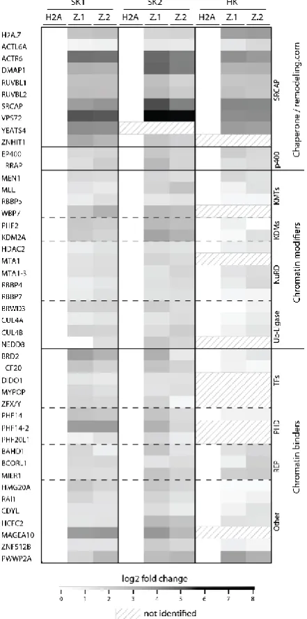
SK-mel147 cell lines (stably expressing GFP-tagged H2A or H2A.Z) [276]. There, more than 40
H2A.Z-specific binding proteins were detected (Figure 1.4.1). In line with the findings of Draker
and co-workers [275], we also identified members of the chaperone complex SRCAP,
chromatin-modifying complexes, PHD finger-containing and PWWP-domain-containing
proteins [276], but also subunits of the p400/TIP60 chaperone complex and many additional
chromatin binders. Interestingly, in comparison to SRCAP we could only enrich the two largest
subunits of the p400/TIP60 complex suggesting rather a transient interaction with H2A.Z e.g.
while removing together with Anp32e the variant upon DNA damage [261]. Moreover, the
detection of histone-modifying complexes such as mixed lineage leukemia (MLL), which was
initially identified in human leukemia [277-280], is a target of chromosomal translocation
[281] and catalyzes the histone modification H3K4me3 [282], supports the idea that H2A.Z
acts as a recruitment platform for distinct proteins [282]. In concordance with that hypothesis,
we additionally identified histone-modifying enzymes like the demethylases PHF2 and KDM2A
[276]. PHF2 harbors an N-terminal plant homeodomain (PHD) binding to H3K4me2/3 and a
Jumonji (JmjC) domain, involved in removing repressive methyl marks (H3K9me1) [283, 284].
KDM2A also harbors a JmjC domain and demethylates mono- and dimethylated Lys 36 of
histone H3 (H3K36me1/2) but not trimethylated Lys 36 [285, 286], that only serves as an
23
Figure 1.4.1 Heatmap of H2A.Z.1- and H2A.Z.2-nucleosome-specific binding proteins. Pull-down
experiments with mononucleosomes derived from HK (HK) and SK-mel147 cells (SK1 and SK2) (each stably expressing GFP-H2A, GFP-H2A.Z.1 (Z.1) and GFP-H2A.Z.2 (Z.2)) were analyzed by label-free qMS-based proteomics and normalized to H2A. SK1 and SK2 represent two biological replicates and were reproduced in HK cells. Log2-fold change was hierarchically clustered and plotted as heatmap (Perseus). Log2 LFQ intensities (fold-change for GFP-H2A.Z.1, GFP-H2A.Z.2 vs GFP_H2A pulldowns) are illustrated for 44 identified proteins specifically enriched for GFP-H2A.Z. White color: no enrichment; black color: strong enrichment in comparison to GFP-H2A. Figure reprinted from [276], with permission of John Wiley and Sons.
Our lab decided to further investigate the vertebrate-specific, PWWP-domain containing
protein PWWP2A, which harbors a unique domain structure and was already discovered in
previous H2A.Z-pull-down studies [275]. PWWP2A has a molecular mass of approximately 82
24
canonical PWWP2A protein and two additional isoforms with most differences located within
their C-terminus. PWWP2A consists of two proline-rich regions “P1” (aa 61 - 146) and “P2” (aa
240 - 291), an internal region “I” [276] (aa 292 - 574) and one serine-rich stretch “S” (aa 575 -
632). In addition, PWWP2A belongs to the PWWP domain family of proteins, as it harbors a
C-terminal PWWP domain “PWWP” (aa 655 - 715) (Figure 1.4.2).
Figure 1.4.2 Primary structure of human PWWP2A. PWWP2A consists of 755 amino acids and
possesses two proline-rich regions P1 (aa 61 - 146) and P2 (aa 240 - 291) (red), an internal region I (aa 292 - 574), which can be further subdivided into IN (brown) and IC (yellow), one serine-rich stretch S (aa 575 - 632) (green) and a C-terminal PWWP domain PWWP (aa 655 - 715) (purple). NCBI reference sequence NP_001124336.1 (NM_001130864.1) or Uniprot identifier Q96N64-1.
PWWP2A locates together with H2A.Z at +1 and -1 nucleosomes at the TSS of highly
transcribed genes [276]. Moreover, PWWP2A does not contain any known enzymatic domains
and might therefore act as an adapter between chromatin-modifying proteins and
H2A.Z-containing chromatin, which is in line with the finding that it is not required for H2A.Z
occupancy and deposition. Like described above, PWWP2A recognizes H2A.Z-containing
nucleosomes at the TSS of actively transcribed genes, but is depleted at non-promoter sites.
However, the exact underlying positioning mechanism remains elusive. Further, the depletion
of PWWP2A leads to the deregulation of gene expression programs and has profound
consequences on cellular mechanisms. In HK cells the knock-down of PWWP2A leads to an
increase in mitotic cells, especially for those in prometaphase. PWWP2A-depleted cells stay
for up to 10 h in mitosis (control cells <1.5 h) while jumping back and forth between two states
25
Furthermore, the depletion of PWWP2A results in developmental defects in frogs (X. laevis
and X. tropicalis). During the developmental stage of the tadpole, the loss of PWWP2A
affected head differentiation and severely impaired proper eye development.These problems
upon PWWP2A depletion in frogs are caused by its fundamental role in neural crest cell
differentiation and/or migration during early developmental stages. Nevertheless, the strong
interaction of PWWP2A with H2A.Z-containing chromatin, thus initiating multiple biological
functions and processes, is likely achieved by its multi-domainous structure and is the main
topic of this thesis.
The special and unique features of PWWP domains are explained in more detail in the next
chapter.
1.5
PWWP domain-containing proteins
The PWWP domain was named after the structural motif “proline (P) – tryptophan (W) –
tryptophan (W) – proline (P)” (90-130 amino acids) that was first discovered in the
PWWP-domain containing Wolf-Hirschhorn syndrome candidate protein 1 (WHSC1) [288] and is
involved in chromatin binding. Thus far, more than 20 different human PWWP
domain-containing proteins have been identified [123]. Despite its high conservation, some variations
of the PWWP-motif can occur like SWWP in DNMT3A/B [289] or PHWP in HDGF [290].
However, PWWP domains that contain a proline residue as first amino acid show higher
stability and are less prone to aggregation [291]. In general, PWWP domains belong together
with Tudor, plant Agenet, chromo and MBT domains to the Tudor domain “Royal Family”
[292], that divergently developed during evolution but share the same ancestor, structurally
characterized by three β-strands [293]. To date, several studies described the
three-dimensional structures of PWWP domains including those of ZMYND11, PSIP1, DNMT3A,
DNMT3B or BRPF1 [294-298]. Interestingly, all these PWWP domains fold in a similar manner:
an N-terminal β-barrel and a C-terminal α-helical substructure [289]. The N-terminally located
β-barrel is conserved and consists of five antiparallel β-strands (β1 – β5), whereby the PWWP
motif is located at the interface of the β1 – β2 arch [289]. In contrast, the C-terminal α-helices
(two to six) are highly variable [289, 298]. In general, the stability of the PWWP domain
structure is based on polar bonds and intramolecular hydrogen interactions, which occur
26
their structural features, PWWP domains fulfill distinct tasks such as protein-protein
interactions, DNA binding or the recognition of posttranslational histone modifications [298].
Indeed, the PWWP domain of DNMT3A directly interacts with the inhibitory factor SALL3
thereby reducing DNMT3A-mediated CpG island methylation [302] and mutations in the
PWWP domain of DNMT3B result in a reduced interaction with SUMO E3 ligase PIAS1 [303].
Another member of PWWP domain-containing protein family is the transcriptional coactivator
PSIP1 (PC4 and SFRS1 interacting protein 1) that was shown to interact via its PWWP domain
with DNA and chromatin, crucial for HIV-1 integration [304, 305]. Likewise, the eukaryotic
mismatch repair protein MSH6 is able to bind non-specifically to double stranded DNA with
its N-terminal PWWP domain [306]. However, for most PWWP domain-containing proteins
the interaction with DNA takes not place in a specific or sequence selective manner but rather
via electrostatic contacts between the phosphate backbone of DNA and the highly positively
charged surface formed by arginine and lysine residues in the PWWP motif [293] (Figure
1.5.1).
Figure 1.5.1 DNA binding interface of HDGF PWWP domain. Residues, directly involved in the
formation of the DNA interaction surface are highlighted in green (left). Electrostatic distribution of the HDGF PWWP surface structure demonstrates an accumulation of highly positively charged residues (blue). Figure reprinted from [293], with permission of Springer Nature.
In addition, the similarities within structure and sequence of PWWP domains and other “Royal
family” member domains suggest a possible role in the recognition of posttranslational
modifications of histones [293]. Indeed, in 2009 the first PWWP domain-containing protein
being able to recognize H4K20me1 was discovered [307] with many further following [293].
Furthermore, a high-throughput mass spectrometry approach unraveled PWWPs ability as a
![Figure 1.1.2 Two current models of the 30 nm chromatin fiber. (Left) Solenoid model of the 30 nm fiber modeled without DNA, proposed by [25]](https://thumb-us.123doks.com/thumbv2/123dok_us/1805161.1233730/19.595.144.451.78.345/figure-current-models-chromatin-fiber-solenoid-modeled-proposed.webp)