Human Memory CD4
ⴙT Cell Immune Responses against
Giardia
lamblia
Christina Skår Saghaug,a,bSteinar Sørnes,bDimitra Peirasmaki,cStaffan Svärd,cNina Langeland,a,b Kurt Hanevika,b
National Centre for Tropical Infectious Diseases, Department of Medicine, Haukeland University Hospital, Bergen, Norwaya; Department of Clinical Science, University of
Bergen, Bergen, Norwayb; Department of Cell and Molecular Biology, Uppsala University, Uppsala, Swedenc
The intestinal protozoan parasiteGiardia lambliamay cause severe prolonged diarrheal disease or pass unnoticed as an asymp-tomatic infection. T cells seem to play an important role in the immune response toGiardiainfection, and memory responses may last years. Recently, TH17 responses have been found in three animal studies ofGiardiainfection. The aim of this study was to characterize the human CD4ⴙT cell responses toGiardia. Peripheral blood mononuclear cells (PBMCs) were obtained from 21 returning travelers with recent or ongoing giardiasis and 12 low-risk healthy controls and stimulatedin vitrowithGiardia lambliaproteins. Production of tumor necrosis factor alpha (TNF-␣), gamma interferon, interleukin-17A (IL-17A), IL-10, and IL-4 was measured in CD4ⴙeffector memory (EM) T cells after 24 h by flow cytometry. After 6 days of culture, activation and proliferation were measured by flow cytometry, while an array of inflammatory cytokine levels in supernatants were measured with multiplex assays. We found the number of IL-17A-producing CD4ⴙEM T cells, as well as that of cells simultaneously pro-ducing both IL-17A and TNF-␣, to be significantly elevated in theGiardia-exposed individuals after 24 h of antigen stimulation. In supernatants of PBMCs stimulated withGiardiaantigens for 6 days, we found inflammation-associated cytokines, including 1L-17A, as well as CD4ⴙT cell activation and proliferation, to be significantly elevated in theGiardia-exposed individuals. We conclude that symptomaticGiardiainfection in humans induces a CD4ⴙEM T cell response of which IL-17A production seems to be an important component.
G
iardia lamblia(synonyms,G. duodenalisandG. intestinalis) isa gastrointestinal protozoan parasite that can infect several different hosts, including humans (1,2). Of the eight recognized genotypes ofG. lamblia, assemblages A and B can infect humans
(1,3).Giardiainfection of humans can produce an acute
symp-tomatic disease with symptoms such as diarrhea with abdominal discomfort leading to weight loss and, at times, malabsorption syndrome (4,5) or be asymptomatic (6). The infection may also become chronic, defined as giardiasis lasting⬎2 months (7).
Eradication of and protection againstGiardiaare likely to be dependent on both B cell-mediated antibody production and T cell-mediated immune responses (8,9). Humans with immuno-deficiencies in the form of common variable immunodeficiency (CVID) and impaired IgA function have an increased risk of de-veloping chronicGiardiainfections (10,11). People living in areas where giardiasis is endemic, who are likely to have experienced numerous encounters withGiardia, are less prone to infection or reinfection, indicating that acquired immunity exists (12,13).
Most of our knowledge regarding the cellular immune re-sponses toGiardiais based on murine models. CD4⫹T cells have been shown to be necessary for immediate responses in mice, as the absence of these cells can lead to poor control ofGiardia. Decreased or nonexistent CD4⫹T cells can give rise to chronic infection, indicating that these cells are crucial for the murine defense against bothG. lambliaandG. muris(9,14). Secretion of the cytokine tumor necrosis factor alpha (TNF-␣) in mice during
G. lambliainfection has been shown to be important for
deter-mining the parasite load and the duration of an infection (15). In mice, clearance ofG. lambliahas been shown to be depen-dent on␣T cells but not on specific polarizations into TH1 or TH2 subsets (9). Gamma interferon (IFN-␥), which is a crucial component of TH1 responses, has, however, been shown to be important for clearance ofG. muris(16). A broad range of
cyto-kines probably secreted by CD4⫹T cells were found in the spleens and mesenteric lymph nodes of mice afterG. lambliainfection (17) and could indicate that a range of THresponses may contrib-ute to protection againstGiardia.
Recently,G. muris-infected mice have been investigated for cytokine transcription patterns during infection. In one study, upregulation of peroxisome proliferator-activated receptor alpha, followed by upregulation of interleukin-17A (IL-17A), was found in the early phase of infection (18). IL-17A was also found to be upregulated in another mouse study, where IL-17A and its recep-tor were important for defense against and eradication of the par-asite and needed for the transport of IgA into the lumen of the intestines (19). Likewise, IL-17A upregulation, in addition to in-creased FoxP3 mRNA levels, was found in proliferating CD4⫹T cells from calves infected withG. lamblia(20). On the basis of these three findings, IL-17A may be linked to protection and per-haps memory responses toGiardia.
Giardia-specific immune responses have also been investigated
in humans. In one study, human intestinal and blood CD4⫹T cells were stimulated withGiardiatrophozoites. IFN-␥was
se-Received15 July 2015 Returned for modification5 August 2015 Accepted10 September 2015
Accepted manuscript posted online16 September 2015
CitationSaghaug CS, Sørnes S, Peirasmaki D, Svärd S, Langeland N, Hanevik K. 2016. Human memory CD4⫹T cell immune responses againstGiardia lamblia. Clin Vaccine Immunol 23:11–18.doi:10.1128/CVI.00419-15.
Editor:P. P. Wilkins
Address correspondence to Christina Skår Saghaug, [email protected]. Supplemental material for this article may be found athttp://dx.doi.org/10.1128 /CVI.00419-15.
Copyright © 2016, American Society for Microbiology. All Rights Reserved.
on August 17, 2020 by guest
http://cvi.asm.org/
creted, and cells were found to be proliferating in response to trophozoites, suggesting that Giardia-specific proliferation of CD4⫹T cells exists in humans as well (21). CD4⫹T cell mem-ory immune responses in peripheral blood mononuclear cells (PBMCs) from humans were investigated 5 years after a large out-break ofGiardiaoccurred (22).Giardia-specific CD4⫹T cell re-sponses were found to be present by the upregulation of surface activation markers (CD25/CD26 and CD45RO/HLA-DR) and higher proliferation rates of T cells inGiardia-exposed persons compared to those in controls.
The relative importance of the cytokines IL-17A, IFN-␥, TNF-␣, IL-4, and IL-10 in the T cell response toGiardiainfection in humans has not been determined. The present study investi-gatedGiardia-specific memory CD4 T cell immune responses in humans with regard to these cytokines by comparing PBMCs from individuals with recent or ongoingGiardiainfections with those from healthy controls presumed to be unexposed.
MATERIALS AND METHODS
Giardia-exposed individuals and low-risk controls.Twenty-one adults with recent or ongoing symptomatic giardiasis were recruited by direct or indirect contact to serve as theGiardia-exposed group. None of these participants had been previously diagnosed with giardiasis. The infection had been acquired by traveling in areas where giardiasis is endemic, and the infections were laboratory confirmed by routine microscopy. The du-ration ofGiardiainfection was defined as the time from symptom onset to the date of successful treatment with antibiotics (i.e., verified by a negative stool sample). The time since giardiasis was defined as the time from successful treatment to the time of sample collection. If study participants had ongoing giardiasis at the time of sample collection, they received metronidazole as a first-line treatment or albendazole in combination with metronidazole as a second-line treatment.
An age- and sex-matched group of individuals with a low risk of ever having had giardiasis was recruited as controls. A low-risk control was defined as someone who had never traveled to an area where giardiasis is highly endemic (a low- or middle-income country), who had no known previous giardiasis or family members with giardiasis, and who had not drunk contaminated water in Bergen, Norway, during the 2004Giardia outbreak.
To ascertain theGiardiainfection status of all of the participants at the time of sample collection, a stool sample was analyzed byGiardia18S small-subunit (SSU) PCR assay according to Verweij et al. (23). The type ofGiardiaassemblage responsible for infection was examined by geno-typing of the triosephosphate isomerase (TPI) gene (24) with minor mod-ifications. All of the study subjects had previously received the BCG vac-cine against tuberculosis. None of the participants in this study had known immunosuppression, ongoing treatment with immunosuppres-sive medication, or autoimmune diseases.
Giardiaantigens and negative and positive controls.Giardia assem-blage A (WB-C6, ATTC 50803) and B (GS/M, ATTC 50581) trophozoites were grown in TYDK medium (Diamond’s TY-S-33 medium supple-mented with bile as described by Keister [25]) at 37°C. The trophozoites were collected by three washing steps with cold, sterile, phosphate-buff-ered saline (PBS), followed by pelleting by centrifugation at 2,500 rpm at 4°C for 5 min. The pellet was resuspended in PBS, snap-frozen and thawed twice in liquid nitrogen, and then sonicated (three times for 30 s each at 50 W). Another centrifugation at 13,000 rpm at 4°C for 15 min was done to remove membrane and cell debris. The protein concentrations in the supernatants containingGiardiasoluble proteins from the assemblage A (SSA) and assemblage B (SSB) fractions were measured with the Direct Detect system (EMD Millipore Corporation, Billerica, MA, USA); diluted in X-vivo 15 serum-free culture medium supplemented withL-glutamine, gentamicin, and phenol red (Lonza, Basel, Switzerland); and stored at
⫺20°C. On the basis of pilot experiments with different SSA and SSB
dilutions, a final concentration of 10g/ml was used for antigen stimula-tion of PBMCs.
X-vivo 15 medium without any additives was used as a negative con-trol. Purified protein derivative (PPD; final concentration, 10g/ml) fromMycobacterium tuberculosis(Statens Serum Institut, Copenhagen, Denmark) served as a protein antigen control. For the cytokine assay only, phorbol 12-myristate 13-acetate (PMA; final concentration, 20 ng/ml) and ionomycin calcium salt (IC; final concentration, 500 ng/ml; Sigma-Aldrich, St. Louis, MO, USA) were used in combination as a positive mitogenic control (added for the last 6 h of the stimulation period). Lipo-polysaccharide (LPS; final concentration, 1g/ml) fromSalmonella en-tericaserovar Typhimurium (Sigma-Aldrich) was used as a positive con-trol in the cytokine assay. Staphylococcal enterotoxin B (SEB; final concentration, 1g/ml) fromStaphylococcus aureus(Sigma-Aldrich) was used as a positive control in the proliferation and surface activation marker assay.
PBMC culture.Venous blood was harvested and placed in BD Vacu-tainer CPT tubes with Na⫹/heparin (Becton Dickinson, Franklin Lakes, NJ, USA), and PBMCs were isolated by density gradient separation. The PBMCs were washed twice in PBS and dissolved in X-vivo medium.
For intracellular cytokine assays, PBMCs (106/well) were cultured with antigens for 24 h and for the last 6 h in the presence of brefeldin A (10
g/ml) (Sigma-Aldrich). To measure proliferation and upregulation of surface activation markers, PBMCs were cultured for 144 h (2⫻105/well) with CellTrace Violet (10l/ml) (Life Technologies, Carlsbad, CA, USA). Cells were cultured in duplicate or triplicate in 96-well V-bottom plates (Sarstedt, Nümbrecht, Germany) at 37°C in 5% CO2for both assays with 200l of X-vivo medium with or without stimulation antigens.
Flow cytometric cytokine assay.After 24 h of stimulation, PBMC cultures from each participant were pooled, mixed, and washed two times with PBS by centrifugation. Dead cells were stained with the LIVE/DEAD Fixable Near-IR Dead Cell Stain kit (Life Technologies) as recommended by the manufacturer. The cells were washed twice with PBS and placed on ice before incubation with 10% normal human serum in PBS at 4°C for 15 min. The cells were washed twice with cold PBS and resuspended in the residual volume in the wells.
The cells were then stained for surface markers with fluorochrome-conjugated antibodies at 4°C in the dark for 30 min to allow gating of the following specific T cell populations: CD3-AF700, CD8a-BV711, CD45RA-BV510 (BioLegend, San Diego, CA, USA), CD4-peridinin chlo-rophyll protein-Cy5.5, CD14-allophycocyanin (APC)-H7, and CD197-phycoerythrin (PE)-CF594 (Becton Dickinson).
Intracellular cytokine staining was done after fixation and permeabi-lization with the BD Cytofix/Cytoperm Fixation/Permeabipermeabi-lization solu-tion kit (Becton Dickinson) according to the manufacturer’s instrucsolu-tions. The cells were stained at 4°C for 30 min in the dark. The intracellular fluorochrome-conjugated antibodies used were TNF-␣–BV421, IL-4 – APC, IL-17A–BV605 (BioLegend), IFN-␥–fluorescein isothiocyanate (FITC), and IL-10 –PE (Becton Dickinson).
Fluorescence minus one (FMO) controls were prepared for all of the intracellular cytokine antibodies. After staining, the cells were washed two times with Perm/Wash, followed by two PBS washing steps, before anal-ysis on a flow cytometer on the same day. A typical cell acquisition con-sisted of 4⫻105(range, 0.12⫻106to 1⫻106) lymphocytes.
Flow cytometric proliferation and activation marker assay.After 144 h of culture, 100l of supernatant from each participant was collected from the wells of the 96-V-well plate containing medium, SSA, and SSB. Supernatants from 19Giardia-exposed individuals and 9 low-risk con-trols were stored at⫺70°C for later multiplex cytokine analysis.
The cells were washed out of the stimulation medium two times with PBS, and duplicate or triplicate cultures of cells from each participant were pooled and mixed. Viability staining, serum incubation, and wash-ing steps before surface stainwash-ing were done accordwash-ing to the flow cytomet-ric cytokine assay protocol.
To separate T cells from the whole cell population, the same surface
on August 17, 2020 by guest
http://cvi.asm.org/
antibodies as used in the cytokine assay were used, except for the CD197 and CD45RA antibodies. To investigate activation and memory cell mark-ers, CD25-APC, HLA-DR–FITC (Becton Dickinson), CD26-PE, and CD45RO-BV605 (BioLegend) were included. FMO controls for activa-tion and memory cell markers were run in each assay.
The cells were stained for 30 min at 4°C in the dark, washed twice, and then fixed with 1% paraformaldehyde in PBS for 30 min on ice. Flow cytometric analysis was done on the same day. A typical cell acquisition consisted of 6⫻104(range, 3⫻104to 10⫻104) lymphocytes.
Supernatant cytokine assays.Levels of IL-1, IL-2 receptor (IL-2R), IL-4, IL-6, IL-9, IL-10, IL-13, IL-17A, IL-22, IFN-␥, TNF-␣, granulocyte-macrophage colony-stimulation factor (GM-CSF), granulocyte-macrophage inflam-matory protein 1 alpha (MIP-1␣), MIP-1, and CD40L were measured in culture supernatants after 144 h of stimulation. These cytokines were analyzed by two multiplex assays (Bio-Plex Pro human Th17 cytokine 10-plex assay and Bio-Plex Pro human cytokine group I 6-plex assay; Bio-Rad Laboratories Inc., Hercules, CA, USA) according to the manu-facturer’s instructions. LPS-stimulated PBMCs were included as positive controls. Concentrations (pg/ml) in the detectable range were used for analysis. For molecules with values⬎50% below the lower limit of detec-tion (IL-4 and IL-22), further analysis was not done.
Background responses in unstimulated cultures were subtracted from those inGiardia-stimulated cultures for each participant in this study as described in reference26.
Flow cytometric analysis.Analysis and gating of flow cytometric data were done for CD4⫹T cells and the effector memory (EM) CD197⫺ CD45RA⫺subpopulation to investigate surface activation, proliferation, and cytokine production, respectively (see Fig. S1 in the supplemental material). To determine the fluorescence properties of cells, a BD LSR Fortessa cell analyzer (Becton Dickinson) was used. To acquire data from cell samples in a flow cytometry standard (FCS) file format, BD FACSDiVa (v. 8; Becton Dickinson) was used. Gating and further flow cyto-metric analysis of the data were performed in FlowJo (v. X10; Tree Star, San Carlos, CA, USA). Polyfunctional EM CD4⫹T cells producing more than one cytokine at the same time were also analyzed with a Boolean gating tool. To further investigate these polyfunctional cells, SPICE (v. 5.0; M. Roederer, Vaccine Research Center, National Institute of Allergy and Infectious Diseases, National Institutes of Health, Bethesda, MD, USA) was utilized. Background responses were always subtracted before statis-tical analysis.
Statistical analysis.TheGiardia-specific immune responses of the Giardia-exposed group and the low-risk healthy controls were compared. To assess the statistical significance of comparisons between the groups and immune responses, the Mann-Whitney U nonparametric test, linear regression with Pearson’s correlation coefficient test, and Fisher’s exact
test were used. For subgroup comparisons within theGiardia-exposed individuals and the low-risk group, comparative analysis with the Kruskal-Wallis test was used and significant results were followed by pair-wise analyses with the Mann-Whitney U nonparametric test. Individuals with giardiasis lasting⬎8 weeks (chronic) were also compared to individ-uals with giardiasis lasting⬍8 weeks to investigate whether there were response differences betweenGiardiainfections lasting⬍8 weeks and those lasting⬎8 weeks.
The statistics software used was IBM SPSS (v. 21; IBM Corp., Armonk, NY). Cytokine combinations displayed in pie charts were compared be-tween the two groups by performing a partial permutation test in SPICE as described by Roederer et al. (26).
Pvalues of⬍0.05 were considered statistically significant.
Ethics.This study was approved by the Regional Committee for Med-ical Research Ethics of Western Norway (2013/1285/REK vest) and per-formed in compliance with the Declaration of Helsinki. Participation in this study was voluntary.
RESULTS
Characteristics of the study groups.Twenty-one Giardia -ex-posed individuals and 12 healthy controls were included in this study. Five individuals in theGiardia-exposed group were unex-pectedly stillGiardiapositive. They were analyzed together with the participants with recentGiardiainfections but also as a sepa-rate group to compare their responses to the recentlyGiardia -exposed individuals and low-risk controls. TheGiardia-exposed individuals with recent infections were further subgrouped ac-cording to the duration of infection (⬍8 or⬎8 weeks). Charac-teristics of the study population are displayed inTable 1.
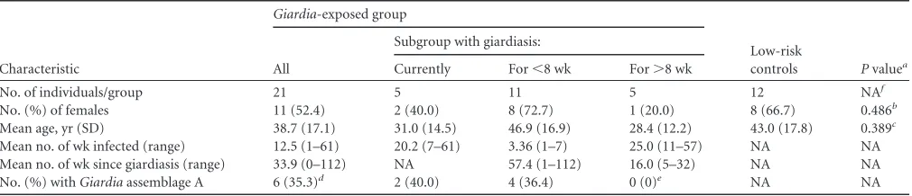
Intracellular cytokine expression. Antigen-activated cyto-kine-producing cells were found to be concentrated in the CD4⫹ CD197⫺ CD45RA⫺ EM T cell population (Fig. 1). These re-sponses reflect a recall response and were therefore the main in-vestigational focus of the cytokine assay. By including only this T cell subpopulation, unspecific responses seen in the CD197⫹ CD45RA⫹population could be avoided.
Analysis of the cytokines TNF-␣, IFN-␥, IL-17A, IL-4, and IL-10 secreted by EM T cells after 24 h showed that the cytokines IL-4 and IL-10 were weakly expressed by all of the cells analyzed when they were stimulated with the SSA and SSB antigens. These two cytokines were therefore not included in the SPICE analysis. SSA responses were generally stronger, with relatively less
unspe-TABLE 1Characteristics of study participants by group including subgroups ofGiardia-exposed individuals
Characteristic
Giardia-exposed group
Low-risk
controls Pvaluea All
Subgroup with giardiasis:
Currently For⬍8 wk For⬎8 wk
No. of individuals/group 21 5 11 5 12 NAf
No. (%) of females 11 (52.4) 2 (40.0) 8 (72.7) 1 (20.0) 8 (66.7) 0.486b
Mean age, yr (SD) 38.7 (17.1) 31.0 (14.5) 46.9 (16.9) 28.4 (12.2) 43.0 (17.8) 0.389c
Mean no. of wk infected (range) 12.5 (1–61) 20.2 (7–61) 3.36 (1–7) 25.0 (11–57) NA NA
Mean no. of wk since giardiasis (range) 33.9 (0–112) NA 57.4 (1–112) 16.0 (5–32) NA NA
No. (%) withGiardiaassemblage A 6 (35.3)d 2 (40.0) 4 (36.4) 0 (0)e NA NA
aPvalues are for comparisons of allGiardia-exposed groups with low-risk controls. b
Fisher’s exact test was used for comparisons of categorical variables.
cThe Mann-Whitney U test was used for comparisons of continuous variables between two groups.
d
TheGiardiaassemblage responsible for infection could not be determined for 4 of the participants by TPI sequencing, and the percentage represents the ratio for 17 individuals. All of the assemblages were detected in the current-giardiasis group.
e
TheGiardiaassemblage of two participants in this subgroup could not be characterized; the other three had assemblage B.
fNA, not applicable.
on August 17, 2020 by guest
http://cvi.asm.org/
cific responses in the low-risk controls than SSB responses, irre-spective of the genotype of the recent or ongoing infection. For simplicity, mainly the SSA responses are presented in the further analyses. Because of the small number of cells available from one control person, only 11 low-risk controls were included in the cytokine analysis.
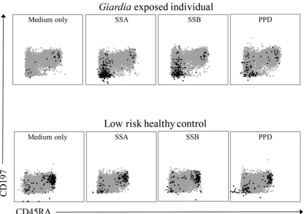
Figure 2shows the T cell cytokine response profiles for IL-17A, TNF-␣and IFN-␥to SSA, including polyfunctional cells, in the two different groups of participants. The relative percentages are expressed in bar graphs, and the proportions of the responses are expressed in pie charts. In the partial permutation analysis, where the distribution of all cytokine-expressing CD4⫹EM T cell pop-ulations was analyzed, the differences between the Giardia -ex-posed group and the controls did not reach statistical significance. However, the total number of IL-17A-positive CD4⫹EM T cells was significantly higher in theGiardia-exposed group than that in low-risk controls for both SSA (0.31 [0.41] versus 0.06 [0.11];P⫽
0.015) and SSB (0.22 [0.30] versus 0.09 [0.21];P⫽0.025). For the responses of the different groups, see Table S2 in the supplemental material. Cells stimulated with SSA showed higher levels of CD4⫹ EM T cells coproducing IL-17A and TNF-␣in theGiardia -ex-posed group than in the low-risk control group.
The IL-17A responses were not dependent upon the assem-blage with which the patient had been infected. A significant pos-itive correlation between the SSA- and SBB-induced IL-17A re-sponses was found (R2⫽0.88 andP⬍0.001) (data not shown).
Interestingly, the PPD internal protein control also showed higher values of cells expressing only IL-17A in the exposed group than in low-risk controls (0.07 [0.14] versus 0 [0];P⫽0.009). However, the total IL-17A expression difference did not reach significance for PPD (0.50 [0.56] versus 0.40 [0.51];P⫽0.60). No correlation between the upregulated IL-17A responses to PPD and the IL-17A responses to SSA and SSB was found (data not shown). IL-17A levels were not different between the groups for other internal positive controls (LPS and PMA-IC). CD4⫹T cells not producing TNF-␣, IL-17A, or IFN-␥
and hence producing IL-10 and/or IL-4 were not significantly differ-ent between the two groups.
Extracellular surface marker expression and proliferation.
To assess the magnitude of CD4⫹T cell activation and proliferation after 144 h of antigen stimulation, the cell markers CD25/CD26 and HLA-DR/CD45RO, proliferation, and a combination of HLA-DR/ CD45RO and proliferation were investigated. The markers CD25/ CD26 alone, and not in combination with HLA-DR/CD45RO, were only slightly upregulated inGiardia-exposed individuals and thus not included in the analysis. The mean percentages of activation and proliferation of CD4⫹T cells fromGiardia-exposed individ-uals and low-risk controls are presented inFig. 3. Because of the low cell numbers obtained from some of theGiardia-exposed in-dividuals, only 18 individuals were included in this analysis and 4 of them had giardiasis at the time of sample collection.
SSA-activated CD45RO⫹/HLA-DR CD4⫹T cell levels were higher inGiardia-exposed individuals than in low-risk controls (3.05 [6.62] versus 0.07 [1.21];P⫽0.007). Further, combining the four surface markers CD25/CD26 and CD45RO/HLA-DR and comparing the levels between theGiardia-exposed individuals and low-risk controls, we found significantly higher responses in SSA-stimulated cells in the exposed group (0.29 [2.42] versus 0.01 [0.24]; P ⫽ 0.016). To further investigate antigen-specific re-sponses, the two surface markers CD45RO⫹HLA-DR⫹were com-bined with proliferating CD4⫹T cells. This analysis revealed sig-nificantly elevated responses in SSA-stimulated cells (2.50 [6.60] versus 0.03 [1.20];P⫽0.006).
Comparison of cellular responses of low-risk controls and individuals with recent giardiasis with those of individuals with acute giardiasis.We wanted to investigate how the cellular re-sponses of individuals with current giardiasis would differ from those of low-risk controls and individuals with recent giardiasis. For the cytokine response, surface marker activation, and prolif-eration data andPvalues, see Tables S1 and S2 in the supplemental material.
FIG 1IL-17A-producing CD4⫹T cells in a CD197⫺CD45RA⫺plot. Flow cytometric plots showing IL-17A-producing cells as black dots in a gray CD4⫹T cell population. The IL-17A-producing cells are concentrated in the CD197⫺CD45RA⫺EM T cell population for the PPD-positive control and in SSA and SSB for theGiardia-exposed individual.
on August 17, 2020 by guest
http://cvi.asm.org/
The number of IL-17A/TNF-␣- and IL-17A-positive EM CD4⫹cells was significantly higher in the current-giardiasis group than in low-risk controls and also significantly higher in individ-uals with current giardiasis than in recentlyGiardia-exposed in-dividuals. Additionally, the IL-17A/TNF-␣-positive EM CD4⫹T cell population was larger in recentlyGiardia-exposed individuals than that in low-risk controls. In SSB-stimulated cells, there were no differences in TNF-␣, IFN-␥, IL-10, or IL-4, but the number of IL-17A-producing cells was significantly higher in the group with current giardiasis than in low-risk controls.
With regard to surface activation marker and proliferation analyses, no significant differences were found between the re-centlyGiardia-exposed group and the current-giardiasis group.
Comparison of individuals with recent giardiasis lasting<8 or>8 weeks.When stimulated with SSA, the percentage of poly-functional EM CD4 T cells responding with the production of both IL-17A and TNF-␣was found to be significantly higher in the subgroup with giardiasis for⬍8 weeks (n⫽11) (0.11 [0.10]) than in the subgroup with giardiasis for⬎8 weeks (n⫽5) (0.03 [0.02];
P⫽0.02). The percentage of IL-17A TNF-␣cells was also signif-icantly higher in the individuals with giardiasis for⬍8 weeks than in the low-risk controls (n⫽11) (0.04 [0.06];P⫽0.03). No other T cell subsets in this subgroup comparison reached significance.
Cytokines in supernatants.A multiplex assay was done to in-vestigate cytokine and chemokine production after 144 h of anti-gen stimulation. IL-1, IL-6, IL-10, IL-17A, IFN-␥, CD40L, TNF-␣, IL-13, GM-CSF, and MIP-1␣levels were significantly higher in SSA-stimulated PBMCs fromGiardia-exposed individ-uals than in those from low-risk controls (Table 2). Levels of IL-17A were also found to be increased in SSB-stimulated cells, and their correlation with the responses to SSA and SSB was therefore tested. IL-17A responses were correlated (R2⫽0.82), and the data were statistically significant (P⬍0.001).
DISCUSSION
In this study, we found that CD4⫹T cell surface activation marker and proliferation responses toGiardialysates were higher in
Gi-ardia-exposed individuals than in low-risk controls. IL-17A
re-sponses were found to be significantly higher inGiardia-exposed individuals than in low-risk healthy controls. Increased IL-17A levels were found both in the flow cytometric assay of EM CD4⫹T cells and in a cytokine analysis of the supernatants of Giardia
lysate-stimulated PBMCs. Levels of cells expressing both IL-17A and TNF-␣were higher inGiardia-exposed individuals than in low-risk controls, in those with current giardiasis than in recent giardiasis, and in those with recent giardiasis with a duration of⬍8 weeks than in those with longer-standing infections. These results indicate the presence ofGiardiaantigen-responsive poly-functional CD4⫹ EM T cells in Giardia-exposed individuals. Higher levels of CD4⫹EM T cells producing only IL-17A were also found in individuals with current giardiasis infections than in in-dividuals with recent giardiasis infections or in low-risk controls. In the supernatant analysis, a broad range of inflammation-asso-ciated cytokines were found to be upregulated in the Giardia -exposed group (IL-17A, MIP-1␣, IL-1, CD40L, TNF-␣, IFN-␥, IL-6, IL-10, IL-13, and GM-CSF).
Although T cells are important in the clearance of aGiardia
infection, neither a specific TH1 nor a specific TH2 polarized re-sponse seems to be necessary for protection againstGiardia infec-tion in mice (9). In our study, both TH1-associated (IFN-␥) and TH2-associated (IL-13) cytokines were found to be upregulated in stimulated PBMC culture supernatants (Table 2), indicating re-dundant mechanisms or that another T cell polarization is more important. Increased IL-17A cytokine levels and IL-17A-positive T cell levels were found in both SSA and SSB assays. Because of these IL-17A responses, TH17 can be speculated to be a CD4⫹T cell polarization important inGiardiaimmune responses in hu-mans. Individuals with current giardiasis infections had higher numbers of cells producing IL-17A than did recently Giardia -infected individuals. This difference could mean that individuals with current infections are in the process of becoming immune, while the IL-17A responses of recentlyGiardia-infected individu-als are disappearing.
The cytokine IL-17A plays a role in proinflammatory responses for the recruitment or activation of different cell types belonging to the innate immune system such as neutrophils and macro-phages (27). Further, IL-17A has been suggested to play an impor-tant role in protection after infection or vaccination, as deficiency in or reduced production of this cytokine can limit full protection
FIG 2Polyfunctional EM CD4⫹T cell cytokine responses to SSA. The bar graph represents the relative mean frequencies of CD197⫺CD45RA⫺CD4⫹T cells producing different combinations of the cytokines IL-17A, TNF-␣, and IFN-␥or not producing any of these cytokines (IL-4⫹and/or IL-10⫹cells). *, P⬍0.05. The pie charts show data from low-risk controls and Giardia-ex-posed individuals. The pie slices represent the different cytokine combinations in the bar graph. The arcs surrounding the pie charts represent the total pro-portions of IL-17A-, TNF-␣-, and IFN-␥-producing cells.
on August 17, 2020 by guest
http://cvi.asm.org/
(28). It has been reported that IL-17A is expressed mainly in CD4⫹CD45RO⫹memory T cells (29), and the main aim of our flow cytometric cytokine analysis was evaluation of responses in the EM T cell (CD197⫺CD45RA⫺) population. The EM CD4⫹T cell population was shown to be a significant source of IL-17A production in previouslyGiardia-exposed individuals, in contrast to controls. Recently, IL-17A was also shown to be associated with currentGiardiainfection in animal studies (18–20). In the first study, IL-17A was suggested to affect mice’s capability to clear
Giardiainfection (18), and mice lacking the IL-17A receptor in
their intestines have higher numbers ofGiardiacysts in feces than do control mice. The present study supports the idea that IL-17A
may be important for rapid clearance ofGiardiaby humans as well, by the observation that individuals with a shorter disease dura-tion had polyfuncdura-tional responsive EM T cells including an IL-17A response. Because of this finding, strong IL-17A responses may con-tribute to a shorter duration ofGiardiainfection in humans than in individuals with generally weaker IL-17A responses, who could have problems eliminatingGiardia. However, the role of IL-17A and how it affects the course of aGiardiainfection need to be inves-tigated further.
In the second mouse study (19), IL-17A upregulation was es-sential for host protection againstGiardia, and mice deficient in this cytokine and its receptor had difficulty eradicating the infec-tion after 1 week. Further, a deficiency in IL-17A led to a defect in IgA transport into the lumen and thus affected the mouse’s ability to clearGiardia.
In a study where calves infected withGiardiawere investigated, upregulation of IL-17A was found to originate from CD4⫹T cells (20), providing evidence that these cells are important in protect-ing cattle againstGiardiainfection. Whether IL-17A has a protec-tive role in humanGiardiainfection has to be investigated further. IL-17A has been shown in other studies to be important for both protection from and eradication of pathogens in association with the induction and stimulation of pathological inflammation dur-ing infection (30,31). Many of the inflammation-related cyto-kines were significantly elevated in PBMC supernatants in the
Giardia-exposed group (Table 2). The two cytokines GM-CSF and
TNF-␣have been shown to be mediators of neutrophil recruit-ment and activation and also cell survival and could originate from TH17 cells (31). The levels of the cytokine IL-1were also increased in the supernatant analysis, and this cytokine has been suggested to be important for the development and maturation of IL-17A-producing cells (31).
FIG 3Activation markers and proliferation of CD4⫹T cells stimulated with SSA. Average percentages of CD4⫹T cells showing proliferation and/or expression of surface activation markers are represented on theyaxis, and the various markers and proliferation measures are presented on thexaxis. *,P⬍0.05. CI, confidence interval.
TABLE 2Analysis of cytokines in supernatant of SSA-stimulated PBMCs cultured for 144 h
Cytokine
Median concn, pg/ml (SD)
Pvalue Low-risk
controls (n⫽9)
Giardia-exposed group (n⫽19)
IL-1 0 (2.5) 4.72 (5.26) 0.007
IL-6 0 (0.0) 265 (554) 0.002
IL-10 0 (5.9) 7.59 (17.8) 0.02
IL-17A 0 (0.0) 11.5 (87.3) ⬍0.001
IFN-␥ 0 (1,525) 566 (3,931) 0.02
sCD40L 0 (0.0) 6.99 (27.0) 0.007
TNF-␣ 28.9 (258) 502 (594) 0.006
IL-9 0 (0.0) 0 (1.7) NSa
IL-13 0 (0.0) 16.4 (86.8) 0.013
GM-CSF 0 (5.4) 11.2 (70.4) 0.011
MIP-1␣ 0 (0.0) 110 (327) 0.002
MIP-1 0 (0.0) 0 (986) NS
aNS, not significant.
on August 17, 2020 by guest
http://cvi.asm.org/
Increased levels of IL-17A were found inGiardia-exposed in-dividuals in our flow cytometric cytokine assay, where the BCG vaccine antigen PPD was used as a positive control. IL-17A re-sponses have previously been detected in CD4⫹ T cells from healthy BCG-vaccinated individuals after 12 h of PPD stimulation (32). Our finding of increased levels of PPD-induced CD4⫹T cells producing only IL-17A inGiardia-exposed individuals could be coincidental or due to polarization of the PPD responses by the ongoing/recent giardiasis in this group. The Giardia-exposed group might also have boosted their PPD responses by recent travel in areas with an increased risk of reexposure to environmen-tal mycobacteria. An alternative hypothesis is that individuals de-veloping symptomatic giardiasis have a tendency toward TH17 polarized responses.
When we compared the responses ofGiardia antigen-stimu-lated T cells inGiardia-exposed individuals and low-risk controls, some of the cells producing IL-17A were also producing TNF-␣. Background responses found in low-risk healthy controls tended to be unspecific and with less polyfunctional EM cells than in individuals with recent or ongoingGiardiainfections. Polyfunc-tional T cells can be linked to better and persistent protection from infection (28) and can be a sign of a good memory response. Poly-functional T cells could therefore prove to be an important marker of protection from and clearance ofGiardia.
Another cytokine responsible for anti-inflammatory re-sponses, IL-10, was also found to be increased in SSA-stimulated cells. The higher levels of this cytokine could mean that SSA may also elicit a regulatory response in addition to inflammatory re-sponses. Regulatory responses toGiardiahave also been found in calves with current infections (20), where FoxP3 was significantly elevated. Regulatory responses may play an important role in gi-ardiasis, and this may account for the negative association be-tweenGiardiainfection and acute diarrhea seen in children in recent studies in countries where giardiasis is endemic (33,34).
Increased levels of T cell markers specific forGiardia-exposed individuals have been shown before (22). Also in the present study, CD45RO/HLA-DR CD4 T cell responses were shown to be significantly elevated in SSA- and SSB-stimulated cells. Prolifera-tion of CD4⫹␣T cells in response toGiardiatrophozoites has been found in a study in which calves were investigated (20). These findings are well in line with the results of the present study and provide evidence that CD4⫹T cells play a key role in the recall immune response toGiardiainfections in calves and humans.
However, the EM CD4⫹T cell responses ofGiardia-exposed individuals measured in this study are subtle, with small popula-tions of cells responding. Subtraction of the unstimulated re-sponses from the stimulated rere-sponses resulted in a number of negative values. A conservative analysis approach was taken in which an equal positive value was adjusted to zero for every neg-ative value adjusted to zero (26).
Other cytokines more closely related to B-cell-mediated anti-body production were also found to be elevated in our study. These cytokines include CD40L, IL-6, and MIP-1␣, and their im-portance for B cells and Ig promotion is described in references28 and35. IL-6 seems to play a crucial role inG. lambliaeradication in mice, as mice deficient in this cytokine could not control the acute phase of infection (36).
The intracellularGiardiaprotein mixtures used in the pres-ent study might not be the best for initiating good relevant anti-Giardia immune responses. The strongest immune
re-sponses toGiardiain humans are probably directed toward the variant-specific surface proteins (VSPs). Only 1 specific type, out of around 250, is expressed at any time point (37). Using semiconserved peptides from VSPs or recombinant Giardia
trophozoites expressing all of the available VSPs could possibly have elicited stronger responses inGiardia-exposed individuals but could also have caused much larger variability, depending on which VSPs were expressed duringGiardiainfection. Responses to theGiardiaantigens were also found in some low-risk controls. The trueGiardiaexposure status of the low-risk controls is hard to ascertain. Only individuals who had not traveled to regions where giardiasis is endemic were recruited, but still it is estimated that 2 to 5% of the population in an industrialized country has had gi-ardiasis or has a current infection (6).
In the flow cytometric assay, some of the selected positive-control antigens were not capable of eliciting good IL-4 and IL-10 responses. In the multiplex assay, where proper controls were used, the IL-4 levels were below the detection limit and could not be analyzed. It is therefore likely that the number of cells producing IL-4 was low. It has been stated that IL-4 is expressed at low levels and consequently can be difficult to measure (29). It remains unclear whether this is a methodological problem or whether TH2 polariza-tions are not important for protection againstGiardia. Better IL-4-and IL-10-eliciting positive controls could have been included in the flow cytometric assay to better interpret these results.
We observed much variability in the response measurements. The time sinceGiardiainfection varied from 0 to⬎2 years, and the age span of the participants and our sample size were relatively small. Also, previousGiardiaexposure(s), host-specific factors, and con-comitant infections/microbiota account for some of the variability.
Concluding remarks.In this study, we found increased num-bers of antigen-specific IL-17A-producing CD4⫹EM T cells after 24 h and in PBMC supernatants after 6 days ofGiardiaSSA and SSB stimulation. Interestingly, levels of responding polyfunc-tional EM CD4⫹T cells simultaneously producing IL-17A and TNF-␣were elevated in the subgroup analysis ofGiardia-exposed individuals with current infections and those infected for ⬍8 weeks. Our findings indicate that TH17 responses are important during acute symptomaticGiardiainfections in humans.
ACKNOWLEDGMENTS
We thank Marianne Enger at the core facility of flow cytometry, Univer-sity of Bergen/Haukeland UniverUniver-sity Hospital, for expertise and advice; Kristin Paulsen Rye at the Section of Haemotology, Department of Clin-ical Science, University of Bergen, for cytokine analysis; Karl Brokstad at the Broegelman Research Laboratory, Department of Clinical Sciences, University of Bergen, for good advice; and all of the laboratory workers who helped with materials and methods. We also thank the study partic-ipants. The staphylococcal enterotoxin B was a gift from Ida Wergeland, Department of Clinical Science, University of Bergen. Martin Kristiansen and Torunn Hjøllo, Department of Clinical Science, University of Bergen, assisted with 18S SSU PCR and TPI sequencing ofGiardia.
C. S. Saghaug, K. Hanevik, and N. Langeland planned and designed the study. C. S. Saghaug carried out the experiments. S. Sørnes, S. Svärd, D. Peirasmaki, and K. Hanevik contributed with reagents, methods, ma-terials, and/or analysis programs. D. Peirasmaki and S. Svärd produced theGiardiaantigens. C. S. Saghaug, K. Hanevik, and N. Langeland ana-lyzed the data. C. S. Saghaug, S. Sørnes, D. Peirasmaki, S. Svärd, K. Han-evik, and N. Langeland wrote the paper.
K. Hanevik has served on an advisory board for Lupin Pharmaceuticals, Inc., Baltimore, MD. The other authors have no conflicts of interest to report.
on August 17, 2020 by guest
http://cvi.asm.org/
The International Committee of Medical Journal Editors form for disclosure of potential conflicts of interest has been submitted by all of the authors.
FUNDING INFORMATION
This study was supported by project grants from the Western Norway Regional Health Authority; the Department of Clinical Science, Univer-sity of Bergen; and the Centre for Tropical Medicine and Imported Infec-tious Diseases (CTID), Division of InfecInfec-tious Diseases, Medical Depart-ment, Haukeland University Hospital, Bergen, Norway. The funding institutions had no influence on data analysis or interpretation of the results.
REFERENCES
1.Adam RD.1991. The biology ofGiardiaspp. Microbiol Rev55:706 –732. 2.Yoder JS, Harral C, Beach MJ.2010. Giardiasis surveillance—United
States, 2006 –2008. MMWR Surveill Summ59:15–25.
3.Stadelmann B, Merino MC, Persson L, Svard SG.2012. Arginine con-sumption by the intestinal parasiteGiardia intestinalisreduces prolifera-tion of intestinal epithelial cells. PLoS One7:e45325.http://dx.doi.org/10 .1371/journal.pone.0045325.
4.Robertson LJ, Hanevik K, Escobedo AA, Morch K, Langeland N.2010. Giardiasis—why do the symptoms sometimes never stop? Trends Parasi-tol26:75– 82.http://dx.doi.org/10.1016/j.pt.2009.11.010.
5.Nash TE, Herrington DA, Losonsky GA, Levine MM.1987. Experimen-tal human infections withGiardia lamblia. J Infect Dis156:974 –984.http: //dx.doi.org/10.1093/infdis/156.6.974.
6.Farthing MJ.1996. Giardiasis. Gastroenterol Clin North Am25:493–515.
http://dx.doi.org/10.1016/S0889-8553(05)70260-0.
7.Escobedo AA, Hanevik K, Almirall P, Cimerman S, Alfonso M.2014. Management of chronicGiardiainfection. Expert Rev Anti Infect Ther 12:1143–1157.http://dx.doi.org/10.1586/14787210.2014.942283. 8.Carlson JR, Heyworth MF, Owen RL.1986. Response of Peyer’s patch
lymphocyte subsets to Giardia muris infection in BALB/c mice. II. B-cell subsets: enteric antigen exposure is associated with immunoglobulin iso-type switching by Peyer’s patch B cells. Cell Immunol97:51–58. 9.Singer SM, Nash TE.2000. T-cell-dependent control of acuteGiardia
lambliainfections in mice. Infect Immun68:170 –175.http://dx.doi.org /10.1128/IAI.68.1.170-175.2000.
10. Oksenhendler E, Gerard L, Fieschi C, Malphettes M, Mouillot G, Jaussaud R, Viallard JF, Gardembas M, Galicier L, Schleinitz N, Suarez F, Soulas-Sprauel P, Hachulla E, Jaccard A, Gardeur A, Theodorou I, Rabian C, Debre P.2008. Infections in 252 patients with common vari-able immunodeficiency. Clin Infect Dis46:1547–1554.http://dx.doi.org /10.1086/587669.
11. Char S, Cevallos AM, Yamson P, Sullivan PB, Neale G, Farthing MJ. 1993. Impaired IgA response toGiardiaheat shock antigen in children with persistent diarrhoea and giardiasis. Gut34:38 – 40.
12. Oyerinde JP, Ogunbi O, Alonge AA.1977. Age and sex distribution of infections withEntamoeba histolyticaandGiardia intestinalisin the Lagos population. Int J Epidemiol6:231–234.
13. Istre GR, Dunlop TS, Gaspard GB, Hopkins RS. 1984. Waterborne giardiasis at a mountain resort: evidence for acquired immunity. Am J Public Health74:602– 604.
14. Heyworth MF, Carlson JR, Ermak TH.1987. Clearance ofGiardia murisinfection requires helper/inducer T lymphocytes. J Exp Med165: 1743–1748.
15. Zhou P, Li E, Shea-Donohue T, Singer SM. 2007. Tumour necrosis factor alpha contributes to protection againstGiardia lambliainfection in mice. Parasite Immunol 29:367–374. http://dx.doi.org/10.1111/j.1365 -3024.2007.00953.x.
16. Roxström-Lindquist K, Palm D, Reiner D, Ringqvist E, Svard SG.2006. Giardiaimmunity—an update. Trends Parasitol22:26 –31.http://dx.doi .org/10.1016/j.pt.2005.11.005.
17. Solaymani-Mohammadi S, Singer SM.2011. Host immunity and patho-gen strain contribute to intestinal disaccharidase impairment following gut infection. J Immunol 187:3769 –3775. http://dx.doi.org/10.4049 /jimmunol.1100606.
18. Dreesen L, De Bosscher K, Grit G, Staels B, Lubberts E, Bauge E, Geldhof P.2014.Giardia murisinfection in mice is associated with a protective interleukin 17A response and induction of peroxisome
prolif-erator-activated receptor alpha. Infect Immun82:3333–3340.http://dx .doi.org/10.1128/IAI.01536-14.
19. Dann SM, Manthey CF, Le C, Miyamoto Y, Gima L, Abrahim A, Cao AT, Hanson EM, Kolls JK, Raz E, Cong Y, Eckmann L.2015. IL-17A promotes protective IgA responses and expression of other potential ef-fectors against the lumen-dwelling enteric parasiteGiardia. Exp Parasitol 156:68 –78.http://dx.doi.org/10.1016/j.exppara.2015.06.003.
20. Grit GH, Van Coppernolle S, Devriendt B, Geurden T, Dreesen L, Hope J, Vercruysse J, Cox E, Geldhof P, Claerebout E.2014. Evaluation of cellular and humoral systemic immune response againstGiardia duode-nalisinfection in cattle. Vet Parasitol202:145–155.http://dx.doi.org/10 .1016/j.vetpar.2014.03.012.
21. Ebert EC.1999. Giardia induces proliferation and interferon gamma pro-duction by intestinal lymphocytes. Gut44:342–346.
22. Hanevik K, Kristoffersen E, Svard S, Bruserud O, Ringqvist E, Sornes S, Langeland N.2011. Human cellular immune response againstGiardia lamblia5 years after acute giardiasis. J Infect Dis204:1779 –1786.http://dx .doi.org/10.1093/infdis/jir639.
23. Verweij JJ, Blange RA, Templeton K, Schinkel J, Brienen EA, van Rooyen MA, van Lieshout L, Polderman AM.2004. Simultaneous de-tection ofEntamoeba histolytica,Giardia lamblia, andCryptosporidium parvumin fecal samples by using multiplex real-time PCR. J Clin Micro-biol42:1220 –1223.http://dx.doi.org/10.1128/JCM.42.3.1220-1223.2004. 24. Sulaiman IM, Fayer R, Bern C, Gilman RH, Trout JM, Schantz PM, Das P, Lal AA, Xiao L.2003. Triosephosphate isomerase gene characteriza-tion and potential zoonotic transmission ofGiardia duodenalis. Emerg Infect Dis9:1444 –1452.http://dx.doi.org/10.3201/eid0911.030084. 25. Keister DB.1983. Axenic culture of Giardia lamblia in TYI-S-33 medium
supplemented with bile. Trans R Soc Trop Med Hyg77:487– 488. 26. Roederer M, Nozzi JL, Nason MC.2011. SPICE: exploration and analysis
of post-cytometric complex multivariate datasets. Cytometry A79:167– 174.http://dx.doi.org/10.1002/cyto.a.21015.
27. Shabgah AG, Fattahi E, Shahneh FZ.2014. Interleukin-17 in human inflammatory diseases. Postepy Dermatol Alergol31:256 –261.http://dx .doi.org/10.5114/pdia.2014.40954.
28. Thakur A, Pedersen LE, Jungersen G.2012. Immune markers and cor-relates of protection for vaccine induced immune responses. Vaccine30: 4907– 4920.http://dx.doi.org/10.1016/j.vaccine.2012.05.049.
29. Lenarczyk A, Helsloot J, Farmer K, Peters L, Sturgess A, Kirkham B. 2000. Antigen-induced IL-17 response in the peripheral blood mononu-clear cells (PBMC) of healthy controls. Clin Exp Immunol122:41– 48.
http://dx.doi.org/10.1046/j.1365-2249.2000.01328.x.
30. Gaffen SL.2008. An overview of IL-17 function and signaling. Cytokine 43:402– 407.http://dx.doi.org/10.1016/j.cyto.2008.07.017.
31. Annunziato F, Romagnani C, Romagnani S.2015. The 3 major types of innate and adaptive cell-mediated effector immunity. J Allergy Clin Im-munol135:626 – 635.http://dx.doi.org/10.1016/j.jaci.2014.11.001. 32. Scriba TJ, Kalsdorf B, Abrahams DA, Isaacs F, Hofmeister J, Black G,
Hassan HY, Wilkinson RJ, Walzl G, Gelderbloem SJ, Mahomed H, Hussey GD, Hanekom WA.2008. Distinct, specific IL-17- and IL-22-producing CD4⫹ T cell subsets contribute to the human anti-mycobacterial immune response. J Immunol180:1962–1970.http://dx .doi.org/10.4049/jimmunol.180.3.1962.
33. Mbae CK, Nokes DJ, Mulinge E, Nyambura J, Waruru A, Kariuki S.2013. Intestinal parasitic infections in children presenting with diarrhoea in outpa-tient and inpaoutpa-tient settings in an informal settlement of Nairobi, Kenya. BMC Infect Dis13:243.http://dx.doi.org/10.1186/1471-2334-13-243.
34. Muhsen K, Cohen D, Levine MM.2014. CanGiardia lambliainfection lower the risk of acute diarrhea among preschool children? J Trop Pediatr 60:99 –103.http://dx.doi.org/10.1093/tropej/fmt085.
35. Smith KA.2014. Toward a molecular understanding of adaptive immu-nity: a chronology, part III. Front Immunol5:29.http://dx.doi.org/10 .3389/fimmu.2014.00029.
36. Bienz M, Dai WJ, Welle M, Gottstein B, Muller N.2003. Interleukin-6-deficient mice are highly susceptible toGiardia lambliainfection but exhibit normal intestinal immunoglobulin A responses against the para-site. Infect Immun 71:1569 –1573. http://dx.doi.org/10.1128/IAI.71.3 .1569-1573.2003.
37. Touz MC, Ropolo AS, Rivero MR, Vranych CV, Conrad JT, Svard SG, Nash TE.2008. Arginine deiminase has multiple regulatory roles in the biol-ogy ofGiardia lamblia. J Cell Sci 121:2930 –2938.http://dx.doi.org/10 .1242/jcs.026963.