Available online: https://edupediapublications.org/journals/index.php/IJR/ P a g e | 3594
Design and Implementation of Bioinformatic Tools to Analyze
Protein Sequences
Sabbavarapu N V Siva Kumar
& Kunjam Nageswara Rao
1
M.Tech CST-Bioinformatics, CS&SE Department, Andhra University, Visakhapatnam
2Associate
Professor, CS&SE Department, Andhra University, Visakhapatnam.
Abstract:
Proteins are one of the important fundamental units of all living cells. Proteins have a large number of functions within all the living organisms. Some of the important functions performed with the help of proteins such as DNA replication, catalysis of metabolic reactions, transportation of molecules from one place to another etc... There are 20 different types of amino acids that can be combined to make a protein sequence. Integrins are cell adhesion molecules that mediate cell-cell, cellextracellular matrix, and cell-pathogen interaction. They play crucial roles in leukocyte trafficking,
migration, immunological synapse formation,
costimulation, and phagocytosis. The studies of integrin molecule structure and function helps in better understanding the various pathological processes such as chronic inflammation, thrombosis and cancer metastasis. The study can help the researchers to discover a new drug to cure all these.To analyze the protein sequence with respect to primary structure, secondary structure and tertiary structure it needs the help of various bioinformatic tools. Most of these tools are web based tools and it requires internet connection to work with them. There are lot of challenges in maintaining proper internet connection all the time. So, developing an offline tool using high performance programming language like julia will be helpful for researchers to work with them at any time irrespective of internet connection.
Keywords
integrin αvβ3; protein analysis; phylogenetic analysis;
biological pathways, offline bioinformatic tools, bioinformatic tools, protparam tool, protein structures
1.Introduction
Proteins plays a crucial role in cell functions. Proteins are large, complex molecules. Proteins are required for the structure, function and regulation of body tissues and organs. Proteins sequences are the combination of 20 amino acids that forms a long
chain. Proteins are the responsible for various functions in our body. For example, Integrin are important members in the family of cell adhesion molecules mediating the signal transmission between extracellular matrix(ECM) and intracellular in many physiological reaction processes. They play an important part in regulating cell adhesion, proliferation, differentiation, metastasis and apoptosis.
Integrin αvβ3 is expressed in many cell types and
combine with various ligands in many cellular activities, involved in many physiological and pathological processes. It is important on tumor angiogenesis [1]. It is capable of identifying the short Arg-Gly-Asp(RGD) peptide motif [2]. The ligands including FN, VN, TSP-1 and vWF, etc. Studies have found that monoclonal antibodies and antagonists of integrin αvβ3 can effectively
inhibit the formation of tumor blood vessels, it may be a new mean of cancer treatment [3].
2.Materials & Methods
A.Tools for prediction of protein structure and function
ProtParam(http://kr.expasy.org/tools/protparam.html )[4], a network analyzer tool to predict protein primary structure, is used to predict the amino acid composition and characterization of the protein, including the molecular weight, extinction coefficient, theoretical PI, half life, aliphatic index, instability index and grand average of hydropathicity(GRAVY)[5]. To predict protein domain, signal peptide, transmembrane region and subcellular localization properties of primary structure Simple Modular Architecture Research Tool(SMART) (http://smart.embl-heidelberg.de/)[6] is a classification scheme used in the identification and analysis of protein domain. SignalP-
4.1(http://www.cbs.dtu.dk/services/SignalP) is used to perform signal peptide prediction[7]. Transmembrane Helices Hidden Markov Model tool TMHMM(http://www.cbs.dtu.dk/services/TMHMM -2.0/) is a used for the prediction of the transmembrane domains. WoLF PSORT
Available online: https://edupediapublications.org/journals/index.php/IJR/ P a g e | 3595
Robson tool GOR4.0 (http://npsa- pbil.ibcp.fr/cgibin/npsa_automat.pl?page=npsa
_gor4.html) is used for the prediction of the protein secondary structure.
ProtParam, a network analyzer about protein primary structure to predict the amino acid composition and characterization of the protein. ProtParam is a web based tool and it requires internet connection to work with it. An Offline tool has been developed using Julia programming language to speed up the process. Following parameters can be calculated using this offline tool.
Molecular Weight: In ProtParam, the molecular weight of protein is calculated by the addition of average isotopic masses of amino acids in the provided protein and the average isotopic mass of one water molecule.
Extinction Coefficient: The extinction coefficient indicates how much light a protein absorbs at a certain wavelength. It is useful to have an estimation of this extinction coefficient of a protein from knowledge of its amino acid composition. From the molar extinction coefficient of tyrosine, tryptophan, and cystine (cysteine does not absorb appreciably at wavelengths >260 nm, while cystine does) at a given wavelength, the extinction coefficient of the native protein in water can be calculated using the following equation:
E(Prot) = Numb(Tyr) * Ext(Tyr) + Numb(Trp) * Ext(Trp) + Numb(Cystine)*Ext(Cystine)
Where (for proteins in water measured at 280 nm): Ext(Tyr) = 1490, Ext(Trp) = 5500, Ext(Cystine) = 125;
The absorbance (optical density) can be calculated using the following formula:
Absorb(Prot) = E(Prot) / Molecular_weight
Theoretical pI: Theoretical pI of of protein is calculated using pKa values of amino acids. The pKa value of Amino acids depends on its side chain. It plays an important role in defining the pH dependent characteristics of a protein.
Half-life: The half-life is a prediction of the time it takes for half of the amount of protein in a cell to disappear after its synthesis in the cell.
Grand average of hydropathicity (GRAVY): The GRAVY value for a protein or a peptide is calculated by adding the hydropathy of each amino acid residues and
dividing by the number of residues in the sequence or length of the sequence. Increasing positive score indicates a greater hydrophobicity.
Aliphatic index: The aliphatic index of a protein is described as the relative volume occupied by the amino acids such as alanine, valine, isoleucine and leucine, which have an aliphatic side chain in their structure. The aliphatic index of a protein is computed using the following equation.
Aliphatic index = X(Ala) + a * X(Val) + b * ( X(Ile) + X(Leu) )
Where X(Ala), X(Val), X(Ile), and X(Leu) are mole percent (100 X mole fraction) of alanine, valine, isoleucine, and leucine.
The coefficients „a‟ and „b‟ are the relative volume of valine side chain (a = 2.9) and of Leu/Ile side chains (b = 3.9) to the side chain of alanine.
Instability Index: The instability index provides an estimation of the stability of your protein in a test tube. A protein with instability index smaller than 40 is predicted as stable, a value above 40 predicts that the protein may be unstable. Instability Index can be computed using the following equation.
i=L-1
II = (10/L) * Sum DIWV(x[i]x[i+1]) i=1
where: L is the length of sequence
DIWV(x[i]x[i+1]) is the instability weight value for the dipeptide starting in position i.
B.Phylogenetic analysis
Using ClustalW, a multiple sequence alignment was performed between the amino acid sequences of αvβ3 in
the different species, and after that to construct a phylogenetic tree for the molecular evolution based on the Neighbor-Joining using Molecular Evolutionary Genentic Analysis tool MEGA 6.0 tool [9].
C.Analysis of biological pathway
The intercomparison results between this species and others will be gotten from the database of Kyoto Encyclopedia of Genes and Genomes (KEGG) biological pathways (http://www.genome.jp/kegg/)
Available online: https://edupediapublications.org/journals/index.php/IJR/ P a g e | 3596
3.Result
A.The Protein Structure of Integrin
αvβ3
The accession number of αv and β3 subunit are P06756
and P05106 respectively in Uniprot database. By using ProtParam, the chemical property of integrin αvβ3 was
gotten and arranged below.
TABLE I.The Result of Primary Structure Analysis of αVβ3
Items
Online Values Offline Values
αv β3 αv β3
The number of amino acids
1048 788 1048 788
The total number of atoms
16245 11981 16245 11981
Theoretical pI 5.45 5.09 5.452536 5.090929
Aliphatic Index 86.15 72.72 86.14902 72.72067
GRAVY -0.220
(hydrophi le)
-0.332 (hydroph ile)
-0.220115 (hydrophi le)
-0.33215 (hydrophi le)
Molecular weight
116051.8 87057.8 116051.8 87057.8
Extinction coefficient
111255/1 10130
86240/8 2740
111255 /110130
86240/82 740
Instability Index
37.69 45.10 37.69012 45.10495
Half-life period (Mammals reticulocyte, out of body)
30 hours 30 hours 30 hours 30 hours
In Table-I the results were compared between existing protparam tool which is in expasy server and offline tool which was developed using high performance programming language “Julia”. The results have been shown that offline tool values and online tool values are equal and even the offline tool generating results faster than online tool because results generated by online tool depends on the speed of internet connection.
The most common types of secondary structures are the α helix and the β pleated sheet. Both structures are held in shape by hydrogen bonds, which form between the carbonyl O of one amino acid and the amino H of another.The secondary structures of αv and β3 were
analyzed with SMART. The results showed that the subunit of αvis consisted with the lamellas occupy
25.38%, the spirals occupy 4.48%, and the rings occupy
70.13%. There is no curly spiral. For the subunit of β3,
the lamellas occupy 6.22%, the spirals occupy 13.83%, and the rings occupy 79.95%. There are four potential curly spirals.
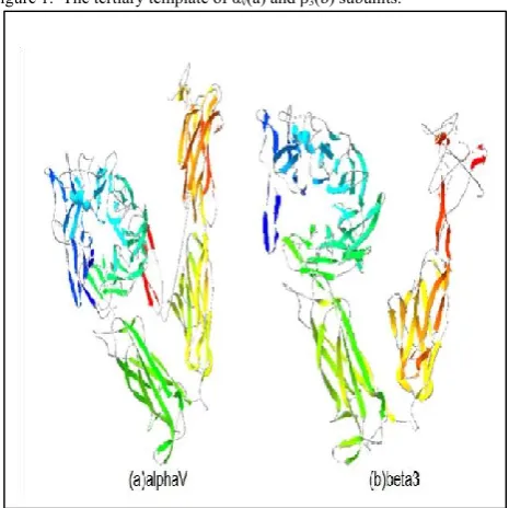
The tertiary structure of related proteins of αvβ3 are
analyzed with Swiss-Model. The tertiary structure model corresponded with αv and β3 is the crystal structure
1L5G(Figure-1). In Figure-1, each secondary structure is signed by different color. Green color indicates lamellas, yellow color indicates spirals, blue and navy-blue combination indicates rings.
Figure 1. The tertiary template of αv(a) and β3(b) subunits.
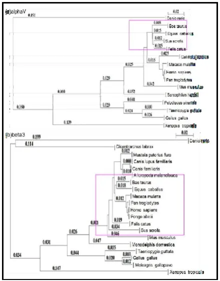
B.The Evolution Analysis of Integrin αvβ3
Using MEGA tool, the protein sequences of different species were aligned with human αv and β3 sequences
and constructed the evolutionary trees, and the relationship was shown in Figure-2. For αv subunit, only
the part within the purple frame is different from this before excluding. For β3 subunit, only the position of Sus
scrofa is different in the evolutionary tree.
In different species, the structural domains are conserved in the process of different species evolution. For αv
subunit, the conservatism of transmembrane is highest. For β3 subunit, the conservatism of integrin tail is highest
Available online: https://edupediapublications.org/journals/index.php/IJR/ P a g e | 3597
Figure 2. The Commonness Analysis of αv(a) and β3(b) in Different
Species.
TABLE II. The Conservatism Analysis Of αV Subunit and β3
Subunit
The name of structural domain
The position of Structural Domain
Conservative average
Conservatism
αv subunit
FG-GAP 1 32-98
57.57 middle
FG-GAP 2 109-170
FG-GAP 3 173-225 FG-GAP 4 237-295 FG-GAP 5 296-357 FG-GAP 6 358-415
FG-GAP 7 419-482
Transmembrane 993-1016 100.00 high
Cytoplasmic 1017-1048 67.00 middle
β3 subunit
PSI 30-76 48.49 low
INB 38-461 79 middle VWFA 135-377 85 high IEGF-1 463-511
89.125 high IEGF-2 512-553
IEGF-3 554-592 IEGF-4 593-629
β tail 634-718 100 high Transmembrane 719-741 96 high Cytoplasmic 742-788 74 high
In following Table-II definition, the average of the mutation is taken from 0 to 50 is low, from 50 to 80 is middle and from 80 to 100 is high. The table contains the locations of protein sequence domains, transmembrane region and subcellular localization of αv and β3 protein sequences and displayed with respective conservatism average and conservatism level for obtaining the target protein sequence.
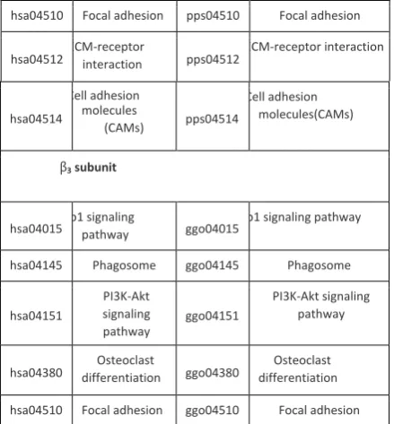
C.Analysis of Biological pathways of Integrin αvβ3 Biological pathways of homologous gene in αv subunit
and β3subunit were analyzed with KEGG. In this study,
human and bonobo are selected to compare.
Table-III contains the information that the biological pathways related to αv subunits of humans is same as the
pathways related to αvsubunits of pan paniscus, only
some biological factors are different. And β3 subunit has
the same situation as αv subunit in the evolution of
similar species. Thus, can conclude that the αv subunit
and β3 subunit in the evolution of similar species are
almost exactly the same, which reflected in its homology between different species. Both αv and β3 subunits
biological pathways are compared between different species and bonobo is the nearest matching species among all other species. The pathway comparison is shown in the following table which clearly illustrates the matching biological pathways of αv and β3 subunits of
homosapiens and pan paniscus.
After analyzing each pathway associated with integrin αvβ3, the results can be concluded that integrin αvβ3
influence all aspects of biological reactions and physiological processes must be through the mutual reaction and extracellular matrix (ECM) to really play its due role, so the research on interaction pathway in ECM-receptor is necessary.
TABLE III. The Pathway Comparison Between Human and Bonobo
hsa3685ˈ Homo sapiens (human)
pps100975589 Pan paniscus (bonobo)
Number of Pathway
Name ofPathway
Number of Pathway
Name ofPathway
αv subunit
hsa04145 Phagosome pps04145 Phagosome
hsa04151
PI3K-Akt signaling pathway
pps04151
Available online: https://edupediapublications.org/journals/index.php/IJR/ P a g e | 3598 hsa04510 Focal adhesion pps04510 Focal adhesion
hsa04512ECM-receptor interaction pps04512ECM-receptor interaction
hsa04514
Cell adhesion molecules
(CAMs) pps04514
Cell adhesion molecules(CAMs)
β3 subunit
hsa04015Rap1 signaling pathway ggo04015Rap1 signaling pathway
hsa04145 Phagosome ggo04145 Phagosome
hsa04151
PI3K-Akt signaling pathway
ggo04151
PI3K-Akt signaling pathway
hsa04380 differentiationOsteoclast ggo04380 differentiationOsteoclast
hsa04510 Focal adhesion ggo04510 Focal adhesion
It showed that the research of integrin and tumor cancer have a high prospect for the future, because from the table get the results that pathways related with integrin αvβ3 can lead to cell proliferation, avoidance of
apoptosis, and sustained angiogenesis. While other physiological outcomes such as the metabolism, transport and translation of glucose, and the stabilization of actin are also likely to be provided for the above three major results. The occurrences of cancer are inextricably linked with those results, because when cells are able to proliferate, evading apoptosis and generate endothelial cell consistently, can think the cells have the ability of infinite proliferation, which is meaned cancerous.
The related molecules such as some protein kinase that SRC, FAK, PI3K, PIP3, PKB/Akt play key roles in the process of biological pathways. Thus, can verify the molecular by experiment, analyzing the position which the drug target on can make an effect of inhibiting the tumor cell proliferation, therefore obtain the effective treatment for cancer.
Activated Akt can not only limit Raf-1, GSA3, P21, P27, FOXO, BAD, Casp, RXR and NUR77, but also promote eNOS(the NO synthetase), GREB, IKK and MDM2, keeping them from entering the nucleus to play normal function, thus affect the process of cell cycle. The PI3K Akt signaling pathway that associated with this pathway and integrin lead to cell survival, the normal process of cell cycle, metabolism, cell proliferation, angiogenesis and DNA repair. As is known to all, the characteristics of cancer cells is unlimited, endless proliferation. Therefore, the study of these signaling pathways has a great significance.
4.Summary
Work shown in this paper is mainly about studying the biological information related to integrin αvβ3, including
the protein structure and function prediction, the system evolution analysis, and the analysis of biological pathways of integrin αvβ3. By using the related biological
informatic tools to understand the physiological processes that integrin αvβ3 involved cancer and other
diseases. It is helpful to provide new insights for the treatment of these diseases. In the process of predicting the chemical properties of protein sequence an offline tool has been developed to save the time by producing the accurate results of the chemical properties of protein sequence without using protparam tool from expasy server that requires internet connection.
5.References
[1] M. Kim, C. V. Carman, and T. A. Springer, "Bidirectional transmembrane signaling by cytoplasmic domain separation in integrins," Science, vol. 301, pp. 1720-1725, 2003.
[2] E. Ruoslahti, "RGD and other recognition sequences for integrins," Annual review of cell and developmental biology, vol. 12, pp. 697-715, 1996.
[3] F. Danhier, A. L. Breton, and V. r. Préat, "RGD-based strategies to target alpha (v) beta (3) integrin in cancer therapy and diagnosis," Molecular pharmaceutics, vol. 9, pp. 2961-2973, 2012.
[4] Protein Identification and Analysis Tools on the ExPASy Server; Gasteiger E., Hoogland C., Gattiker A., Duvaud S., Wilkins M.R., Appel R.D., Bairoch A.; (In) John M. Walker (ed): The Proteomics Protocols Handbook, Humana Press (2005). pp. 571-607.
[5] A. Sudhakar, H. Sugimoto, C. Yang, J.
Lively, M. Zeisberg, and R. Kalluri, "Human tumstatin and human endostatin exhibit distinct antiangiogenic activities mediated by v3 and 51 integrins," Proceedings of the National Academy of Sciences, vol. 100, pp. 47664771, 2003.
[6] I. Letunic, R. R. Copley, S. Schmidt, F. D.
Ciccarelli, T. Doerks, J. Schultz, et al., "SMART 4.0: towards genomic data integration," Nucleic acids research, vol. 32, pp. D142-D144, 2004.
[7] J. D. Bendtsen, H. Nielsen, G. von Heijne, and S. Brunak, "Improved prediction of signal peptides: SignalP 3.0,"
Journal of molecular biology, vol. 340, pp. 783795, 2004. [8] P. Horton, K.-J. Park, T. Obayashi, N. Fujita, H. Harada, C. Adams-Collier, et al., "WoLF PSORT: protein localization predictor," Nucleic acids research, vol. 35, pp. W585W587, 2007.
[9] S. Kumar, M. Nei, J. Dudley, and K. Tamura, "MEGA: a biologist-centric software for evolutionary analysis of DNA and protein sequences,"
Briefings in bioinformatics, vol. 9, pp. 299306, 2008.
[10] M. Kanehisa and S. Goto, "KEGG: kyoto encyclopedia of genes and genomes," Nucleic acids research,