C
LINICAL ANDV
ACCINEI
MMUNOLOGY, Nov. 2011, p. 1865–1871
Vol. 18, No. 11
1556-6811/11/$12.00
doi:10.1128/CVI.05318-11
Copyright © 2011, American Society for Microbiology. All Rights Reserved.
Studies on Porcine Circovirus Type 2 Vaccination of
5-Day-Old Piglets
䌤
K. C. O’Neill,
1H. G. Shen,
1K. Lin,
1M. Hemann,
1N. M. Beach,
2X. J. Meng,
2P. G. Halbur,
1and T. Opriessnig
1*
Department of Veterinary Diagnostic and Production Animal Medicine, College of Veterinary Medicine, Iowa State University,
1600 S. 16th Street, Ames, Iowa 50011,
1and Department of Biomedical Sciences and Pathobiology, Center for
Molecular Medicine and Infectious Diseases, College of Veterinary Medicine, Virginia Polytechnic Institute and
State University, Blacksburg, Virginia
2Received 25 July 2011/Returned for modification 22 August 2011/Accepted 12 September 2011
Porcine circovirus type 2 (PCV2) vaccines have become widely used since they became available in 2006. It
is not uncommon for producers to use PCV2 vaccines in pigs younger than what is approved by manufacturers.
The objective of this study was to determine the efficacy of a chimeric and a subunit PCV2 vaccine administered
at 5 or 21 days of age. Forty-eight PCV2-naïve piglets were randomly divided into six groups of eight pigs each.
Vaccination was done at day 5 or day 21, followed by triple challenge with PCV2, porcine parvovirus (PPV), and
porcine reproductive and respiratory syndrome virus (PRRSV) at day 49. Vaccinated pigs seroconverted to
PCV2 approximately 14 days postvaccination and had a detectable neutralizing antibody response by 21 days
postvaccination regardless of age at vaccination. At day 49, the pigs vaccinated with the chimeric vaccine had
significantly higher levels of neutralizing antibodies than the pigs vaccinated with the subunit vaccine. After
challenge, vaccinated pigs had significantly decreased levels of PCV2 viremia and a decreased prevalence and
severity of microscopic lesions compared to the positive-control group, which had severe lymphoid lesions
associated with abundant PCV2 antigen, compatible with PCV-associated disease. The results of this study
indicate that, under the conditions of this study, vaccination of PCV2-naïve pigs at day 5 or day 21 resulted in
development of a detectable humoral immune response and provided reduction or complete protection against
PCV2 viremia and PCV2-associated lesions after triple challenge with PCV2, PPV, and PRRSV.
Porcine circovirus (PCV) is a circular, single-stranded,
non-enveloped DNA virus (46) that can be separated into two main
types: PCV type 1 (PCV1) and PCV type 2 (PCV2). PCV1 is
not associated with disease or lesions in pigs and is commonly
considered nonpathogenic (47). PCV2 is linked with a variety
of clinical disease manifestations collectively referred to as
PCV-associated disease (PCVAD), including systemic disease
or postweaning multisystemic wasting syndrome (PMWS) (16),
respiratory disease (17), and enteric disease (20) in growing
pigs.
PCV2 is prevalent worldwide, and most herds are
seropos-itive (5, 6, 47). From 2005 to 2006, PCVAD became
increas-ingly problematic in North America, leading to high
produc-tion losses for producers (19). Aggressive vaccinaproduc-tion
programs initiated in 2006 have substantially decreased the
prevalence and severity of PCVAD (14, 21).
U.S. pork producers now have several choices of approved
commercial vaccines. Two of the commercial PCV2 vaccines
commonly used in the United States are a subunit vaccine and
a chimeric vaccine. One of the subunit vaccines (Ingelvac
CircoFLEX; Boehringer Ingelheim Vetmedica) is licensed for
use in pigs at 3 weeks of age or older, provides protection
beginning 2 weeks postvaccination, and has at least a 17-week
duration of immunity. The inactivated chimeric PCV2 vaccine
(formerly Suvaxyn PCV2 from Fort Dodge Animal Health,
Inc., and now reformulated as Fostera PCV from Pfizer
Ani-mal Health, Inc.) is also licensed for use in pigs 3 weeks of age
or older. According to the manufacturer, this product provides
protection against PCV2 challenge 3 weeks (two-dose
applica-tion) or 6 weeks (one-dose applicaapplica-tion) postvaccination for up
to 4 months duration. This product was voluntarily removed
from the market in May 2010 due to concerns regarding the
inactivation process (13) and was reintroduced to the market in
August 2011.
In the field, coinfections heavily influence the severity and
outcome of PCVAD. Some of the most severe field case
re-ports of PCVAD describe coinfection of pigs with PCV2,
por-cine parvovirus (PPV), and porpor-cine reproductive and
respira-tory syndrome virus (PRRSV) (7, 8). PPV has been shown to
cause stillbirths and mummification in breeding herds but is
generally considered nonpathogenic in growing pigs (22);
how-ever, when pigs are coinfected with PCV2 and PPV, this can
lead to severe PCVAD in a portion of the pigs (1, 8, 22).
PRRSV has become endemic and is known to cause abortions
in the breeding herd and pneumonia in growing pigs (39).
When found combined with PCV2 in the field or when pigs are
experimentally coinfected with PRRSV and PCV2, disease and
lesions are often quite severe (2, 18, 40).
A common concern when evaluating a vaccination program,
besides the efficacy of the vaccine, is the appropriate timing of
vaccination to provide maximal protection for the pig and
convenience of use for the pork producer. Vaccines are
com-monly labeled for use at day 21 or older. Many pig farm
* Corresponding author. Mailing address: Department of
nary Diagnostic and Production Animal Medicine, College of
Veteri-nary Medicine, Iowa State University, 1600 S. 16th Street, Ames, IA
50011. Phone: (515) 294-1137. Fax: (515) 294-3564. E-mail: tanjaopr
@iastate.edu.
䌤
Published ahead of print on 21 September 2011.
1865
on August 17, 2020 by guest
http://cvi.asm.org/
managers prefer to vaccinate pigs at day 2 to day 5, which is
when they are handling piglets for other reasons. There are
concerns, ongoing discussions, and debate over whether the pig
has a sufficiently mature immune system at this age and if
passively acquired antibodies interfere with vaccination.
Therefore, if vaccination against pathogens such as PCV2 is
proven to be effective in pigs less than 1 week of age, this
ultimately could lead to substantial changes in vaccination
pro-tocols on many farms.
The objective of this study was to determine the efficacy of
two commercial PCV2 vaccines, an inactivated chimeric
vac-cine and a subunit vacvac-cine, at day 5 and day 21, in a triple
challenge model with PCV2, PPV, and PRRSV. The triple
challenge model was used to mimic field conditions where
coinfections with PCV2, PPV, and PRRSV are commonly
ob-served (7, 8, 35, 36).
MATERIALS AND METHODS
Animals and housing.Forty-eight conventional cross-bred pigs were derived from six sows from a breeding herd known to be free of PCV2, PRRSV, and PPV as determined by routine serology conducted monthly. At 4 days of age, while still on the dam, all pigs were ear tagged and randomly assigned to one of six treatment groups within each litter so that at least one pig from each sow was in a given treatment group. The pigs were weaned at approximately 14 days of age and transported to the research facility. Upon arrival at the Iowa State University Livestock Infectious Disease Isolation Facility, the pigs were separated into four rooms: one room for the negative-control group, one room for the positive-control group, one room for both groups receiving the inactivated chimeric vaccine, and one room for both groups receiving the subunit vaccine. Pigs were housed in pens on a concrete floor that was cleaned once daily. Each room had separate ventilation systems and one nipple drinker. The vaccinated pigs were separated in two pens placed on opposite sides of the room based on timing of vaccination at day 5 or day 21. All pigs were fed an age-appropriate diet free of animal proteins (excluding whey) and antibiotics (Natures Made; Heartland Co-op, Cambridge, IA).
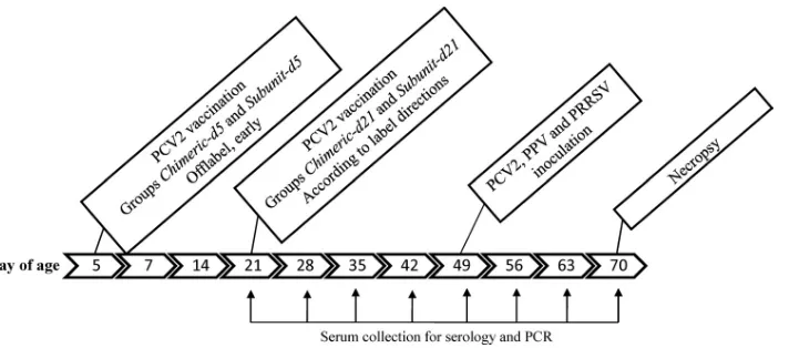
Experimental design.The study was approved by the Iowa State University Institutional Animal Care and Use Committee (IACUC 11-09-6831-S) and the Institutional Biosafety Committee (IBC 09-I-0030-A). The 48 pigs were ran-domly divided into groups of 8 pigs. The timeline of the experiment is summa-rized in Fig. 1. At day 5, 16 pigs were vaccinated with one of two PCV2 vaccines: an inactivated chimeric vaccine (chimeric-d5) or a viral subunit vaccine (subunit-d5). Similarly at day 21, 16 pigs were vaccinated with the inactivated chimeric vaccine (chimeric-d21) or a subunit vaccine (subunit-d21). Upon arrival to the research facility, blood was collected at weekly intervals until termination of the project at week 10. The blood samples were collected in serum separator tubes
(Becton Dickinson vacutainer; 8.5 ml) and centrifuged at 2,000⫻gfor 10 min at 4°C, and the serum was separated into two aliquots and stored at⫺80°C until testing. All pigs, except for the negative-control group, were inoculated with PPV, PRRSV, and PCV2b at day 49, and all pigs were euthanized for necropsy at day 70.
Clinical evaluations.Upon arrival at the research facility the pigs were indi-vidually examined and then monitored daily for clinical signs of disease, such as inappetence, lethargy, lameness, and respiratory disease.
Vaccination.The inactivated chimeric vaccine used in this study was Suvaxyn PCV2 (serial number 1861229A; Fort Dodge Animal Health, Inc.). The subunit vaccine was Ingelvac CircoFLEX (serial number 309-136; Boehringer Ingelheim Vetmedica). Each of the pigs in the vaccinated groups received 2 ml of Suvaxyn PCV2 or 1 ml of Ingelvac CircoFLEX vaccine intramuscularly into the right neck via a 0.77-mm 22-gauge needle. Vaccination was done at day 5 or day 21.
PCV2b, PPV, and PRRSV inoculation.All pigs, excluding the negative-control group, were inoculated at day 49 with PCV2b, PPV, and PRRSV.
PCV2 inoculation.The PCV2 inoculum consisted of PCV2b isolate NC-16845 (32), which was propagated on PK-15 cells to a titer of 104.5
50% tissue culture infective doses (TCID50) per ml. PCV2 inoculation was done by administering 1 ml of the inoculum intramuscularly into the right neck and slowly dripping 2 ml of the inoculum intranasally (1 ml per nostril) while the pig was held in the upright position.
PPV inoculation.The PPV inoculum consisted of a tissue homogenate con-taining strain NADL-8 at a titer of 106.0
TCID50per ml (25). PPV inoculation was done by slowly dripping 1 ml of inoculum intranasally while the pig was held in the upright position.
PRRSV inoculation. The PRRSV inoculum consisted of PRRSV isolate ATCC VR2385 (15). PRRSV was propagated on MARC-145 cells to the seventh passage at a titer of 105.0TCID
50per ml. PRRSV inoculation was done by slowly dripping 2.5 ml of inoculum intranasally while the pig was held in the upright position.
Serology. (i) PCV2. All pig sera, from day 21 to day 70, were tested for anti-PCV2 antibodies by a PCV2 capsid protein-based enzyme-linked immu-nosorbent assay (ELISA) as previously described (28). A sample-to-positive (S/P) ratio of greater than or equal to 0.2 was considered positive. A fluorescent focus neutralization (FFN) assay was performed on serum samples collected 21 days after vaccination for all vaccinated pigs and at the day of challenge (day 49) for all pigs for the detection of neutralizing antibodies, using a previously de-scribed method (37).
(ii) PPV.The anti-PPV IgG antibodies were detected in serum from day 49 and day 70 via a hemagglutination inhibition (HI) assay, as previously described (26).
(iii) PRRSV.All pig sera from day 49 and day 70 were tested for anti-PRRSV antibodies by ELISA (PRRS X3Ab test; IDEXX Laboratories Inc., Westbrook, MA) according to the manufacturer’s instructions. An S/P ratio of 0.4 was used as the minimum positive cutoff value.
Quantitative real-time PCR assays. (i) Total nucleic acid extraction.All day 49, day 56, day 63, and day 70 serum samples were extracted using a total nucleic acid extraction kit (MagMAX viral isolation kit; Applied Biosystems, Foster City,
FIG. 1. Experimental design. All serum samples collected were tested for the presence of PCV2 antibody. Samples from day of age 49, 56, 63,
and 70 were tested for the presence and amount of PCV2 DNA and PPV DNA. Samples from day 56, day 63, and day 70 were tested for the
presence and amount of PRRSV RNA. Samples from day 49 and day 70 were tested serologically for PRRSV and PPV.
on August 17, 2020 by guest
http://cvi.asm.org/
CA) with the KingFisher Flex magnetic particle processor extraction system (Thermo Fisher Scientific, Waltham, MA).
(ii) PCV2.PCV2 viremia was determined by the detection of the presence and amount of viral DNA in serum samples from all pigs on day 49, day 56, day 63, and day 70 via quantitative PCR using the same primers and probes as previously described (42). This was done in a 7500 fast real-time PCR system (Applied Biosystems, Foster City, CA). A final 25-l volume containing 2.5l of extracted DNA was processed under the following thermocycler conditions: 2 min at 50°C and 10 min at 95°C, followed by 40 cycles of 15 s at 95°C and 1 min at 60°C.
(iii) PPV.Viremia for PPV was determined by detection of the presence and amount of PPV DNA in serum samples collected on day 49, day 56, day 63, and day 70 via quantitative real-time PCR as previously described (42). The final volume of the reaction mixture was 25l, which consisted of 12.5l of com-mercially available master mix (TaqMan Universal PCR master mix; PE Applied Biosystems), 2.5l of DNA from either sample extraction or standard, 1l (0.4
M) of each primer, and 0.5 l (0.2M) of the probe. The thermocycler conditions were as follows: 2 min at 50°C and 10 min at 95°C, followed by 40 cycles of 15 s at 95°C and 1 min at 60°C.
(iv) PRRSV.Quantitative real-time reverse transcription-PCR (RT-PCR) for PRRSV viremia was performed on serum samples collected on day 56, day 63, and day 70 using the TaqMan NA and EU PRRSV reagents (Applied Biosystem) as previously described (42). PRRSV RNA presence and quantity were identified with real-time RT-PCR by utilizing TaqMan NA and EA PRRSV reagents (Applied Biosystems) with a final volume of 25l, containing 12 l of 2⫻ multiplex RT-PCR buffer, 2.5l of 10⫻PRRSV primer probe mix, 1.25l of 20⫻multiplex enzyme mix, 0.75l of nuclease-free water, and 8l of either PRRSV RNA from the previous extraction or standards. The thermocycler conditions were as follows: 10 min at 45°C and 10 min at 95°C, followed by 40 cycles of 15 s at 95°C and 70 s at 60°C.
Necropsy.All pigs were humanely euthanized with an overdose of pentobar-bital sodium (Fatal Plus; Vortech Pharmaceuticals, Dearborn, MI) at day 70. The total amount of macroscopic lung lesions was estimated and scored (0 to 100% of the lung affected) as previously described (15). The sizes of lymph nodes (score range from 0 to 3: 0 [normal], 1 [two times the normal size], 2 [three times the normal size], and 3 [four times the normal size]) were estimated as described previously (29). Sections of lung, heart, liver, lymph nodes (tracheobronchial, superficial inguinal, external iliac, mediastinal, and mesenteric), spleen, kidney, ileum, colon, tonsil, and thymus were collected, placed into 10% neutral buffered formalin, and routinely processed for histological examination.
Histopathology and immunohistochemistry.Microscopic examination of tis-sues was done by a veterinary pathologist who was blinded to the treatment groups. Lung sections were scored for presence and severity of interstitial pneu-monia, with scores ranging from 0 to 6 (0 [normal]; 6 [severe diffuse]) (15). Sections of heart, liver, kidney, ileum, and colon were evaluated for the presence of lymphohistiocytic inflammation and scored on a scale of 0 (none) to 3 (severe). Lymphoid tissues, including lymph nodes, tonsil, and spleen, were evaluated for the presence of lymphoid depletion, with scores ranging from 0 (normal) to 3 (severe lymphoid depletion) and scores for histiocytic replacement of follicles ranging from 0 (normal) to 3 (severe) (34).
Immunohistochemistry (IHC) for PCV2 was performed on formalin-fixed, paraffin-embedded tissue sections by using a rabbit polyclonal antibody as pre-viously described (45). Tissues evaluated included tonsil, spleen, lymph nodes (mesenteric, mediastinal, tracheobronchial, external inguinal, and subiliac), and thymus. PCV2 antigen scoring was performed in a blinded fashion, and scores ranged from 0 (no signal) to 3 (more than 50% of lymphoid follicles containing cells with PCV2 antigen staining) (34).
The overall PCV2-associated lesion scores were determined as previously described (34). A combined scoring system for each lymphoid tissue that ranged from 0 to 9 (lymphoid depletion score, 0 to 3; histiocytic replacement score, 0 to 3; PCV2 IHC score, 0 to 3) was used. The scores (lesions and PCV2 IHC) of the seven lymphoid tissues (lymph node pool⫻5, spleen, and tonsil) were added together and divided by 7. The lymph node pool consisted of one section each of tracheobronchial, superficial inguinal, external iliac, mediastinal, and mesenteric lymph nodes. Pigs were grouped into four categories based on overall micro-scopic lymphoid lesion score: normal (score of 0), mild (score of 1 to 3), mod-erate (score of 4 to 6), and severe (score of 7 to 9). A pig was diagnosed with PCVAD if the mean lymphoid microscopic lesion severity score was severe (score of 7 to 9). The mean group overall lymphoid score was calculated and compared between groups.
Statistical analysis.The data were statistically analyzed by performing a one-way analysis of variance (ANOVA) with JMP software version 9.0.0 (SAS Insti-tute, Cary, NC). The significance level wasP⬍0.05, followed by pairwise testing using the Tukey-Kramer adjustment to identify the groups that were different.
All real-time PCR data were log10transformed prior to analysis. The percentage of reduction of PCV2 viremia in vaccinated groups compared to the nonvacci-nated positive-control group was calculated as follows: 100⫺[(100⫻mean log10 genomic copies per ml of serum in vaccinated animals)/(mean log10genomic copies per ml of serum in positive-control animals)]. Nonrepeated measures, such as histopathology data, were assessed using a nonparametric Kruskal-Wallis ANOVA. If a nonparametric ANOVA test was significant (P⬍0.05), then Wilcoxon tests were used to assess the differences of pairs of groups. Differences in incidence were evaluated by using Fisher’s exact test.
RESULTS
Clinical disease.
After challenge, triple-challenged pigs in all
groups developed mild to severe respiratory disease
character-ized by sneezing, increased respiratory rates, and clear nasal
discharge. A portion of the triple-challenged pigs also became
lethargic.
Seroconversion against PCV2, PPV, and PRRSV. (i) PCV2.
The negative-control pigs remained seronegative until
termi-nation of the study (Fig. 2A). Seroconversion to PCV2 in the
vaccinated groups was similar for the day 5 (Fig. 2B) and day
21 (Fig. 2C) groups. By 14 days postvaccination, 2/8 subunit-d5,
3/8 subunit-d21, 7/8 chimeric-d5, and 8/8 chimeric-d21 animals
had seroconverted; by 21 days after vaccination all vaccinated
pigs except 2/8 subunit-d21 animals had seroconverted; by 28
days after vaccination all vaccinated pigs were seropositive for
PCV2. There was a trend to lower levels of detectable
anti-PCV2 IgG in pigs vaccinated with the subunit vaccine
com-pared to those vaccinated with the chimeric vaccine, and this
was independent of age of vaccination (Fig. 2B and C). The
mean amounts of neutralizing antibody levels 21 days
postvac-cination were similar in pigs vaccinated at day 5 (mean group
log
10titers of 1.84
⫾
0.16) (
⫾
standard error [SE]) and day 21
(1.56
⫾
0.12); however, there was a significant difference when
the data were analyzed by product (2.01
⫾
0.14 for the
chime-ric vaccine and 1.39
⫾
0.11 for the subunit vaccine).
As expected, when the data were evaluated by day of age
rather than by days after vaccination, vaccination at day 5
resulted in significantly (
P
⬍
0.05) higher anti-PCV2 IgG levels
from day 21 until day 42; however, there were no differences
between the day 5- and day 21-vaccinated groups thereafter
(Fig. 2A). At day 21, anti-PCV2 IgG was detected in 25% (2/8)
of the subunit-d5 pigs and 87.5% (7/8) of the chimeric-d5 pigs.
The prevalence of seropositive pigs was 100% at day 28 for the
day 5-vaccinated pigs and 18.8% (3/16) for the day
21-vacci-nated pigs. All pigs in these groups were seropositive for PCV2
by day 42. Regardless of timing of vaccination, the chimeric
vaccine induced significantly (
P
⬍
0.05) higher levels of
neu-tralizing antibodies at day 49 than the subunit vaccine, with
mean group log
10titers of 2.38
⫾
0.18 for the chimeric vaccine
compared to 1.82
⫾
0.12 for the subunit vaccine.
Positive-control pigs started to seroconvert by day 63 (62.5%; 5/8 pigs)
and day 70 (75%; 6/8 pigs) as detected by ELISA.
(ii) PPV.
All groups were negative for anti-PPV antibodies
on the day of challenge (day 49), and the nonchallenged
neg-ative controls remained negneg-ative until day 70. All pigs
chal-lenged with PPV seroconverted by day 70; however, 2/8
posi-tive-control pigs had noticeably lower titers (1:2,048) than all
other pigs (1:4,096 to
⬎
16,384). Overall, the mean group PPV
titers of the PPV-challenged animals were not different among
treated groups (data not shown).
V
OL. 18, 2011
COMPARISON OF PCV2 VACCINATION AT 5 AND 21 DAYS OF AGE
1867
on August 17, 2020 by guest
http://cvi.asm.org/
1868
on August 17, 2020 by guest
(iii) PRRSV.
All pigs in all groups were negative for
anti-PRRSV IgG on the day of challenge (day 49), and the
non-challenged negative controls remained negative until day 70.
The majority of the pigs challenged with PRRSV
serocon-verted by day 70, with the exception of 2/8 positive-control pigs.
Overall, the mean group anti-PRRSV IgG S/P ratios of the
PRRSV-challenged pigs were not different among groups
(data not shown).
PCV2, PPV, and PRRSV viremia. (i) PCV2.
All pigs were
negative for PCV2 DNA at the day of challenge (day 49), and
the negative-control pigs remained negative for PCV2 DNA in
serum until termination of the study at day 70. The prevalence
and the log
10mean group amount of PCV2 DNA in the
chal-lenged groups are summarized in Table 1. All vaccinated
groups had significantly (
P
⬍
0.05) smaller amounts of PCV2
DNA in serum than the positive-control group. When the data
were divided based on age of vaccination, no evidence of an
effect of age at vaccination on PCV2 viremia was seen.
How-ever, pigs vaccinated with the chimeric vaccine had significantly
lower mean amounts of PCV2 genomic copies in serum
sam-ples on day 63 (
P
⫽
0.021) and day 70 (
P
⫽
0.03) than those
vaccinated with the subunit vaccine. After challenge, PCV2
viremia was reduced by 75.4% to 100% in the vaccinated
groups compared to the positive-control group.
(ii) PPV.
All pigs were negative for PPV DNA at the day of
challenge (day 49), and the negative-control pigs remained
negative until the termination of the study. The prevalence of
PPV DNA positive pigs at day 56 was 100% for subunit-d5 and
s
ubunit-d21, and it was 88.9% (7/8) for the chimeric-d5,
chi-meric-d21, and the positive-control groups. The overall
prev-alence rate of PPV DNA-positive animals was 68.8% (33/48)
by day 63 and 20.8% (10/48) by day 70, with no significant
differences among groups.
(iii) PRRSV.
All pigs were negative for PRRSV RNA at the
day of challenge (day 49), and the negative-control pigs
re-mained PCR negative throughout the study. PRRSV RNA was
detected in all PRRSV-challenged pigs on day 56, day 63, and
day 70 without significant differences in the mean group
PRRSV RNA levels among the challenged groups, regardless
of vaccination status.
Gross lesions.
There were no visible gross lesions in the
noninfected control pigs. A portion of the triple-challenged
pigs, regardless of vaccination status, had moderate to severe
mottled, tan-colored, consolidated areas of lung tissue
involv-ing up to 51% of the lung surface. A portion of the pigs had
lymph nodes that were up to three times the normal size. There
were no significant differences in gross lesions between
chal-lenged pigs.
Microscopic lesions and presence of PCV2 antigen in
tis-sues.
The majority of the pigs developed mild to severe
inter-stitial pneumonia lesions characterized by thickening of
alve-olar septa by macrophages and lymphocytes and mild to severe
type 2 pneumocyte hypertrophy and hyperplasia. The mean
group interstitial pneumonia scores ranged from 3.0
⫾
0.1 to
3.6
⫾
0.4 in the triple-challenged groups and were significantly
higher (
P
⬍
0.05) than for the negative-control group (0.8
⫾
0.1). Lymphoid lesions, if present, were characterized by mild
to severe lymphoid depletion and mild to severe histiocytic
replacement of lymphoid follicles.
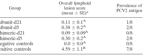
The prevalence rates of PCV2 antigen and overall
lym-phoid lesion scores for the different groups are summarized
in Table 2. The majority of vaccinated pigs had no
remark-able lesions and were considered normal. Individual
vacci-nated pigs (7/32) had an overall lesion score of 1 or 2. In the
positive-control group, 25% (2/8) of the pigs had
micro-scopic lesions compatible with PCVAD associated with
abundant amounts of PCV2 antigen and an overall lymphoid
score of 9; 37.5% (3/8) of the pigs had moderate lymphoid
lesions; the remaining 37.5% (3/8) of the pigs had no to mild
lymphoid lesions.
DISCUSSION
The main objective of this study was to determine the
effi-cacy of PCV2 vaccination at an earlier age than recommended
FIG. 2. (A) Mean group PCV2 ELISA S/P ratios (
⫾
SE) on serum collected from piglets vaccinated at day of age 5 (d5) or 21 (d21) or
nonvaccinated and challenged with PCV2, PPV, and PRRSV at day 49, which corresponds to 44 days after vaccination for day 5 piglets and 28
days after vaccination for day 21 piglets. An S/P ratio of 0.2 or greater was considered seropositive. Significant differences among groups on a
certain day are indicated by different letters (A, B, and C). (B) Comparison of subunit-d5 and chimeric-d5 pigs at different days postvaccination.
Significant differences among groups on a certain day are indicated by an asterisk. (C) Comparison of subunit-d21 and chimeric-d21 pigs at
different days postvaccination. Significant differences among groups on a certain day are indicated by an asterisk.
TABLE 1. Prevalence and mean log
10PCV2 DNA in pigs
challenged with PCV2 at age 49 days
Group
Prevalence (mean level⫾SE)aof log
10 PCV2 DNA on day:
56 63 70
Subunit-d21
1/8 (0.7
⫾
0.7)
A3/8 (1.6
⫾
0.8)
A,B1/8 (0.5
⫾
0.5)
ASubunit-d5
0/8 (0.0)
A4/8 (2.3
⫾
0.9)
A3/8 (1.5
⫾
0.7)
AChimeric-d21
1/8 (0.6
⫾
0.6)
A0/8 (0.0)
B0/8 (0.0)
AChimeric-d5
0/8 (0.0)
A0/8 (0.0)
B0/8 (0.0)
APositive controls 7/8 (4.1
⫾
0.6)
B8/8 (7.1
⫾
0.3)
C8/8 (6.1
⫾
0.6)
BaDifferent superscript capital letters (A, B, and C) within a column indicate significantly (P⬍0.05) different amounts of group mean PCV2 DNA.
TABLE 2. Lymphoid depletion score and prevalence of PCV2
antigen in lymphoid tissues as determined by IHC
Group
Overall lymphoid lesion score (mean⫾SE)a
Prevalence of PCV2 antigen
Subunit-d21
0.11
⫾
0.1
A1/8
Subunit-d5
0.38
⫾
0.2
A2/8
Chimeric-d21
0.09
⫾
0.09
A0/8
Chimeric-d5
0.30
⫾
0.2
A2/8
Negative controls
0.0
⫾
0.0
A0/8
Positive controls
4.59
⫾
1.1
B7/8
aSignificant differences among groups are indicated by different superscript capital letters (A and B).
V
OL. 18, 2011
COMPARISON OF PCV2 VACCINATION AT 5 AND 21 DAYS OF AGE
1869
on August 17, 2020 by guest
http://cvi.asm.org/
by the vaccine manufacturers. Several research groups have
studied the efficacy of commercial PCV2 vaccines in pigs
sin-gularly infected with PCV2 (30, 31) or in pigs concurrently
infected with multiple pathogens (33, 42). In all previous
stud-ies, vaccination was done according to the manufacturer’s label
instructions. To our knowledge, this is the first controlled
ex-perimental study to test the efficacy of commercial vaccines
used at less than 3 weeks of age in a manner not approved by
the manufacturer; however, this regimen mimics what is
com-monly now done in the field in the United States. Many
pro-ducers prefer to vaccinate with a single-dose PCV2 product
while piglets are undergoing castration, iron shots, tail docking,
and teeth clipping between 2 and 5 days of age. However, there
is concern that the immune system may not be mature enough
to effectively respond to the vaccinations, potentially resulting
in decreased vaccine efficacy and duration of immunity. To
evaluate the benefits and shortcomings of early vaccination,
this study entailed use of piglets blocked by litter and randomly
assigned to early vaccination (day 5), regular vaccination (day
21), or no-vaccination (positive- and negative-control) groups.
After challenge, PCV2 viremia and associated lesions were
similarly reduced in all vaccinated pigs regardless of timing of
vaccination, indicating that day 5 pigs are capable of mounting
a protective immune response. Vaccinated pigs were protected
from development of PCV2-associated lesions independent of
timing of vaccination, further indicating that both day 5 and
day 21 vaccination protocols with either vaccine were effective.
The pig immune system is unique in many ways that may be
responsible for its ability to develop protective immunity from
early vaccination. These factors include the full-length
com-plementarity-determining region 3 (CDR3) of the heavy chain
of immunoglobulin (4), limited genetic combinatorial
preim-mune repertoire development (4), and the absence of true
gene conversion sometimes seen in other species (44). The
above-mentioned characteristics of the pig immune system
combined with the results of this study demonstrate that the
5-day-old suckling pig is indeed capable of mounting a
protec-tive immune response against PCV2 challenge.
The current study was done in PCV2-naïve pigs; however,
under field conditions most pigs will be seropositive due to the
ubiquitous nature of PCV2 and high levels of anti-PCV2
an-tibodies in colostrum. Interference with vaccination against
swine influenza virus associated with the presence of passively
acquired antibodies has been documented (3, 23, 27, 38);
how-ever, evidence of passive antibody interference with PCV2
vaccination has not been confirmed under experimental
con-ditions (30). Furthermore, PCV2 vaccines have been highly
effective in the field, and almost all pigs are seropositive to
PCV2 at the time of PCV2 vaccination (9, 19, 21, 41). In
experimental PCV2 challenge models, outcomes are often
sim-ilar between vaccine treatment groups (11, 24), and
conclu-sions often lack power. Passively acquired antibodies in many
instances decrease PCV2 viremia and prevent the development
of clinical disease under controlled experimental conditions.
PCV2 viremia and expression of clinical disease are often the
main outcomes used for vaccine efficacy comparisons;
how-ever, when using animals with maternally derived anti-PCV2
antibodies, a much larger sample size may be required to
demonstrate differences. Although the antibody-negative
sta-tus of the pigs in the current study did not necessarily mimic
what occurs with the majority of pigs in the field, studies
performed in PCV2 antibody-free and PCV2 virus-free pigs
are an important first step to increasing our understanding of
potential advantages and disadvantages of early vaccination
regimens.
To determine if there were differences in the efficacy of one
vaccine over another, two different products were used side by
side in this study. Several previous studies had been performed
to determine the efficacy of PCV2 subunit vaccines and
chi-meric PCV2 vaccines (10, 12, 43). In these studies, vaccinated
animals were shown to have strong antibody responses
associ-ated with decreased PCV2 viremia after challenge. Similarly, in
our study the vaccinated animals, regardless of the type of
PCV2 vaccine used, all developed a detectable antibody
re-sponse and protective immunity as evidenced by significantly
decreased PCV2 viremia and a decreased incidence and
sever-ity of lesions compared to the positive-control group. However,
pigs vaccinated with the chimeric product had a stronger
anti-PCV2 IgG response that was independent of age at vaccination
and a lower prevalence of PCV2 viremic animals at day 63 and
day 70 than pigs vaccinated with the subunit product.
More-over, and similar to a previous study using single-dose
vacci-nation (42), vaccivacci-nation with the chimeric product was
associ-ated with production of a stronger neutralizing antibody
response than vaccination with the subunit vaccine.
In summary, under the conditions of this study, vaccination
with chimeric or subunit PCV2 vaccines at 5 or 21 days of age
induced a protective immune response in PCV2-naïve pigs as
demonstrated by development of anti-PCV2 antibodies and
reductions of PCV2 viremia and PCV2-associated lesions in a
triple challenge model with PCV2, PPV, and PRRSV.
ACKNOWLEDGMENTS
This study was funded by the National Pork Board Pork Checkoff
Dollars.
We thank Shayleen Schalk for assistance with the animal work.
REFERENCES
1.Allan, G. M., et al. 1999. Experimental reproduction of severe wasting disease by co-infection of pigs with porcine circovirus and porcine parvovi-rus. J. Comp. Pathol.121:1–11.
2.Allan, G. M., et al.2000. Experimental infection of colostrum deprived piglets with porcine circovirus 2 (PCV2) and porcine reproductive and re-spiratory syndrome virus (PRRSV) potentiates PCV2 replication. Arch. Vi-rol.145:2421–2429.
3.Blaskovic, D., et al. 1970. Experimental infection of weanling pigs with A-swine influenza virus. 3. Immunity in piglets farrowed by antibody-bearing dams experimentally infected a year earlier. Bull. World Health Organ.
42:771–777.
4.Butler, J. E.2009. Isolator and other neonatal piglet models in developmen-tal immunology and identification of virulence factors. Anim. Health Res. Rev.10:35–52.
5.Dulac, G. C., and A. Afshar.1989. Porcine circovirus antigens in PK-15 cell line (ATCC CCL-33) and evidence of antibodies to circovirus in Canadian pigs. Can. J. Vet. Res.53:431–433.
6.Edwards, S., and J. J. Sands.1994. Evidence of circovirus infection in British pigs. Vet. Rec.134:680–681.
7.Ellis, J., et al.2004. Porcine circovirus-2 and concurrent infections in the field. Vet. Microbiol.98:159–163.
8.Ellis, J. A., et al.2000. Coinfection by porcine circoviruses and porcine parvovirus in pigs with naturally acquired postweaning multisystemic wasting syndrome. J. Vet. Diagn. Invest.12:21–27.
9.Fachinger, V., R. Bischoff, S. B. Jedidia, A. Saalmu¨ller, and K. Elbers.2008. The effect of vaccination against porcine circovirus type 2 in pigs suffering from porcine respiratory disease complex. Vaccine26:1488–1499. 10.Fenaux, M., T. Opriessnig, P. G. Halbur, F. Elvinger, and X. J. Meng.2004.
A chimeric porcine circovirus (PCV) with the immunogenic capsid gene of the pathogenic PCV type 2 (PCV2) cloned into the genomic backbone of the
on August 17, 2020 by guest
http://cvi.asm.org/
nonpathogenic PCV1 induces protective immunity against PCV2 infection in pigs. J. Virol.78:6297–6303.
11.Fort, M., et al.2008. Porcine circovirus type 2 (PCV2) vaccination of con-ventional pigs prevents viremia against PCV2 isolates of different genotypes and geographic origins. Vaccine26:1063–1071.
12.Fort, M., et al.2009. One dose of a porcine circovirus 2 (PCV2) sub-unit vaccine administered to 3-week-old conventional piglets elicits cell-mediated immunity and significantly reduces PCV2 viremia in an experimental model. Vaccine27:4031–4037.
13.Gagnon, C. A., N. Music, G. Fontaine, D. Tremblay, and J. Harel.2010. Emergence of a new type of porcine circovirus in swine (PCV): a type 1 and type 2 PCV recombinant. Vet. Microbiol.144:18–23.
14.Gillespie, J., T. Opriessnig, X. J. Meng, K. Pelzer, and V. Buechner-Maxwell.
2009. Porcine circovirus type 2 and porcine circovirus-associated disease. J. Vet. Intern. Med.23:1151–1163.
15.Halbur, P. G., et al.1995. Comparison of the pathogenicity of two US porcine reproductive and respiratory syndrome virus isolates with that of the Lelystad virus. Vet. Pathol.32:648–660.
16.Harding, J., and E. Clark.1997. Recognizing and diagnosing postweaning multisystemic wasting syndrome (PMWS). Swine Health Prod.5:201–203. 17.Harms, P. A., P. G. Halbur, and S. D. Sorden.2002. Three cases of porcine
respiratory disease complex associated with porcine circovirus type 2 infec-tion. J. Swine Health Prod.10:27–30.
18.Harms, P. A., et al.2001. Experimental reproduction of severe disease in CD/CD pigs concurrently infected with type 2 porcine circovirus and porcine reproductive and respiratory syndrome virus. Vet. Pathol.38:528–539. 19.Horlen, K. P., et al.2008. A field evaluation of mortality rate and growth
performance in pigs vaccinated against porcine circovirus type 2. J. Am. Vet. Med. Assoc.232:906–912.
20.Kim, J., Y. Ha, K. Jung, C. Choi, and C. Chae.2004. Enteritis associated with porcine circovirus 2 in pigs. Can. J. Vet. Res.68:218–221.
21.Kixmo¨ller, M., et al.2008. Reduction of PMWS-associated clinical signs and co-infections by vaccination against PCV2. Vaccine26:3443–3451. 22.Krakowka, S., et al.2000. Viral wasting syndrome of swine: experimental
reproduction of postweaning multisystemic wasting syndrome in gnotobiotic swine by coinfection with porcine circovirus 2 and porcine parvovirus. Vet. Pathol.37:254–263.
23.Loeffen, W. L., P. P. Heinen, A. T. Bianchi, W. A. Hunneman, and J. H. Verheijden.2003. Effect of maternally derived antibodies on the clinical signs and immune response in pigs after primary and secondary infection with an influenza H1N1 virus. Vet. Immunol. Immunopathol.92:23–35.
24.Lyoo, K., et al.2011. Comparative efficacy of three commercial PCV2 vac-cines in conventionally reared pigs. Vet. J.189:58–62.
25.Mengeling, W. L., T. T. Brown, P. S. Paul, and D. E. Gutekunst.1979. Efficacy of an inactivated virus vaccine for prevention of porcine parvovirus-induced reproductive failure. Am. J. Vet. Res.40:204–207.
26.Mengeling, W. L., J. F. Ridpath, and A. C. Vorwald.1988. Size and antigenic comparisons among the structural proteins of selected autonomous parvo-viruses. J. Gen. Virol.69:825–837.
27.Mensik, J., and J. Pokorny.1971. Development of antibody response to swine influenza virus in pigs. I. The influence of experimental infection of pregnant sows on serum antibody production by their progeny during post-natal development. Zentralbl. Veterinarmed. B18:177–189.
28.Nawagitgul, P., et al.2002. Modified indirect porcine circovirus (PCV) type 2-based and recombinant capsid protein (ORF2)-based enzyme-linked im-munosorbent assays for detection of antibodies to PCV. Clin. Diagn. Lab. Immunol.9:33–40.
29.Opriessnig, T., et al. 2006. Evidence of breed-dependent differences in susceptibility to porcine circovirus type-2-associated disease and lesions. Vet. Pathol.43:281–293.
30.Opriessnig, T., A. R. Patterson, J. Elsener, X. J. Meng, and P. G. Halbur.
2008. Influence of maternal antibodies on efficacy of porcine circovirus type 2 (PCV2) vaccination to protect pigs from experimental infection with PCV2. Clin. Vaccine Immunol.15:397–401.
31.Opriessnig, T., et al.2010. Comparison of the effectiveness of passive (dam) versus active (piglet) immunization against porcine circovirus type 2 (PCV2) and impact of passively derived PCV2 vaccine-induced immunity on vacci-nation. Vet. Microbiol.142:177–183.
32.Opriessnig, T., et al.2008. Differences in virulence among porcine circovirus type 2 isolates are unrelated to cluster type 2a or 2b and prior infection provides heterologous protection. J. Gen. Virol.89:2482–2491.
33.Opriessnig, T., H. G. Shen, N. Pal, S. Ramamoorthy, and P. G. Halbur.2011. A live-attenuated chimeric PCV2 vaccine is transmitted to contact pigs but is not upregulated by concurrent infection with PPV and PRRSV and is efficacious in a PCV2a-PRRSV-PPV challenge model. Clin. Vaccine Immu-nol.18:1261–1268.
34.Opriessnig, T., et al.2004. Experimental reproduction of postweaning mul-tisystemic wasting syndrome in pigs by dual infection withMycoplasma hyo-pneumoniaeand porcine circovirus type 2. Vet. Pathol.41:624–640. 35.Pallare´s, F. J., et al.2002. Porcine circovirus type 2 (PCV-2) coinfections in
US field cases of postweaning multisystemic wasting syndrome (PMWS). J. Vet. Diagn. Invest.14:515–519.
36.Pogranichniy, R. M., K. J. Yoon, P. A. Harms, S. D. Sorden, and M. Daniels.
2002. Case-control study on the association of porcine circovirus type 2 and other swine viral pathogens with postweaning multisystemic wasting syn-drome. J. Vet. Diagn. Invest.14:449–456.
37.Pogranichniy, R. M., et al.2000. Characterization of immune response of young pigs to porcine circovirus type 2 infection. Viral Immunol.13:143–153. 38.Renshaw, H. W.1975. Influence of antibody-mediated immune suppression on clinical, viral, and immune responses to swine influenza infection. Am. J. Vet. Res.36:5–13.
39.Rossow, K. D.1998. Porcine reproductive and respiratory syndrome. Vet. Pathol.35:1–20.
40.Rovira, A., et al.2002. Experimental inoculation of conventional pigs with porcine reproductive and respiratory syndrome virus and porcine circovirus 2. J. Virol.76:3232–3239.
41.Segale´s, J., et al.2009. A genetically engineered chimeric vaccine against porcine circovirus type 2 (PCV2) improves clinical, pathological and viro-logical outcomes in postweaning multisystemic wasting syndrome affected farms. Vaccine27:7313–7321.
42.Shen, H. G., et al.2010. Comparison of commercial and experimental por-cine circovirus type 2 (PCV2) vacpor-cines using a triple challenge with PCV2, porcine reproductive and respiratory syndrome virus (PRRSV), and porcine parvovirus (PPV). Vaccine28:5960–5966.
43.Shen, H. G., et al.2008. Protective immunity against porcine circovirus 2 by vaccination with ORF2-based DNA and subunit vaccines in mice. J. Gen. Virol.89:1857–1865.
44.Sinkora, M., et al. 2003. Antibody repertoire development in fetal and neonatal piglets. VI. B cell lymphogenesis occurs at multiple sites with differences in the frequency of in-frame rearrangements. J. Immunol.170:
1781–1788.
45.Sorden, S. D., P. A. Harms, P. Nawagitgul, D. Cavanaugh, and P. S. Paul.
1999. Development of a polyclonal-antibody-based immunohistochemical method for the detection of type 2 porcine circovirus in formalin-fixed, paraffin-embedded tissue. J. Vet. Diagn. Invest.11:528–530.
46.Tischer, I., H. Gelderblom, W. Vettermann, and M. A. Koch.1982. A very small porcine virus with circular single-stranded DNA. Nature295:64–66. 47.Tischer, I., W. Mields, D. Wolff, M. Vagt, and W. Griem.1986. Studies on
epidemiology and pathogenicity of porcine circovirus. Arch. Virol.91:271– 276.