R E S E A R C H A R T I C L E
Open Access
Differential effect of surgical manipulation
on gene expression in normal breast tissue
and breast tumor tissue
Inge Søkilde Pedersen
1,5,6*†, Mads Thomassen
2†, Qihua Tan
2, Torben Kruse
2, Ole Thorlacius-Ussing
3,5,6,
Jens Peter Garne
4and Henrik Bygum Krarup
1,5Abstract
Background:Gene expression profiles of normal and tumor tissue reflect both differences in biological processes taking place in vivo and differences in response to stress during surgery and sample handling. The effect of cold (room temperature) ischemia in the time interval between surgical removal of the specimen and freezing is described in a few studies. However, not much is known about the effect of warm (body temperature) ischemia during surgery.
Methods: Three women with primary operable breast cancer underwent in situ biopsies from normal breast and tumor tissue prior to radical mastectomy. Ex vivo biopsies from normal and tumor tissue were collected immediately after surgical excision. The putative effects on gene expression of malignancy (tumor versus normal), surgical manipulation (post- versus pre-surgical) and interaction between the two (differences in effect of surgical manipulation on tumor and normal samples) were investigated simultaneously by Generalized Estimating Equation (GEE) analysis in this self-matched study.
Results:Gene set enrichment analysis (GSEA) demonstrates a marked difference in effect of surgical manipulation on tumor compared to normal tissue. Interestingly, a large proportion of pathways affected by ischemia especially in tumor tissue are pathways considered to be specifically up regulated in tumor tissue compared to normal. Conclusion:The results of this study suggest that a large contribution to this differential expression originates from altered response to stress in tumor cells rather than merely representing in vivo differences. It is important to bear this in mind when using gene-expression analysis to deduce biological function, and when collecting material for gene expression profiling.
Keywords:Specimen handling, Ischemia, Gene expression, Cell cycle, Cancer
Introduction
Several multigene classifiers are currently commercially available for breast cancer, among them MammaPrint (Agendia, The Netherlands), Oncotype DX (Genomic Health, USA) and PAM50 (Nano String, USA). However, caution should be taken interpreting gene signatures, when sampling conditions are suboptimal (i.e. prolonged
delay of sample preservation after surgery), since sample handling and preservation methods affect gene expres-sion. This has been shown previously by snap freezing aliquots of breast tumor tissue at different time points after surgical removal in a few studies. De Cecco et al. (2009) investigated breast tumor tissue from 11 patients. For each patient 4 aliquots were analyzed, 1 was frozen immediately and the remaining left at room temperature for 2, 6 and 24 h respectively. No major effect on RNA integrity was observed. However, expression levels of 461 genes were significantly altered. Borgan et al. (2011) observed significantly altered expression of 1788 mRNAs and 56 miRNAs in a study with delayed freezing of 0.5,
* Correspondence:[email protected]
†Inge Søkilde Pedersen and Mads Thomassen contributed equally to this
work.
1Molecular Diagnostics, Clinical Biochemistry, Aalborg University, Aalborg, Denmark
5Clinical Cancer Research Center, Aalborg University Hospital, Aalborg, Denmark Full list of author information is available at the end of the article
1, 2, 3 and 6 h when compared to the breast tumor tis-sue frozen immediately post surgery. Aktas et al. (2014) investigated the difference between preservation of breast tumor tissue in RNAlater and freezing on dry ice in prechilled vials, they found expression of 481 tran-scripts to be significantly altered. In addition, they observed an effect of delayed preservation on 41 tran-scripts. In a previous study on the same samples, com-paring preservation methods, it was concluded that RNAlater improves RNA yield and quality (Hatzis et al., 2010). Recently the sparse literature on the effect of cold ischemia has been reviewed (Grizzle et al.,2016).
As pointed out by Grizzle et al., warm ischemia may be the more important variable, however this is not easily controlled and data on the effect of warm ischemia is rarely available (Grizzle et al., 2016) . To our knowledge no study has investigated the effect of surgical manipula-tion on gene expression profiles in breast tissue. In animal studies a difference between effect of ischemic stress at room temperature and body temperature has been shown (Almeida et al., 2004). In addition, a study investigating gene expression in histologically benign prostate tissue be-fore and after surgery demonstrated substantial gene ex-pression changes as a result of surgical manipulation with a median warm ischemia time of 28 min during surgery (Lin et al., 2006). Albeit, the effect is likely to be at least partly contributable to ischemia, other factors may also affect gene expression. The best studied example being anesthesia (Lucchinetti et al., 2007). Since environmental changes cause the cell to adjust its regulatory mechanisms which in turn alters the way it reacts to external input, tumor and normal cells are not only different per se, they could also be expected to react differently when exposed to stress such as surgical manipulation. The current study has been designed to investigate the effect of surgical ma-nipulation in both normal breast tissue and breast tumor tissue by comparing gene expression profiles before and after surgery using a self-matched study design for in-creased statistical power.
Materials and methods
Patient material
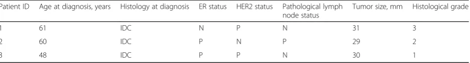
Participants were 3 women with primary operable breast cancer. None of the participants had received anti-cancer treatment prior to surgery. Time between biopsy was ap-proximately 1 h in the 3 cases. Clinical and pathological characteristics of the patients are summarized in Table1. The study protocol was approved by the Research Ethics Committee of the North Denmark Region (N-20090029). All participants gave written informed consent.
Tissue collection
Biopsies from normal and tumor tissue were taken in situ immediately after induction of anesthesia (byintra-venous
injection of fentanyl and propofol) prior to radical mastec-tomy. All tumors in this study were well defined and easily palpable. Biopsies from normal tissue were taken as far from the tumor as possible. After surgical excision 4 biop-sies were taken ex vivo, 3 from normal (to test for vari-ation among replicate samples) and 1 from tumor tissue. Origin (tumor or normal) of the biopsies was confirmed by macroscopic inspection. All biopsies were snap frozen in liquid nitrogen and subsequently kept at−80 °C until RNA extraction.
RNA isolation and expression profiling
Total RNA was extracted using RNeasy Micro kit (Qiagen, Hilden, Germany) according to manufacturer’s instruc-tions. RNA concentration and purity was determined by UV spectrometry on a NanoDrop 2000 (Thermo Scien-tific, Waltham, Massachusetts, USA). Amplified RNA was synthesized from 300 ng total RNA using the Messa-geAmpTM III RNA amplification kit and fragmented ac-cording to manufacturer’s instructions (Ambion, Austin, TX, USA). Fragmented amplified RNA was hybridized to Human Genome U133 Plus 2.0 GeneChip® (Affymetrix, Santa Clara, California, USA), washed and scanned as rec-ommended by the manufacturer. Data are available from Gene Expression Omnibus (http://www.ncbi.nlm.nih.gov/ geo; accession no. GSE71053).
Data analysis and statistics
The affy package (www.bioconductor.org), implemented in the statistical programming language R version 3.1.1 was applied for initial data analysis. Robust multi-array average expression measure (rma) (Irizarry et al., 2003) was applied to perform background correction, quantile normalization, and expression index calculation of all microarrays. Only perfect match probes were used for data analysis.
Hierarchical clustering of genes was carried out using MultiExperiment Viewer (MeV; http://www.tm4.org) with Pearson correlation as distance metric and average linkage.
ranked data and the preranking option in GSEA. The GSEA method takes the entire rank of genes into ac-count allowing identification of pathways not only repre-sented among most differentially regulated genes, but also pathways supported by a larger number of moder-ately differentially expressed genes. The Reactome path-way collection from MSigDB was used for all GSEA analyses. For the ease of presentation of data a more stringent FDR cut-off of 0.005 was emploid for the GSEA analyses. Out of 674 gene sets, 486 passed a cri-terion of at least 15 genes represented in the data set.
In order to compare our results with a previous publi-cation investigating the effect of cold ischemia on gene expression in tumor tissue, GSEA analysis was used to compare tumor tissue before and after surgical manipu-lation. Genes were in this analysis ranked according to differentially expression in tumor post-surgery compared to pre-surgery, using the statistical parameterd, derived from Significance Analysis of Microarray (SAM). Path-ways with a FDR value below 0.0005 were considered significant.
Results
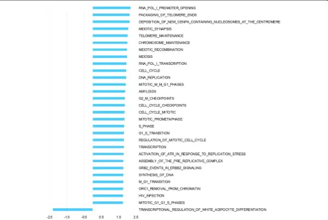
Hierarchical clustering of pre surgery samples using all genes (Fig1a) shows a combined effect of individual and tissue type, with no distinct clustering of tumor and nor-mal. Post surgery there is a more pronounced effect of tissue origin with tumor samples clustering separately from normal samples (Fig1b). The 3 biopsies taken from normal tissue post surgery cluster together for each pa-tient, as expected for tissue of the same tissue type han-dled identically. In order to analyze the interaction of ischemia (pre/post-surgery) and tissue (tumor/normal) GEE was applied. This analysis of the interaction identi-fied 3179 differentially expressed genes. Top 50 up- and down-regulated genes are listed in additional material (Additional file1and Additional file2). Pathway analysis of interaction, including all genes ranked by GEE and performed with GSEA, revealed significant up-regulation of 29 pathways and down-regulation of 1 pathway (Fig2). Among the pathways specifically upregulated in tumor as a consequence of surgical manipulation were several cell cycle related pathways. Enrichment plots and infor-mation on pathways significantly affected by interaction are provided as additional material (Additional file3and Additional file4).
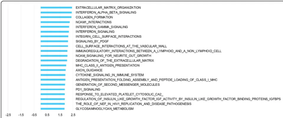
The analysis of ischemia, i.e. surgical manipulation, on tissue regardless of malignancy state identified 667 differ-entially expressed genes Top 50 up- and down-regulated genes are listed in additional material (Additional file 5 and Additional file 6). GSEA demonstrated 6 pathways upregulated after ischemia, including pathways involved in adipocyte differentiation, lipid mobilization, cell interac-tions and immune system pathways (Fig 3). Enrichment plots and information on pathways significantly affected by surgical manipulation are provided as additional material (Additional file7and Additional file8).
The comparison of tumor tissue (pre- and post-sur-gery) and normal tissue (pre- and post-surpost-sur-gery) identi-fied a total of 6166 differentially expressed genes. Top 50 up- and down-regulated genes are listed in additional material (Additional file 9and Additional file 10). Path-way analysis demonstrated 22 pathPath-ways that were upreg-ulated in tumor tissue compared to normal tissue
(Fig4). Among these pathways are pathways
characteris-tic for invasive tumors such as extracellular matrix deg-radation, PDGF signaling and immune system pathways. Enrichment plots and information on pathways signifi-cantly affected by tissue type are provided as additional material (Additional file11and Additional file12).
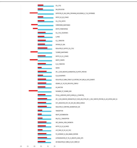
We next wanted to perform validation of our findings in external data sets. Since no data of warm ischemia are available, we used the data set from Aktas et al.’s study of cold eschemia in tumor tissue (Aktas et al., 2014) . This provided a strong validation of observed effect in tumor tissue (Fig 5) with 30 of 36 pathways being af-fected in the same direction, and 1 of these pathways (REGULATION_OF_MITOTIC_CELL_CYCLE) had a nominalp-value of less than 0.05.
Discussion
Our study reveals a remarkable effect of surgical ma-nipulation, such as warm ischemia and anesthesia. It is not possible from the current study to deduce the spe-cific contribution of these variables. In our hierarchical clustering analysis it is only for the post-surgery samples that tumor samples cluster separately from normal sam-ples, indicating a differential effect of surgical manipula-tion on gene expression profiles in normal and tumor tissue. Indeed GSEA analysis identifies several pathways which are differentially affected by warm ischemia be-tween tumor and normal tissue. This is in keeping with
Table 1Patient cilincal ad pathological charcteristics and details of sample collection
Patient ID Age at diagnosis, years Histology at diagnosis ER status HER2 status Pathological lymph node status
Tumor size, mm Histological grade
1 61 IDC N P N 31 3
2 60 IDC P N P 29 2
3 48 IDC P P N 30 1
observations of cell type specific response to environ-mental stress such as hypoxia (Chi et al., 2006) and a likely consequence of different pconditioning as a re-sult of the different milieu of tumor cells compared to normal. Interestingly, immune system pathways are up-regulated both as a result of surgical manipulation and
when comparing tumor and normal tissue. This is in agreement with what has already been documented in the literature. (Grigoryev et al.,2006; Bottai et al.,2017; Liu et al., 2017). However, immune system pathways are not significantly affected in the interaction analysis, indicating activation of a similar immune response in Fig. 1Hierarchical clustering dendrograms. Dendrograms based on hierarchical clustering of all genes on pre (a) and post (b) surgery samples. Patient samples are numbered Pt_1, Pt_2 and Pt_3, N for normal, T for tumor and pre/post for pre surgery and post-surgery respectively
tumor and normal tissue as a consequence of surgical manipulation.
Pathways preferentially enriched in tumor compared to normal tissue as a result of surgical manipulation are pathways involved in cell cycling. Despite it being well documented for neurons to enter cell cycling in re-sponse to ischemia (Love, 2003), this is a somewhat counterintuitive response for actively replicating cells. However, when restricting analysis to the effect of surgi-cal manipulation on tumor tissue, 1 of the cell-cycle re-lated pathways up regure-lated in tumor tissue as a result of surgery was found to be significantly up regulated as a result of 40 min cold ischemia of breast tumor tissue post surgery in a previous study by Aktas et al. (Aktas et al.,2014). Furthermore, there is a significant correlation between direction of the effect of warm and cold ische-mia, with 30 out of 36 pathways affected in the same dir-ection in both studies, providing strong validation of the observed effect on cell cycle pathways of ischemia on tumor tissue. Differences between this study and
previous studies could be explained both by a differential effect of cold and warm ischemia and differences in baseline, since up regulation of different genes have been shown to peak at different time points after tissue resec-tion (Spruessel et al., 2004). In previous studies baseline has been within minutes of surgery, effectively the end-point of the current study. For the purpose of the current study it was of utmost importance to ensure im-mediate preservation of baseline samples. In other stud-ies samples were pathologically evaluated before mincing into smaller fragments and dividing into aliquots for freezing at different time points (Aktas et al.,2014; Hat-zis et al.,2010). This procedure minimizes intra-tumoral variation. However, it introduces a median delay prior to stabilization of 40 min, and hence would not be suitable in our set up. On the other hand, lack of control over tissue composition adds some degree of uncertainty to our results. It is also important to keep in mind that the three tumors in this study represent three different histopathological entities (Table 1). This could Fig. 3Pathway analysis of the effect of surgical manipulation (warm ischemia). Pathway analysis was performed with GSEA using ranking of genes according to the time term from GEE. NES depicted for all Reactome pathways significantly altered in samples collected post-surgery compared to samples collected pre-surgery irrespective of whether it was normal or tumor tissue
potentially affect the analysis and may explain the lack of clustering of tumor samples pre-surgery. Interestingly, our results suggest a distinct response of this heteroge-neous group to surgical manipulation.
In order to elucidate differences in gene expression be-tween tumor and normal tissue in addition to what is
evident from hierarchical clustering, GSEA was carried out and 22 pathways displayed differential representa-tion. This shows a difference between normal and tumor samples when it comes to pathways involved in organization and degradation of extracellular matrix and interferon signaling.
Fig. 5Comparison of GSEA on effect of warm and cold ischemia on tumor tissue. NES depicted in blue for all Reactome pathways significantly altered in tumor tissue collected post-surgery compared to pre-surgery (warm ischemia). To illustrate concordance with previously published data
by Aktas et al. (2014) NES from tumor tissue subjected to 40 min cold ischemia post surgery are represented by red bars. One pathway had a
Speculatively, it may not be surprising that the dif-ferences between normal and tumor tissue in vivo are less pronounced than the differences after being sub-jected to ischemic stress. After all, the cells have to carry out more or less the same functions in order to survive. Pronounced difference in reaction to changes in external environment could putatively be what constitutes the main difference between normal cells and tumor cells. Activation of cell cycling genes dur-ing stress could be an acquired response of a cancer cell no longer reacting as expected to environmental signals. Similar to what is seen in cell cultures where normal cells stop growing when reaching confluence whereas tumor cells keep dividing.
Reported differential expression of pathways between tumor and normal tissue almost exclusively refers to post surgery samples. This has been proven to work well for several multigene classifiers. Our results open the possibil-ity that differences observed in post-surgical specimens not necessarily mirrors differences present in vivo, and whereas difference in response to stress may be an equally good measurement for classifying tumors into subgroups, it requires standardized control over sampling conditions and information about surgical procedures. Furthermore, when it comes to investigating biological differences of tumor and normal cells and considering possible thera-peutical targets, caution should be taken deducing in vivo differences based on gene expression profiles from tissue sampled after surgery.
Conclusions
The current study is to our knowledge the first study to simultaneously investigate the effect of surgical manipula-tion, malignancy state and the interaction of these two pa-rameters on gene expression profiling in breast tissue. The self-matched design gives added statistical power com-pared to a case-control design, and even with a limited number of samples we are able demonstrate an effect on a large number of pathways and a differential response be-tween tumor and normal tissue. The majority of pathways affected primarily in tumor tissue are pathways that have previously been implicated in oncogenesis. It is of great interest that the well documented up regulation of gene sets involved in cell cycling may in fact primarily reflect al-tered reaction to surgical manipulation/ischemic stress of tumor cells compared to normal cells rather than reflect-ing the state of cells in the body.
Additional files
Additional file 1:Top 50 up-regulated genes (interaction). The top 50 genes up-regulated in the GEE interaction analysis. (PDF 30 kb)
Additional file 2:Top 50 down-regulated genes (interaction). The top 50 genes down-regulated in the GEE interaction analysis. (PDF 31 kb)
Additional file 3:GSEA enrichment plots–interaction. GSEA enrichment plots of pathways significantly affected by interaction. (TIF 3730 kb)
Additional file 4:Significantly affected pathways–interaction. Information on rank and enrichment scores of significantly affected pathways in the GSEA analysis on interaction. (PDF 27 kb)
Additional file 5:Top 50 up-regulated genes (surgical manipulation). The top 50 genes up-regulated in the GEE surgical manipulation analysis. (PDF 31 kb)
Additional file 6:Top 50 down-regulated genes (surgical manipulation). The top 50 genes down-regulated in the GEE surgical manipulation analysis. (PDF 30 kb)
Additional file 7:GSEA enrichment plots–surgical manipulation. GSEA enrichment plots of pathways significantly affected by surgical manipulation. (TIF 1173 kb)
Additional file 8:Significantly affected pathways - surgical manipulation. Information on rank and enrichment scores of significantly affected pathways in the GSEA analysis on surgical manipulation. (PDF 9 kb)
Additional file 9:Top 50 up-regulated genes (tissue type). The top 50 genes up-regulated in the GEE tissue type analysis. (PDF 35 kb)
Additional file 10:Top 50 down-regulated genes (tissue type). The top 50 genes down-regulated in the GEE tissue type analysis. (PDF 35 kb)
Additional file 11:GSEA enrichment plots–tissue type. GSEA enrichment plots of pathways significantly affected by tissue type. (TIF 3588 kb)
Additional file 12:Significantly affected pathways - tissue type. Information on rank and enrichment scores of significantly affected pathways in the GSEA analysis on tissue type. (PDF 21 kb)
Abbreviations
FDR:False Discovery Rate; GEE: Generalized Estimating Equation; GSEA: Gene Set Enrichment Analysis; MeV: MultiExperiment Viewer; rma: Robust multi-array average expression measure; SAM: Significance Analysis of Micromulti-array
Acknowledgements
We thank technician Anne Bentzen-Petersen for her assistance in collecting the samples and Bettina Andersen for help with the figures.
Funding
This work was supported by“Det Obelske Familiefond”[grant number
29190941].
Availability of data and materials
Data are available from Gene Expression Omnibus (http://www.ncbi.nlm.nih.gov/ geo; accession no. GSE71053).
Authors’contributions
ISP and HBK conceived of the study, designed and coordinated it. ISP drafted the manuscript. MT and QT carried out data analysis and contributed in drafting of the manuscript. TK were involved in data interpretation and revised the manuscript. OTU and JPG were involved in study design and revised the manuscript. JPG collected samples. All authors read and approved the manuscript.
Ethics approval and consent to participate
The study protocol was approved by the Research Ethics Committee of the North Denmark Region (N-20090029). All participants gave written informed consent.
Consent for publication
Not applicable.
Competing interests
Publisher’s Note
Springer Nature remains neutral with regard to jurisdictional claims in published maps and institutional affiliations.
Author details
1Molecular Diagnostics, Clinical Biochemistry, Aalborg University, Aalborg, Denmark.2Department of Clinical Genetics, Odense University Hospital, Odense, Denmark.3Department of Gastrointestinal Surgery, Aalborg University Hospital, Aalborg, Denmark.4Department of Breast Surgery, Aalborg University Hospital, Aalborg, Denmark.5Clinical Cancer Research Center, Aalborg University Hospital, Aalborg, Denmark.6Department of Clinical Medicine, Aalborg University, Aalborg, Denmark.
Received: 27 September 2018 Accepted: 29 October 2018
References
Aktas B, et al. Global gene expression changes induced by prolonged cold ischemic stress and preservation method of breast cancer tissue. Mol Oncol. 2014;8(3):717–27.
Almeida A, Paul Thiery J, Magdelénat H, Radvanyi F. Gene expression analysis by real-time reverse transcription polymerase chain reaction: influence of tissue handling. Anal Biochem. 2004;328(2):101–8.
Borgan E, et al. Ischemia caused by time to freezing induces systematic microRNA and mRNA responses in cancer tissue. Mol Oncol. 2011;5(6):564–76. Bottai G, et al. Integrated MicroRNA–mRNA profiling identifies Oncostatin M as a
marker of mesenchymal-like ER-negative/HER2-negative breast Cancer. Int J Mol Sci. 2017;18(1):194.
Chi J-T, et al. Gene expression programs in response to hypoxia: cell type specificity and prognostic significance in human cancers. PLoS Med. 2006; 3(3):e47.
De Cecco L, et al. Impact of biospecimens handling on biomarker research in breast cancer. BMC Cancer. 2009;9:409.
Grigoryev DN, Liu M, Cheadle C, Barnes KC, Rabb H. Genomic profiling of kidney ischemia-reperfusion reveals expression of specific Alloimmunity-associated genes: linking“immune”and“nonimmune”injury events. Transplant Proc. 2006;38(10):3333–6.
Grizzle WE, Otali D, Sexton KC, Atherton DS. Effects of cold ischemia on gene expression: a review and commentary. Biopreserv Biobank. 2016;14(6):548–58. Hatzis C, et al. Effects of tissue handling on rna integrity and microarray
measurements from resected breast cancers. J Natl Cancer Inst. 2010;103(24): 1871–83.
Irizarry RA, et al. Exploration, normalization, and summaries of high density oligonucleotide array probe level data. Biostatistics. 2003;4(2):249–64. Lin DW, et al. Influence of surgical manipulation on prostate gene expression:
implications for molecular correlates of treatment effects and disease prognosis. J Clin Oncol. 2006;24(23):3763–70.
Liu JC, Zacksenhouse M, Eisen A, Nofech-Mozes S, Zacksenhaus E. Identification of cell proliferation, immune response and cell migration as critical pathways in a prognostic signature for HER2+:ERα- breast cancer. PLoS One. 2017;12(6): e0179223.
Love S. Apoptosis and brain ischaemia. Prog Neuro-Psychopharmacol Biol Psychiatry. 2003;27(2):267–82.
Lucchinetti E, et al. Gene regulatory control of myocardial energy metabolism predicts postoperative cardiac function in patients undergoing off-pump coronary artery bypass graft surgery: inhalational versus intravenous anesthetics. Anesthesiology. 2007;106(3):444–57.