R E S E A R C H A R T I C L E
Open Access
NaCl pretreatment attenuates
H.pylori
-induced
DNA damage and exacerbates proliferation of
gastric epithelial cells (GES-1)
Ying Xu
1, Ying Yan
1, Ming-xiao Hou
2and Yun-en Liu
2*Abstract
Background:BothH. pyloriinfection and high salt (NaCl) diet are risks of gastric cancer, however, the interaction pattern of the two is not very clear. Our objective was to investigate the effects of NaCl-pretreatedH. pylorion DNA damage and proliferation of gastric epithelial cell (GES-1).
Methods:GES-1 cells were co-cultured withH.pylorior NaCl-pretreatedH. pylori(with 30% NaCl) for 24 h. The morphological changes of all cells were observed by inverted phase contrast microscopy and transmission electron microscopy. Oxidative DNA damage was examined by immunofluorescence. Alterations in mitochondrial membrane potential and apoptosis rate were detected by flow cytometry and western blot, and expression of Ki-67, PCNA and P21 were evaluated using the immunocytochemical staining.
Results:GES-1 cells co-cultured with NaCl-pretreatedH.pyloriexhibited morphological changes and oxidative DNA damage. Although no significant disruption of the mitochondrial membrane potential (ΔΨm) and apoptotic rate were observed compared with control groups, there were significant decreased in Bax and Caspase3 proteins and increased in Bcl-2 protein in GES-1 cells infected withH. pylori30when compared with GES-1 cells cultured withH. pylori. In addition, we found a proliferative effect on GES-1 cells with an increased expression of Ki-67 and PCNA as well as a decreased p21 expression, through which the cells may acquire the potential for malignant transformation.
Conclusion:NaCl-pretreatedH. pyloripossessed the ability to cause cell injury and promote proliferation in gastric epithelial cells.
Keywords:NaCl-pretreatment,H. pylori, Gastric epithelial cells, Apoptosis, Proliferation
Background
Epidemiological evidence suggested a positive association between high salt or andH. pyloriinfection in gastric dis-eases [1]. Some contaminated foods or water, including those with very high NaCl concentrations may serve as res-ervoirs for the transmission ofH.pylori[2].H. pylorihas to conquer a tumultuous environment before colonizing the gastric mucosa to cause consequent gastroduodenal dis-eases. Thus, it is important to investigate the survivabil-ity and self-regulation of H.pylori exposed to high salt concentrations. In our preliminary studies, we found that H. pylori can tolerate salt concentrations and
correspondingly change biological characteristics to sur-vive. However, it has never been shown whether H.pylori pretreated with high salt concentrations will retain the ability to cause cell oxidative damage and how its effect on proliferation of the gastric epithelial cells changes
in vitro.
Among the pathogenic mechanisms relevant to gastric carcinogenesis and correlated withH. pylori infection, it has been demonstrated that the production of reactive oxygen species (ROS) and subsequent damage to DNA may be quite important. 8-hydroxy-2’-deoxyguanosine (8-OHdG) is a specific product of DNA oxidative dam-age and commonly recognized as a biomarker of en-dogenous and exogenous oxidative DNA damage [3,4].
H. pylori-associated inflammation is indicated by in-creased levels of oxidative DNA damage, and inin-creased * Correspondence:lye9901@163.com
2Emergency Medicine Department of General Hospital of Shenyang Military Command, Laboratory of Rescue Center of Severe Wound and Trauma PLA, 83 Wenhua Road, Shenhe District, Shenyang l10016, China
Full list of author information is available at the end of the article
© 2015 Xu et al; licensee BioMed Central. This is an Open Access article distributed under the terms of the Creative Commons Attribution License (http://creativecommons.org/licenses/by/4.0), which permits unrestricted use, distribution, and
occurrences of apoptosis and proliferation, which seems to provide the mechanistic link betweenH. pylori infec-tion and gastric carcinogenesis [5]. There is ample evi-dence that ROS can induce cell proliferation, apoptosis and, at high doses, necrotic cell death. Oxidative DNA damage of cells can lead to mitochondrial transmem-brane potential (ΔΨm) collapse [6] which is considered as an initial and irreversible step towards apoptosis [7]. In addition, other studies have demonstrated thatH. pylori plays a critical role in the evolution of gastritis to gastric carcinoma. During this process, increased proliferation of gastric epithelial cells due toH. pylori infection has been observed [8-10]. Ki67 antigen is an important biomarker for the assessment of tumor cell proliferation [11]. PCNA is a major biological index of cell proliferation, which can objectively reflect the proliferation of tumor cells [12]. The p21 protein is a cyclin-dependent kinase(cdk) inhibi-tory protein that functions as a cell cycle regulator to block the transition from G1 phase to S phase, thus sup-pressing cell proliferation [13].H. pyloriinfection may be an initiating step in gastric carcinogenesis through pro-moting proliferation of gastric epithelial cells along with changes in proliferation-related proteins.
The aim of this study was to identify the ability of H.
pylori pretreated with high salt to cause cell oxidative damage and describe its biological effects on the prolif-eration of GES-1 cells.
Results
Morphological changes in GES-1 cells infected with
H. pylori
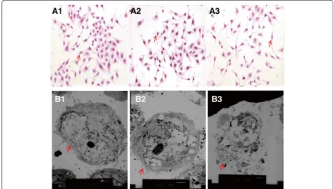
GES-1 control cells presented with a polygonal or fusiform shape, a regular appearance and clear edge (Figure 1A1). After 24 h co-culture withH.pyloriandH.pylori30(
H.pyl-ori pretreated with 30% NaCl), GES-1 cells transformed from multiangular to round or irregular shapes of various sizes, with disrupted cell walls and cytoplasmic leakage (Figure 1A2-A3). Transmission electron microscopy re-vealed that the GES-1 cells co-cultured with H. pylori30 and H. pylori for 24 h were characterized by the loss of microvilli, karyorrhexis and vacuolization of the cytoplasm (Figure 1B1-3).
NaCl pretreatment attenuatedH. pylori- induced oxidative DNA damage
Immunofluorescence showed oxidative DNA damage in GES-1 cells after co-culture with H. pylori30. Fluores-cent expression was strong in the nucleus, cytoplasm and cell membrane. The expression level of 8-OHdG was significantly increased in GES-1 cells infected with
H. pyloriandH. pylori30. In addition, we also found that the expression of 8-OHdG in GES-1 cells cultured with
H. pylori30
was significantly decreased when compared with GES-1 cells cultured withH. pylori (Figure 2A-D, P < 0.05).
Figure 1The effect ofH. pylori30on GES-1 cell morphology.Control GES-1 cells(A1, B1); GES-1 cells co-cultured withH.pylori30(A2, B2); GES-1 cells co-cultured withH. pylori(A3, B3)(HE×200; TEM×4000).
Changes in the mitochondrial membrane potential of GES-1 infected withH. pylori30
The results showed that GES-1 cells co-cultured withH.
pylori30 were not depolarized; however, the cell popula-tion shifted to left, indicating no apparent disruppopula-tion of the mitochondrial membrane potential (ΔΨm). No sig-nificant change was detected in mitochondrial mem-brane potential of GES-1 cells infected withH. pylori30 by flow cytometry, compared with that of the GES-1 control groups (Figure 3A-C, p > 0.05).
NaCl pretreatment attenuatedH. pylori-induced apoptosis
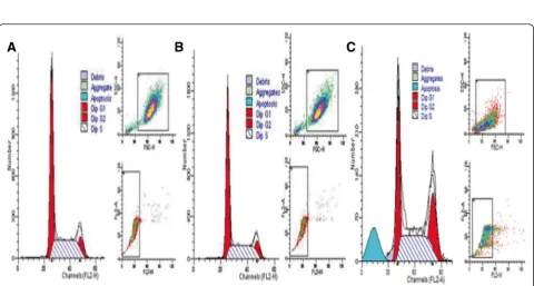
After co-culturing withH. pylori30, flow cytometry showed that apoptotic rate was not significantly increased in GES-1 cells infected withH. pylori30compared with GES-1 cells. However, the apoptotic rate was increased in GES-1 cells infected with H. pylori compared with GES-1 cells in-fected withH. pylori30(Figure 4A-C, p > 0.05).
To analyze further the effect of high salt pretreatment on H. pylori-induced apoptosis, we also detected estab-lished protein markers of apoptosis including Bax, Bcl-2
and Caspase3 proteins using Western blot. The results suggested that there were significant increased in Bax and Caspase3 proteins and decreased in Bcl-2 protein in GES-1 cells infected withH. pyloriand H. pylori30. How-ever, there were significant decreased in Bax and Caspase3 proteins and increased in Bcl-2 protein in GES-1 cells in-fected with H. pylori30when compared with GES-1 cells cultured withH. pylori. These results demonstrated that high salt pretreatment attenuatedH. pylori-induced apop-tosis (Figure 5A-D, P < 0.05).
NaCl pretreatment exacerbatedH. pylori-induced proliferation of gastric epithelial cells
Immunohistochemical staining showed that after co-culturing with H. pylori30, the expression of Ki-67 and PCNA was significantly increased in GES-1 cells (p < 0.05), compared with that in control cells. The expression of Ki-67 and PCNA was mostly located to the nuclei of experimental GES-1 cells compared with the control groups (Figure 6A-B). The expression levels of p21 was significantly decreased in GES-1 cells compared
Figure 3Changes in mitochondrial membrane potential (ΔΨm). A: GES-1 cells (negative control group);B: GES-1 cells co-cultured with
H. pylori30;C: GES-1 cells co-cultured withH. pylori.D: statistic data. *p> 0.05, compared to control;§p> 0.05, compared toH. pylori.
Figure 4Cell cycle and apoptosis analysis by flow cytometry.Cell numbers were calculated according to DNA content of G0/G1, S, and G2/M phases.A: negative control GES-1 cells;B: GES-1 cells co-cultured withH. pylori30;C: GES-1 cells co-cultured withH. pylori(PI staining).
to negative controls. The expression of p21 was mostly lo-cated to the cytoplasm in the GES-1 cells after co-culture withH. pylori30(Figure 6C).
Discussion
H. pylori was found to survive and adapt to high salt stress through alterations in biological characteristics of viability, morphology and expression of virulence factors. Besides, high salt (NaCl) diet is closely associated with high incidence of gastric illness [14,15]. The risk of gas-tric cancer in high salt diet population is significantly higher than that in low salt diet population [16]. The pre-vious experiments showed that high salt solution resulted in gastric mucosal hyperemia, erosion and ulceration in rats [17]. High salt diet is found to be closely linked with
H. pylori infection, which causes gastric mucosal injury and temporarily damages mucous barrier [18]. Too much salt intake increases H. pyloricolonization in gastric mu-cosa and promotes mumu-cosa malignant transformation [19]. The synergy ofH. pylori and 10% high salt diet in-duces gastric mucosa to express inflammatory cytokines iNOS and Cox-2 and aggravates mucosal damage in MGs [20]. High salt has been widely used to kill microbes in preservation of foods because of the difference in osmo-lality between the outside and inside of bacterial mem-brane. In the present study, we initially observed the
morphological changes of GES-1 cells after co-culturing with H. pylori that had been pretreated with high concentration NaCl. The GES-1 cells morphologically transformed from multiangular to round or irregular shapes, and occurred in various sizes, with disrupted cell walls, cytoplasmic vacuoles and cytoplasmic leak-age. These findings suggested that H. pylori30 retained the ability to damage GES-1 cells.
H. pyloricould stimulate gastric epithelial cells to gen-erate a large number of reactive oxygen species (ROS), the accumulation of which can cause oxidative damage to lipids, proteins and DNA [21]. 8-OHdG is a product of oxidative DNA damage and serves as an established marker of oxidative stress related to carcinogenesis [22]. In the current study, we found that the expression level of 8-OHdG was significantly increased in GES-1 cells infected with H. pylori and H. pylori30. In addition, we also found that the expression of 8-OHdG in GES-1 cells cultured withH. pylori30was significantly decreased when compared with GES-1 cells cultured with H.
pyl-ori. The results suggested that high salt pretreatment attenuatedH. pylori- induced oxidative DNA damage.
Cell proliferation and apoptosis are essential events in the cellular turnover of gastric tissue. Previously published reports have suggested that mucosal oxidative damage due to H. pylori infection was associated with increased
inflammatory cell infiltration, enhanced apoptosis and cell proliferation [23]. Although no significant disruption of the mitochondrial membrane potential (ΔΨm) and apop-totic rate were observed compared with control groups in this study, there were significant decresed in Bax and Cas-pase3 proteins and increase in Bcl-2 protein in GES-1 cells infected withH. pylori30when compared with GES-1 cells cultured with H. pylori. These results indicated that NaCl pretreatment attenuated H. pylori-induced apoptosis. Previous studies generally thought that bac-teria might not be able to survive in 30% of saturated sodium chloride solution, but Loh et al. [24] found whenH. pyloriwas cultured with various concentrations (0.25% through 2%) of salt in media, theH. pylori26695
cagA transcription and expression increased in a salt concentration-dependent manner. Rogers et al. [25] found
that after high salt pretreatment, G27 H. pylori strains gradually stopped reproduction, and ultimately died. The results suggested there wasH. pyloristrain heterogeneity for tolerance to high salt environment. Some H. pylori strains were more fragile to high salt environment. Our previous findings [26] showed that 30% of sodium chloride induced the biologically characteristic alter-ations ofH. pyloriwith deceased ATP level and reduced virulence gene transcription and expression, but with increased urease activity and cellular colonizing potential. Thus, we are considered that NaCl pretreatment attenu-atedH. pylori-induced apoptosis might be associated with the reduced virulence ofH. pylori.
Ki67 is a nuclear proliferation-associated antigen, the expression of which is strictly associated with cell prolif-eration. It is present during all active phases of the cell
Figure 6Expression of Ki-67, PCNA and P21 protein in GES-1 cells. A: Ki67 antigen was mostly located in the nuclei of GES-1 cells. The intensity of Ki67 was higher in theH. pylori30group (A2) than in the control group (A1) andH.pylorigroup (A3).B: PCNA antigen was mostly located in the nuclei of GES-1 cells; The intensity of PCNA was higher in theH. pylori30group (B2) than in the control group (B1) andH. pylori group (B3).(C)Expression of P21 was mostly located in the cytoplasm of GES-1 cells; the expression intensity of P21 was lower in theH. pylori30 group (C2) than in the control group (C1) andH. pylorigroup (C3) (Immunohistochemical staining × 200).
cycle (G1, S, G2, and mitosis), but is absent from resting cells (G0). Ki67 protein expression also has a positive correlation with the activity of tumor proliferation and prognosis; therefore, it is considered to be a reliable cel-lular marker to determine the growth fraction of a given cell population [27]. Our study found that the expression of Ki-67 in GES-1 cells was significantly increased after co-culturing with H. pylori30. Proliferating cell nuclear antigen (PCNA), a known cofactor of DNA polymeraseδ, is closely associated with cell proliferation [28]. PCNA forms a homotrimer that circles the DNA and operates as a scaffold to assemble a multitude of proteins re-quired for DNA unwinding and synthesis, cell cycle progression and chromatin structure maintenance [29]. The p21 protein exists in a quaternary complex with a cyclin, a cyclin-dependent protein kinase (CDK) and PCNA. The p21 protein has been found to mediate p53-induced growth arrest triggered by DNA damage by inhi-biting p53 activation to decrease cell proliferation [30]. The p21 protein blocks the initiation of DNA-replication by inhibiting CDK complexes [31] and blocks the action of PCNA. In the present study, we found that NaCl-pretreatedH. pylorican specifically affect the proliferation of GES-1 cells, while increasing PCNA expression and de-creasing P21 expression, which suggested thatH. pylori30 may enhance cell proliferation through the PCNA/P21 signal pathway.
The DNA oxidative damage and increased prolifera-tion of GES-1 cells observed in our study may be due to the release of CagA or other virulence factors from H.
pylori, which stimulated the corresponding receptors in GES-1 and led to the production of inflammatory mediators and the formation of oxygen free radicals and consequently promoted cell proliferation. Virulence fac-tor CagA and urease can induce cell proliferation. Yan et al. found that CagA+ H. pylori culture filtrates in-duced DNA damage in human gastric epithelial cells
in vitro, and accelerated human gastric epithelial cell proliferation and altered their morphology [32]. In our preliminary study, the level of CagA protein increased with NaCl concentrations. Abnormal regulation ofH.
pyl-oriand CagA expression in response to high salt concen-trations may be a potential factor in the development of gastric carcinoma although the molecular mechanisms re-main unclear.
Conclusion
In conclusion, GES-1 cells co-culturing with NaCl-pretreated H. pylori exhibited oxidative DNA damage accompanied with morphological alterations and cell proliferation. Our study provided in vitroevidence that
H. pylori can adapt to high salt environmental stress and delay pathogenecity, which may facilitate its trans-mission and infection in gastric carcinogensis.
Materials and methods
Culture and pretreatment ofH. pyloriwith high-salt
H. pylori (L301 strains), which is positive for CagA and VacA, was kindly provided by the Third Laboratory of Cancer Institute, China Medical University. The bacteria were grown on brain heart infusion agar supplemented with 7% sheep blood, 0.4% BBLTM IsoVitaleXTM Enrich-ment, 0.08% amphotericin B, 0.2% vancomycin and 0.5% trimethoprim (Sigma,USA), and incubated under micro-aerophilic conditions at 37°C and 95% humidity. Media routinely used for H.pylori culture contain physiological concentrations of NaCl. In our study, the media used for the experimental group were supplemented with 30% NaCl (H.pylori30) [26].
Co-culture of GES-1 withH. pylori
Co-culture of GES-1 withH. pyloriwas done as described previously [33]. Briefly, GES-1 was grown in RPMI 1640 media containing 10% fetal bovine serum, 100 U/ml peni-cillin, and 100μg/ml streptomycin at 37°C in 5% CO2.H.
pyloricells in the exponential phase were added to GES-1 cells (200:1 forH. pylorito GES-1 cells) for 24 h. The ex-perimental group was a co-culture of GES-1 andH. Pylori cells, whereas GES-1 cells co-cultured with or withoutH.
pylori was separately defined as a positive control group and negative control group, respectively.
Cell morphology
The morphology of GES-1 cells from the three groups co-cultured with or without H. pyloriwas observed by light microscopy. GES-1 cells were placed on chamber slides, fixed with acetone at 4°C and stained with hematoxylin and eosin (HE) for morphologic observation. The ultra-structure of GES-1 cells was observed by transmission electron microscopy (H-600, Olympus, Japan).
Oxidative DNA damage in GES-1 cells detected by immunofluorescence
Immunofluorescence was done as described previously [34]. Briefly, GES-1 cells were washed twice with PBS and fixed on slides with acetone at 4°C. The slides were incubated in bovine serum albumin for 30 min and with mouse anti-8-OHdG antibody (1:20, Santa Cruz, USA) overnight at 4°C, and then incubated with goat anti mouse IgG antibodies (Santa Cruz, USA) at 37°C for 1 h. The cells were observed by upright fluorescence microscopy. The total gray value average was obtained by measuring five randomly selected fields per slide with a micro ELISA reader (Bio-Tek, USA).
Alterations in mitochondrial membrane potential of GES-1 cells detected by flow cytometry
co-culture with H. pylori. Briefly, 1x10⁶cells were incu-bated with 2×10⁸H. pylori30 or H. pyloricells for 24 h. The cells were resuspended, washed in PBS twice, and incubated with 5μM rhodamine 123 at 37°C for 30 min. The cells were then analyzed using a FACStar flow cyt-ometer (BD, USA) and alterations in mitochondrial membrane potential were quantified using ModFit soft-ware [35].
Cell cycle and apoptosis rate detection by flow cytometry
The apoptosis rate was detected by flow cytometry. Briefly, after co-culturing withH. pyloriorH. pylorifor 24 h, 1 × 10⁶ GES-1 cells were ingested by trypsin and washed with PBS (pH 7.3-7.4) three times, then fixed with 70% ethanol. After incubation for 20 min, cells were stained with 50 mg/L propidium iodide.
Protein markers of apoptosis detection by Western blot
Western blotting was done as described previously [36]. Briefly, approximately 20μg (20μL) protein per gel well was loaded and resolved by 10% sodium dodecyl sulfate polyacrylamide gel electrophoresis (SDS-PAGE). The SDS-PAGE gel was transferred to polyvinylidene difluor-ide membranes. The membranes were incubated in tris-buffered saline (TBS) containing 3% non-fat dry milk and a specific proportion of the primary antibodies for Bax, Bcl-2 and Caspase3 proteins(Santa cruz, USA) overnight at 4°C. The blot was then washed and incubated with goat anti-mouse IgG conjugated to peroxidase (Santa cruz, USA). Antibody binding was detected by chemolumi-nescence staining using the ECL detection kit (Bio-Rad). The density of each band was quantified by densitom-etry of Bandscan 5.0 software.
Immunocytochemistry assay for protein expression of biomarkers related to cell proliferation
Expression of proliferation-related proteins was evaluated using the immunocytochemical staining [37]. In brief, 5x10⁵GES-1 cells were cultured on glass cover slips over-night, and co-cultured with 1×10⁸ H. pylori or H. pylori cells for 24 h. The cells were fixed with 4% paraformalde-hyde in PBS for 15 min followed by permeabilization with 0.5% Triton X-100 in PBS for 3 min. The cells were incu-bated with mouse anti-Ki 67, anti-PCNA or P21 antibody (Santa cruz, USA) at 4°C overnight. The cells were washed three times with PBS and incubated with biotinylated rabbit anti-mouse IgG secondary antibody for 30 min. A positive staining signal was developed by incubating the cells in 3,3-diaminobenzidine (DAB) solution for 10 min at RT in the dark. Cells from five randomly selected fields were immunostained on cover slips and micro-scopically reviewed.
Statistical analysis
Statistical analysis was performed using SPSS version 18.0 software. Results were expressed as the mean ± standard deviation and compared between groups using the Stu-dent t-test. A p value < 0.05 (two-sided) was considered statistically significant.
Competing interests
The authors declare that they have no competing interests.
Authors’contributions
YX carried out the Bacterial and cell culture, immunofluorescence, flow cytometry, Western blot, and drafted the manuscript. YY carried out the mmunohistochemica. MH was responsible for statistical analysis of data. YL provided funding and modified the articles, and submitted the manuscript. All authors read and approved the final manuscript.
Acknowledgments
This work was supported by the grants of Liaoning Province Key Scientific and Technological Project (No. 20141173), and PLA foundation of China during the Twelfth Five-year Plan Period (CSY12J002).
Author details
1Radiation oncology Department of General Hospital of Shenyang Military Command, Shenyang l10016, China.2Emergency Medicine Department of General Hospital of Shenyang Military Command, Laboratory of Rescue Center of Severe Wound and Trauma PLA, 83 Wenhua Road, Shenhe District, Shenyang l10016, China.
Received: 11 September 2014 Accepted: 6 February 2015
References
1. Kato S, Tsukamoto T, Mizoshita T. High salt diets dose-dependently promote gastric chemical carcinogenesis in Helicobacter pylori-infected Mongolian gerbils associated with a shift in mucin production from glandular to surface mucous cells. Int J Cancer. 2006;119:1558–6.
2. Azuma T. Helicobacter pylori CagA protein variation associated with gastric cancer in Asia. J Gastroenterol. 2004;39:97–103.
3. Ishii Y, Umemura T, Kanki K. Possible involvement of NO-mediated oxidative stress in induction of rat forestomach damage and cell proliferation by combined treatment with catechol and sodium nitrite. Arch Biochem Biophys. 2006;447:127–35.
4. Kuroiwa Y, Ishii Y, Umemura T. Combined treatment with green tea catechins and sodium nitrite selectively promotes rat forestomach carcinogenesis after initiation with N-methyl-N’-nitro-N-nitrosoguanidine. Cancer Sci. 2007;98:949–57.
5. Uehara T, Ma D, Yao Y. H.pylori infection is associated with DNA damage of Lgr5-positive epithelial stem cells in the stomach of patients with gastric cancer. Dig Dis Sci. 2013;58:140–9.
6. Mendivil PM, Velez P, Jimenez DM. TPEN induces apoptosis independently of zinc chelator activity in a model of acute lymphoblastic leukemia and ex vivo acute leukemia cells through oxidative stress and mitochondria caspase-3 - and AIF-dependent pathways. Oxid Med Cell Longev. Epub 2012 Dec 23. Volume 2012, Article ID 313275,14 pages.
7. Liu Y, Liu JH, Chai K. Inhibition of c-Met promoted apoptosis, autophagy and loss of the mitochondrial transmembrane potential in oridonin-induced A549 lung cancer cells. J Pharm Pharmacol. 2013;65:1622–42.
8. Ashktorab H, Dashwood RH, Dashwood MM, Zaidi SI, Hewitt SM. H.pylori-induced apoptosis in human gastric cancer cells mediated via the release of apoptosis-inducing factor from mitochondria. Helicobacter. 2008;13:506–17. 9. Iijima K, Koike T, Sekine H, Abe Y, Asanuma K, Uno K. Sustained epithelial
proliferation in a functionally irreversible fundic mucosa after Helicobacter pylori eradication. J Gastroenterol. 2009;44:47–55.
10. Yan F, Cao H, Chaturvedi R, Krishna U, Hobbs SS, Dempsev PJ. Epidermal growth factor receptor activation protects gastric epithelial cells from Helicobacter pylori-induced apoptosis. Gastroenteroloqy. 2009;136:1297–307. 11. Gudlaugsson E, Klos J, Skaland I. Prognostic comparison of the proliferation
markers (mitotic activity index, phosphohistone H3, Ki67), steroid receptors,
HER2, high molecular weight cytokeratins and classical prognostic factors in T1-2N0M0breast cancer. Pol J Pathol. 2013;64:1–8.
12. Paulo MS, Santos FT, Rocha PG. Immunoexpression of proliferating cell nuclear antigen (PCNA) in spleen of splenectomized rats with preservation of inferior pole, submitted to hyperbaric oxygenation. Acta Cir Bras. 2013;28:691–5.
13. Al-Khalaf HH, Aboussekhra A. p16(INK4A) positively regulates p21(WAF1) expression by suppressing AUF1-dependent mRNA decay. PLoS One. 2013;8:e70133.
14. Xu X, Liu Z, Fang M, Yu H, Liang X, Li X. Helicobacter pylori CagA induces ornithine decarboxylase upregulation via Src/MEK/ERK/c-Myc pathway: implication for progression of gastric diseases. Exp Biol Med. 2012;237(4):435–41.
15. Lopez-Carrillo L, Camargo MC, Schneider BG, Sicinschi LA, Hernandez-Ramirez RU, Correa P. Capsaicin consumption, Helicobacter pylori CagA status and IL1B-31C > T genotypes: a host and environment interaction in gastric cancer. Food Chem Toxicol. 2012;50(6):2118–22.
16. Murata A, Fujino Y, Pham TM, Kubo T, Mizoue T, Tokui N. Prospective cohort study evaluating the relationship between salted food intake and gastrointestinal tract cancer mortality in Japan. Asia Pac J Clin Nutr. 2010;19(4):564–71.
17. Takahashi M, Hasegawa R. Enhancing effects of dietary salt on both initiation and promotion stages of rat gastric carcinogenesis. Princess Takamatsu Symposia. 1985;16:169–82.
18. Ox JG, Dangler CA, Taylor NS, King A, Koh TJ, Wang TC. High-salt diet induces gastric epithelial hyperplasia and parietal cell loss, and enhances Helicobacter pylori colonization in C57BL/6 mice. Cancer Res. 1999;59(19):4823–8.
19. Nozaki K, Tsukamoto T, Tatematsu M. Effect of high salt diet and Helicobacter pylori infection on gastric carcinogenesis. Nihon Rinsho. 2003;61(1):36–40.
20. Toyoda T, Tsukamoto T, Hirano N, Mizoshita T, Kato S, Takasu S. Synergistic upregulation of inducible nitric oxide synthase and cyclooxygenase-2 in gastric mucosa of Mongolian gerbils by a high-salt diet and Helicobacter pylori infection. Histol Histopathol. 2008;23(5):593–9.
21. Yuan L, Dietrich AK, Nardulli AM. 17β-Estradiol alters oxidative stress response protein expression and oxidative damage in the uterus. Mol Cell Endocrinol. 2013;382:218–26.
22. Tanaka M, Takano H, Fujitani Y. Effects of exposure to nanoparticle-rich diesel exhaust on 8-OHdG synthesis in the mouse asthmatic lung. Exp Ther Med. 2013;6:703–6.
23. Noguchi K, Kato K, Moriya T. Analysis of cell damage in Helicobacter pylori-associated gastritis. Pathol Int. 2002;52:110–8.
24. Loh JT, Torres VJ, Cover TL. Regulation of Helicobacter pylori cagA expression in response to salt. Cancer Res. 2007;67(10):4709–15. 25. Rogers AB, Taylor NS, Whary MT, Stefanich ED, Wang TC, Fox JG.
Helicobacter pylori but not high salt induces gastric intraepithelial neoplasia in B6129 mice. Cancer Res. 2005;65(23):10709–15.
26. Xu Y, Jing JJ, Gong YH, Xu Q, Zhang WL, Piao Y. Changes in biological and virulent characteristics of Helicobacter pylori exposed to high salt. Asian Pac J Cancer Prev. 2011;12(10):2637–41.
27. Fukase K, Kato M, Kikuchi S. Effect of eradication of Helicobacter pylori on incidence of metachronous gastric carcinoma after endoscopic resection of early gastric cancer: an open-label, randomised controlled trial. Lancet. 2008;372:392–7.
28. Wang SC, Yu M, Li YH. Cyclosporin A promotes proliferating cell nuclear antigen expression and migration of human cytotrophoblast cells via the mitgen-activated protein kinase-3/1-mediated nuclear factor-κB signaling pathways. Int J Clin Exp Pathol. 2013;6:1999–2010.
29. Andersen PL, Xu F, Xiao W. Eukaryotic DNA damage tolerance and translesion symthesis through covalent modifications of PCNA. Cell Res. 2008;18:162–73.
30. Neise D, Sohn D, Stefanski A. The p90 ribosomal S6 kinase (RSK) inhibitor BI-D1870 prevents gamma irradiation-induced apoptosis and mediates senescence via RSK- and p53-independent accumulation of p21(WAF1/CIP1). Cell Death Dis. 2013;4:e859.
31. Chen JY, Lin JR, Tsai FC. Dosage of Dyrk1a shifts cells within a p21-cyclin D1 signaling Map to control the decision to enter the cell cycle. Mol Cell. 2013;52:87–100.
32. Yan YG, Zhao G, Ma JP, Cai SR, Zhan WH. Effects of different Helicobacter pylori culture filtrates on growth of gastric epithelial cells. World J Gastroenterol. 2008;14:3745–9.
33. Wang F, Luo LD, Pan JH, Huang LH, Lv HW, Guo Q, et al. Comparative genomic study of gastric epithelial cells co-cultured with Helicobacter pylori. World J Gastroenterol. 2012;8(48):7212–24.
34. Kang D, Gong Y, Zhu Y, Li A, Dong N, Piao Y, et al. The Biological Activity of
H. PyloriSlyD in Vitro. Helicobacter. 2013;18:347–55.
35. Zhang B, Bian W, Pal A, He Y. Macrophage apoptosis induced by aqueous C60 aggregates changing the mitochondrial membrane potential. Environ Toxicol Pharmacol. 2014;39(1):237–46.
36. Zhang H, Cai C, Fang W, Wang J, Zhang Y, Liu J. Oxidative damage and apoptosis induced by microcystin-LR in the liver of Rana nigromaculata in vivo. Aquat Toxicol. 2013;140:11–8.
37. Shirazi MS, Heidari B, Shirazi A, Zarnani AH, Jeddi-Tehrani M, Rahmati-Ahmadabadi M, et al. Morphologic and proliferative characteristics of goat type a spermatogonia in the presence of different sets of growth factors. J Assist Reprod Genet. 2014;31(11):1519–31.
Submit your next manuscript to BioMed Central and take full advantage of:
• Convenient online submission
• Thorough peer review
• No space constraints or color figure charges
• Immediate publication on acceptance
• Inclusion in PubMed, CAS, Scopus and Google Scholar
• Research which is freely available for redistribution


