INTRODUCTION
High mobility group box 1 (HMGB1) is a DNA-binding protein that has been well characterized as a prototypical dam-age-associated molecular pattern mole-cule (1–3). The initial characterization of HMGB1 as a late-acting mediator of lipopolysaccharide (LPS)- (4) or sepsis-induced (5) lethality in mice was further affirmed in patients with sepsis and sep-tic shock (6,7). Extracellular HMGB1
acti-vates inflammatory responses in immune cells, endothelial cells and enterocytes (4,8,9). While much attention has been focused on the function of extracellular HMGB1, the mechanisms of HMGB1 re-lease in sepsis have received little consid-eration (10).
Poly(ADP-ribose) polymerases (PARPs; also known as ADP-ribosyl transferases [ARTDs]) are a family of enzymes found in eukaryotes and prokaryotes that
gener-ate ADP-ribose modifications onto accep-tor proteins. PARP1 is the most abundant isoform of the PARP enzyme family and, upon activation by genotoxic stimuli, cleaves nicotinamide adenine dinu-cleotide (NAD+) into nicotinamide (NAM), resulting in the formation of ADP-ribose moieties; these moieties cova-lently attach to various acceptor proteins, including PARP itself. The continued acti-vation of PARP leads to depletion of its substrate, NAD+and, consequently, adenosine-5′-triphosphate (ATP), energy failure and cell death (11–20). The benefits conferred by pharmacological inhibitors of poly(ADP-ribosyl)ation in several ex-perimental disease models, including sep-sis, further emphasize the potential im-portance of PARP1 as a pharmacological target (21–44). In addition to posttransla-tional poly(ADP-ribosyl)ation of nuclear proteins, PARP1 also regulates
transcrip-Regulates LPS-Mediated High Mobility Group Box 1 Secretion
Thomas D Walko III,
1Valentina Di Caro,
1Jon Piganelli,
2Timothy R Billiar,
3Robert SB Clark,
4and
Rajesh K Aneja
41
Department of Critical Care Medicine, 2Department of Immunology, 3Department of Surgery, and 4Departments of Critical Care Medicine and Pediatrics, University of Pittsburgh School of Medicine and Children’s Hospital of Pittsburgh, Pittsburgh,
Pennsylvania, United States of America
Pathophysiological conditions that lead to the release of the prototypic damage-associated molecular pattern molecule high mobility group box 1 (HMGB1) also result in activation of poly(ADP-ribose) polymerase 1 (PARP1; now known as ADP-ribosyl trans-ferase 1 [ARTD1]). Persistent activation of PARP1 promotes energy failure and cell death. The role of poly(ADP-ribosyl)ation in HMGB1 release has been explored previously; however, PARP1 is a versatile enzyme and performs several other functions includ-ing cross-talk with another nicotinamide adenine dinucleotide- (NAD+) dependent member of the Class III histone deacetylases
(HDACs), sirtuin-1 (SIRT1). Previously, it has been shown that the hyperacetylation of HMGB1 is a seminal event prior to its secretion, a process that also is dependent on HDACs. Therefore, in this study, we seek to determine if PARP1 inhibition alters LPS-mediated HMGB1 hyperacetylation and subsequent secretion due to its effect on SIRT1. We demonstrate in an in vitro model that LPS treat-ment leads to hyperacetylated HMGB1with concomitant reduction in nuclear HDAC activity. Treattreat-ment with PARP1 inhibitors mit-igates the LPS-mediated reduction in nuclear HDAC activity and decreases HMGB1 acetylation. By utilizing an NAD+-based mech-anism, PARP1 inhibition increases the activity of SIRT1. Consequently, there is an increased nuclear retention and decreased extracellular secretion of HMGB1. We also demonstrate that PARP1 physically interacts with SIRT1. Further confirmation of this data was obtained in a murine model of sepsis, that is, administration of PJ-34, a specific PARP1 inhibitor, led to decreased serum HMGB1 concentrations in mice subjected to cecal ligation and puncture (CLP) as compared with untreated mice. In conclusion, our study provides new insights in understanding the molecular mechanisms of HMGB1 secretion in sepsis.
Online address: http://www.molmed.org doi: 10.2119/molmed.2014.00156
tion (45–53). Ditsworth et al., in an in vitro DNA-alkylating damage model, sug-gested that PARP1 activity may play a role in the nuclear-to- cytosolic transloca-tion of HMGB1 (54). Similarly, Davis et al., demonstrated that the release of HMGB1 to the extracellular milieu requires not only direct protein–protein interaction with PARP1, but also PARP1 enzymatic activity, which results in ribosyl)ation of HMGB1 (55). However, PARP1 is a versatile enzyme and per-forms several other functions including cross-talk with another NAD+-dependent member of the Class III histone deacety-lases (HDACs), sirtuin-1 (SIRT1) (56–58). In this study, we aim to define the role of PARP1 in the modulation of mediated HMGB1 expression, more spe-cifically, the effect of PARP1-SIRT1 cross-talk on HMGB1 acetylation. We show in an in vitromodel that LPS treatment leads to hyperacetylated HMGB1with concomi-tant reduction in nuclear HDAC activity. Treatment with PARP1 inhibitor mitigates the LPS- mediated reduction in nuclear HDAC activity and preserves the HDAC activity by increasing SIRT1 activity.
MATERIALS AND METHODS
Cell Culture
The human acute monocytic leukemia cell line THP-1 was purchased from American Type Culture Collection (ATCC #TIB-202; Manassas, VA, USA). For all experiments, cells first underwent a dif-ferentiation step by treatment with inter-feron (IFN)-γ(100 U/mL; Pierce Biotech-nology, Rockford, IL, USA) for 18 h. THP-1 cells were cultured in RPMI-1640 medium containing 10% fetal bovine serum (FBS), 1% penicillin/streptomycin, 0.35% β-mercaptoethanol (2-ME), and 2% glutamine, 10 mmol/L 4-(2-hydroxyethyl)-1-piperazine ethanesulfonic acid (HEPES) (pH 7.2).
Mouse fibroblasts from a strain geneti-cally deficient in PARP1 and fibroblasts from the corresponding wild-type (WT) controls were created by immortalization by a standard 3T3 protocol (59). Cell monolayers were grown at 37°C in 5%
CO2air in Dulbecco’s modified Eagle medium (DMEM) (Gibco [Thermo Fisher Scientific Inc., Waltham, MA, USA) con-taining 10% FBS, penicillin (100 U/mL), and streptomycin (100μg/mL).
Studies were performed in tissue cul-ture plates (Becton, Dickinson and Com-pany [BD], Franklin Lakes, NJ, USA) at a density of 1.0 × 10⁶cells. Where indi-cated, THP-1 cells were stimulated with 10 μg/mL LPS (Escherichia coli, serotype O55:B5, Sigma-Aldrich, St. Louis, MO, USA). Where indicated, cells were pre-treated with PARP1 inhibitor, 5 dihy-droxyisoquinoline (DIQ, 100 μmol/L; Sigma-Aldrich), EB-47 (50 and
100 nmol/L; Calbiochem, Billerica, MA, USA), SRT1720 (0.01 and 0.1 μmol/L; Calbiochem) or NAM (5 and 10 mmol/L; Sigma-Aldrich)
Bone Marrow–Derived Macrophage Cultures
Isolation of murine bone marrow– derived macrophages has been described previously (60). Briefly, bone marrow cells were obtained from 129S1/SvImJ, PARP1–/–(Jackson Laboratory, Bar Har-bor, ME, USA), mice and were cen-trifuged in lymphocyte separation me-dium (Mediatech Inc., Herndon, VA, USA). Cells harvested from the interface were cultured in 75-cm2flasks in Eagle MEM (EMEM; Mediatech Inc.) supple-mented with 10% FBS and 10 ng/mL granulocyte-macrophage colony-stimulat-ing factor (GM-CSF) (Thermo Scientific [Thermo Fisher Scientific]). After a 24-h adherence step (Day 2), which allowed for the removal of mature monocytes and fi-broblasts from the bone marrow, nonad-herent cells from each flask were trans-ferred to a second flask and were supplemented with medium containing GM-CSF (10 ng/mL). The cells were again supplemented with GM-CSF (10 ng/mL) on d 4. After a total of 7 d in culture, macrophages were removed en-zymatically with the neutral protease, dis-pase (Worthington Biochemical Corp., Lakewood, NJ, USA), and collected by gentle scraping. The cells were resus-pended in complete EMEM and exposed
to experimental conditions. Where indi-cated, stimulation was performed with 10 μg/mL LPS (Escherichia coli, serotype O55:B5, Sigma-Aldrich) for 18 h.
Nuclear Protein Extraction
All nuclear protein extraction proce-dures were performed on ice with ice-cold reagents. Cells were washed twice with phosphate-buffered saline (PBS) and harvested by scraping. Cells were pelleted in 1.5 mL of PBS at 268 ×gfor 5 min. The pellet was washed twice with PBS and resuspended in lysis buffer (10 mmol/L Tris-HCl, pH 7.8, 10 mmol/L KCl, 1 mmol/L ethylenegly-coltetraacetic acid [EGTA], 5 mmol/L MgCl2, 1 mmol/L dithiothreitol (DTT), and 0.5 mmol/L phenylmethylsulfonyl fluoride [PMSF]). The suspension was in-cubated on ice for 15 min and 10% Non-idet P-40 was added. This was followed by centrifugation at 4°C at 268 ×gfor 5 min. The supernatant was discarded and the cell pellet was dissolved in ex-traction buffer (20 mmol/L Tris-HCl, pH 7.8, 32 mmol/L KCl, 0.2 mmol/L EGTA, 5 mmol/L MgCl2, 1 mmol/L DTT, 0.5 mmol/L PMSF and 25% v/v glycerol) was added to the nuclear pellet and in-cubated on ice for 15 min. Nuclear pro-teins were isolated by centrifugation at 13,148 ×gat 4°C for 10 min. Protein con-centrations of the resultant supernatants were determined using the bicinchoninic acid assay (Pierce Biotechnology [Thermo Fisher Scientific]).
Real-Time Reverse Transcriptase-PCR Analysis
The mixture was used in a TaqMan qPCR reaction, according to the manufacturer’s instructions: 10 μL TaqMan Universal PCR Master Mix, 1 μL of each Gene Ex-pression Assay mix, 1 μL cDNA template and 7 μL of water (Applied Biosystems [Thermo Fisher Scientific]). HDAC qPCR primers used were as follows: HDACI-Hs02621185_s1; HDAC2- Hs00231032_m1; HDAC3- Hs00187320_m1 (Life Technolo-gies [Thermo Fisher Scientific]). Human ACTB was used as the endogenous con-trol. PCR reactions were performed in an Applied Biosystems thermocycler 7300 Real-Time PCR System (Thermo Fisher Scientific). After an initial denaturing step of 95°C for 2 min, the following steps were repeated for 40 cycles: a denaturing step of 95°C for 30 s, annealing step of 54°C for 30 s, elongation step of 68°C for 45 s. Each sample was assayed in duplicate and the Ct values were averaged. Lastly, the stan-dard ΔΔCt relative quantification method was used to calculate mRNA levels for all HDACs, normalized to ACTB.
SiRNA-Mediated Inhibition of PARP Expression
In this experiment, cells were incubated for 6 h and transfected with 20 nm siRNA duplexes using Lipofectamine2000 (Invitrogen [Thermo Fisher Scientific]) ac-cording to the manufacturer’s instructions. Commercially available stealth small inter-ference RNA (siRNA) sequence for PARP1 (accession number NM_001618) and con-trol sequences were obtained (Life Tech-nologies [Thermo Fisher Scientific]; cata-log numbers 4390824 and 4390846). For these experiments, we used differentiated THP-1 cells. Studies were performed in 12-well plates (BD) at a density of 5 × 10⁶cells. All the experiments were per-formed 18 h after transfection. The effi-ciency and specificity of siRNA gene knockdown of PARP1 was determined by real time PCR for PARP1 mRNA and Western blotting for PARP1 expression.
Western Blot Analysis
Concentrated supernatant, whole cell lysates or nuclear extracts were mixed with Laemmli buffer (20% glycerol, 10%
β-mercaptoethanol, 5% sodium dodecyl sulfate (SDS), 0.2 mol/L Tris-HCl, pH 6.8, and 0.4% bromphenol blue). After boiling for 10 min, the samples were subjected to 10% SDS-polyacrylamide gel electrophore-sis. Proteins were separated on 85% to 16% polyacrylamide gels and subsequently transferred to polyvinylidene difluoride (PVDF) membranes (GE Healthcare, Buckinghamshire, UK). For immunoblot-ting, membranes were blocked with 5% nonfat dried milk in PBS for 1 h. Primary polyclonal rabbit antibody to SIRT1 (1:1000, Santa Cruz Biotechnology, Dallas, TX, USA), primary polyclonal mouse HMGB1 antibody (1:1000; R&D Systems, Minneapolis, MN, USA), β-actin (1:5000, Sigma-Aldrich), polyclonal mouse PAR and PARP1antibody (1:1000, Trevigen, Gaithersburg, MD, USA) were used for Western blot. After washing twice with PBS containing 0.5% Tween 20 (PBST), secondary antibody (horseradish peroxidase-conjugated rabbit anti-mouse immunoglobulin G) (Sigma-Aldrich) was applied at 1:10,000 dilution for 1 h. Blots were washed in PBST thrice for 10 min, incubated in Enhanced Chemilumines-cence Reagent (GE Healthcare, Bucking-hamshire, UK), and used to expose X-ray film (GE Healthcare).
HDAC and SIRT1 Activity
Histone deacetylase, histone acetyl-transferase and SIRT1 activity were mea-sured with the Fluor-de-Lys-green fluori-metric HDAC assay kit, HAT and SIRT1 activity kit (Enzo Life Sciences, Farming-dale, NY, USA) according to the manu-facturer’s instructions.
Serum HMGB1 Concentrations We measured serum HMGB1 using a commercially available enzyme-linked im-munoassay (ELISA) according to the man-ufacturer’s instructions (ST51011, Shino-Test Corporation, Kanagawa, Japan). We used the normal range standard curve: the limit of detection was 1.0 ng/mL.
Proximity Ligation Assay
PLAs were carried out using a Duolink II detection kit (Olink Bioscience, Uppsala,
Sweden) as described previously. The cells are prepared as cytospin slides and two primary antibodies selected from two different host species, rabbit mono-clonal SIRT1 antibody (Santa Cruz Biotechnology) and PARP1mouse mono-clonal antibody (Enzo Life Sciences), are used together with the Duolink species-specific secondary antibodies. These sec-ondary antibodies contain unique DNA strands that template the hybridization of added oligonucleotides. When in close proximity (<40 nm), the oligonucleotides are ligated by a ligase to form a circular template. This template, still anchored to the antibody, is subsequently amplified and detected using complementary la-beled oligonucleotide probes. Detection was performed with a fluorescent mi-croscopy for bright-field detection. The resulting distinct spots are derived from single-molecule protein interaction events, which were visualized.
Immunofluorescence Confocal Microscopy
times in PBS. The cells were mounted using gelvatol [23 g of poly(vinyl alcohol) 2000, 50 mL glycerol and 0.1% sodium azide to 100 mL of PBS] and then viewed on a confocal scanning fluorescence mi-croscope (Fluoview 1000; Olympus, Malvern, NY, USA).
Immunoprecipitation
Whole cell lysates (300 μg) that had been harvested in RIPA buffer (10 mmol/L Tris-HCl, pH 8.0, 1.0 mmol/L ethylenedi-aminetetraacetic acid (EDTA), 0.5 mmol/L EGTA, 1% Triton X-100, 0.1% sodium de-oxycholate, 0.1% SDS, 140 mmol/L NaCl, with 1.0 mmol/L fresh PMSF), were pre-cleared for 1 h with Protein G DynaBeads (Thermo Fisher Scientific). Samples were then incubated with normal, non-specific mouse IgG (20 μL, Santa Cruz Biotechnol-ogy; sc-2343), mouse monoclonal anti-HMGB1 antibody (R&D Systems; catalog # MAB1690), rabbit monoclonal SIRT1 an-tibody (Santa Cruz Biotechnology), or rabbit polyclonal anti-acetylated-lysine antibody (Cell Signaling Technology, Dan-vers, MA, USA; catalog # 9441S), overnight at 4°C. The next day, the samples were ap-plied to a Protein G DynaBead reaction, and incubated at 4°C to bind the primary antibody. The effluent was reserved, and the Protein G beads were washed three times with sample wash buffer. Samples were then eluted in denatured conditions in 100 μL of 2× SDS sample buffers and analyzed by Western blot as previously described in this manuscript, using a 10% polyacrylamide gel.
Cecal Ligation and Puncture (CLP) Model
Twenty WT mice, 4–6 wks old were sub-jected to CLP. Mice were anesthetized with a mixture of buprenorphine (0.1 mg/kg) and pentobarbital (90 mg/kg) by subcu-taneous and intraperitoneal (IP) injec-tions. Mice were treated with N-(6-oxo-5, 6-dihydro-phenanthridin-2-yl)-N, N-dimethyacetamide HCl (PJ34) (Sigma-Aldrich) 10 mg/kg 3h prior to CLP. Under aseptic conditions, cecal ligation was performed via a short (1 cm) midline incision and cecum is delivered into the
wound and ligated 1.0 cm from the tip using a single 4-0 silk suture. The cecum is punctured through and through (that is, front wall and back wall) with a single pass of a 20-gauge hypodermic needle. The cecum was then gently squeezed to extrude a small amount of feces from the perforation sites. The cecum was re-turned to the peritoneal cavity and the la-parotomy was closed with 4.0 silk su-tures. The animals were returned to their cages with free access to food and water. Mice were euthanized at 24 h after CLP and blood samples were collected for measuring HMGB1 concentrations.
RESULTS
PARP1 Inhibition (Chemical or Genetic) Decreases LPS-Mediated HMGB1 Secretion
We first investigated the effect of PARP1 inhibition on LPS-mediated HMGB1 secretion and determined dose response inhibition of PARP1 in THP-1 cells. We treated THP-1 cells with a com-petitive inhibitor of PARP1, 10 to 300μmol/L (1,5-dihydroxyisoquinoline [DIQ]) and achieved ~80% inhibition of PARP1 activity with 300 μmol/L of DIQ (data not shown). Next, we examined the effect of PARP1 inhibition on HMGB1 production; THP-1 cells were treated with varying concentrations of DIQ for 45 min. Subsequently, they were exposed to 10 μg/mL LPS for 18 h and supernatant HMGB1 levels were as-sessed by immunoblotting. As expected, cells incubated with LPS demonstrated increased HMGB1 secretion as com-pared with naïve cells (Figure 1A). How-ever, when the cells were pretreated with DIQ, LPS-induced HMGB1 secre-tion was significantly attenuated (see Figure 1A). To confirm that PARP1 is responsible for the decrease in HMGB1 supernatant levels, we repeated the ex-periment with a new potent PARP1 in-hibitor, EB-47. Similar to DIQ, pretreat-ment with EB-47 (50 and 100 nmol/L) led to a significant decrease in mediated HMGB1 secretion (Figure 1B). Together, these results indicate that
chemical inhibition of PARP1 inhibits LPS-induced HMGB1 secretion.
To validate the specificity of PARP1 protein in LPS-mediated HMGB1 secre-tion, we examined this process in bone marrow–derived macrophages obtained from PARP1–/–and WT mice. Bone marrow–derived macrophages were har-vested from WT and PARP1–/–mice and were pretreated with LPS for 18 h and su-pernatant HMGB1 levels were assessed by immunoblotting. Following LPS stimu-lation, WT macrophages demonstrated a significant increase in supernatant HMGB1 levels as compared with un-treated macrophages. In contrast, PARP1–/–macrophages exposed to LPS demonstrated no detectable HMGB1 lev-els in the supernatant (Figure 1C). Taken together, the results of our experiments suggest that either chemical inhibition or genetic deletion of PARP1 results in a de-crease in LPS-mediated HMGB1 secretion.
To further verify that the decrease in LPS-mediated HMGB1 secretion is not a compensatory response to the missing PARP1 gene, we employed posttranscrip-tional gene silencing technology by RNA interference. Cells were transfected with siRNA for 12 h, exposed to LPS, and su-pernatant was collected at 18 h for mea-suring HMGB1 concentrations. After LPS exposure, naïve cells demonstrated a sig-nificant increase in supernatant HMGB1 concentrations. The supernatant HMGB1 concentration in cells transfected with nontarget siRNA was comparable to naïve cells after LPS exposure (Figure 1D). PARP1 siRNA-transfected cells that were exposed to LPS demonstrated a signifi-cant decrease in supernatant HMGB1 concentrations as compared with naïve or nontarget siRNA transfected cells (see Figure 1D). Taken together, these data support the view that both genetic and chemical inhibition of PARP1 gene in-hibits LPS-induced HMGB1 secretion.
Serum HMGB1 Levels Are Decreased with PARP1 Inhibition in a Murine Model of Sepsis
in vivomodel of sepsis. CLP is a reliable and clinically relevant model of sepsis that is widely used. The median serum HMGB1 concentrations were signifi-cantly higher in the CLP mice as com-pared with the sham group (160.0 ± 26 ng/mL versus 11.8 ± 3.9 ng/mL; P≤ 0.05) respectively (Figure 1E). Mice treated with PJ34 demonstrated a signifi-cant decrease in serum HMGB1 levels as compared with untreated mice (62.4 ± 22.7 versus 160.0 ± 26 ng/mL; P≤ 0.05) respectively. Together, these data support the view that inhibition of PARP1 de-creases HMGB1 both in vitroand in vivo models of sepsis.
PARP1 Inhibition Has No Significant Effect on LPS-Mediated HMGB1 Transcription
Considering the significant decrease in HMGB1 secretion after PARP1 inhibi-tion, we next sought to define the mech-anisms involved in greater detail. Using
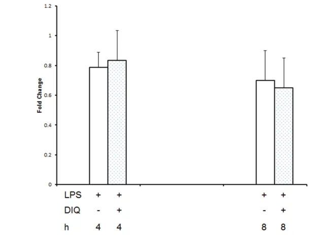
real time RT-PCR we examined the effect of PARP1 inhibition on LPS-mediated HMGB1 mRNA expression at 3 and 6 h after exposing THP-1 cells to LPS.
HMGB1 mRNA levels in THP-1 demon-strated no significant change at 3 and 6 h after stimulation with LPS as com-pared with naïve cells (Figure 2).
DIQ-Figure 1. (A,B) Representative autoradiograph of Western blot analysis for supernatant HMGB1 levels in THP-1 cells treated with LPS in the presence or absence of DIQ (300μmol/L), a PARP1 inhibitor (Figure 1A). Experiment was repeated by treating THP-1 cells with EB-47, a new potent PARP1 inhibitor and supernatant HMGB1 concentrations were analyzed (Figure 1B). THP-1 cells were treated with LPS (10 μg/mL) for 18 h. The gel is representative of three experiments with similar results. (C) Representative autoradiograph of Western blot analysis for super-natant HMGB1 levels in bone marrow–derived macrophages from WT and PARP1–/–treated with LPS. The gel is representative of three
ex-periments with similar results. (D) Representative Western blot analysis for supernatant HMGB1 concentrations in naïve, nontarget siRNA and PARP1 siRNA transfected THP-1 cells treated with LPS. Cells were treated with LPS (10 μg/mL) for 18 h. The gel is representative of three experiments with similar results. (E) PJ-34 (10 mg/kg, IP, n = 10), or saline (IP, n = 10) was administered 3 h prior to CLP. CLP was performed and blood was collected 24 h later. Serum HMGB1 concentrations were measured by ELISA.
Figure 2.RT-PCR of HMGB1 expression in THP-1 cells treated with LPS in the presence or ab-sence of DIQ (300 μmol/L), a PARP1 inhibitor. THP-1 cells were treated with LPS (10 μg/mL) for 3 or 6 h. Error bars indicate mean ± SEM
A
C
E
B
treated cells demonstrated a slight de-crease in HMGB1 mRNA as compared with LPS-stimulated cells, but this was not significant either. These data suggest that the pharmacological inhibition of PARP1 activity has no appreciable effect on HMGB1 gene expression as evi-denced by steady-state HMGB1 mRNA levels.
Effect of PARP1 Inhibition on the Compartmentalization of HMGB1 after LPS Stimulation in THP-1 Cells
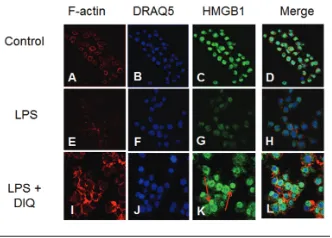
After LPS exposure, activation of monocytes leads to relocalization of HMGB1 from the nucleus into cytoplas-mic organelles that belong to the en-dolysosomal compartment (61), this is a seminal event prior to HMGB1 release into the extracellular compartment. We investigated the intracellular localization of HMGB1 in LPS-treated THP-1 cells with immunofluorescence. In the basal state, HMGB1 staining was present both in the nuclear and cytoplasmic compart-ments (Figure 3C), however, following LPS stimulation, the cells demonstrated morphological changes with decreased nuclear and cytoplasmic staining for
HMGB1 as compared with naïve cells. In contrast, cells that were pretreated with a PARP1 inhibitor demonstrated a signifi-cant increase in nuclear staining for HMGB1 (Figure 3K). Taken together, these findings are consistent with the view that DIQ pretreatment leads to ac-cumulation of HMGB1 in the nucleus and prevents the relocation of HMGB1 from nucleus to the extracellular com-partment with LPS treatment.
PARP1 Inhibition Decreases LPS-Mediated HMGB1 Acetylation
The secretion of HMGB1 occurs via a nonclassic secretory pathway, and hyper-acetylation of several lysine residues dis-tributed throughout the molecule is a vital step prior to its release. As a next step, we sought to determine whether DIQ pretreatment alters the hyperacetyla-tion status of HMGB1, thereby leading to a decrease in LPS-mediated HMGB1 se-cretion. In this experiment, we immuno-precipitated nuclear lysates with antibod-ies against HMGB1, and these
immunoprecipitated proteins were im-munoblotted with acetyl lysine antibody. As shown in Figure 4A, acetylated HMGB1 was immunoprecipitated effi-ciently with HMGB1 antibody, and no
signal was observed when IgG was used as a control for immunoprecipitation (Fig-ure 4, panel 1, lane 1 versus lane 5). In ad-dition, DIQ pretreatment alone had no ap-preciable effect (Figure 4, panel 1, lane 3 versus lane 2). Cells incubated with LPS demonstrated an increase in acetylated HMGB1 as compared with unstimulated cells. Coincubation with DIQ demon-strated a significant inhibition of LPS-me-diated acetylation of HMGB1 (Figure 4, panel 1, lane 4 versus lane 2). The blot was then stripped and reprobed for HMGB1to demonstrate equal HMGB1 loading in lanes 1–4 (Figure 4, panel 2).
To confirm the results obtained by HMGB1 immunoprecipitation, we con-ducted the reverse experiment by im-munoprecipitating whole cell lysates with an acetyl lysine antibody and sub-sequent analysis of the immunoprecipi-tate for HMGB1 by immunoblotting. Whole cell lysates of cells incubated with LPS demonstrated a reduction in acety-lated HMGB1 as compared with controls, as some acetylated HMGB1 may have been secreted at 8 h (Figure 4, panel 3, lane 2). Cells pretreated with a PARP1 hibitor alone demonstrated a slight in-crease in acetylated HMGB1 content as compared with LPS treated cells (Figure 4,
Figure 3.Representative confocal images of THP-1 cells demonstrate the nuclear and cytoplasmic distribution of HMGB1 after LPS exposure in the absence (E–H) or presence of DIQ (300 μmol/L) (I–L). The cells were exposed to 10 μg/mL LPS in the absence or presence of 300 μmol/L DIQ. In these panels, the green staining indicates the presence of HMGB1 and the red stain-ing indicates the presence of F-actin. The nuclear stain DRAQ5 is blue. Red arrows point to an increase in nuclear staining for HMGB1 with PARP1 inhibition.
panel 3, lane 3 versus lane 2). Analogous to our finding above, whole cell lysates pretreated with a PARP1 inhibitor prior to LPS exposure demonstrated a de-crease in acetylated HMGB1 as com-pared with untreated cells. The blot was then stripped and reprobed for acetyl ly-sine to demonstrate equal acetyl lyly-sine loading in lanes 1–4 (Figure 4, panel 4). Taken together, these results suggest that the decrease in LPS-mediated HMGB1 secretion with PARP1 inhibition is related to a decrease in HMGB1 acety-lation leading to nuclear retention of HMGB1 and decreased secretion.
DIQ Pretreatment Abrogates LPS-Mediated Reduction in HDAC Activity
On the basis of the results in Figure 4, we next sought to determine the en-zymes that play a role in acetylation of HMGB1. We examined the effect of PARP1 inhibition on the HAT activity of CBP/p300, transcriptional coactivators that can acetylate intranuclear histones and were unable to discern any effect (data not shown). Next, we investigated histone deacetylases, a family of en-zymes that can efficiently deacetylate nuclear proteins by removing acetyl groups from lysine residues.
Accord-ingly, for these experiments, we exam-ined the nuclear HDAC activity in THP-1 cells pretreated with DIQ in the presence or absence of LPS by using a colorimet-ric assay. As shown in Figure 5, LPS-treated THP-1 cells demonstrated a 20%
and 30% reduction in nuclear HDAC ac-tivity as compared with naïve cells at 4 and 8 h respectively, and this reduction in HDAC activity persisted at 16 h after exposure to LPS. DIQ pretreatment abro-gates LPS-mediated reduction in HDAC activity and, furthermore, maintains it at 100% to 120% as compared with naïve cells. Taken together, our results suggest that LPS-mediated suppression in HDAC activity leads to hyperacetylation and subsequent HMGB1 release; in con-trast, DIQ pretreatment preserves the HDAC activity leading to decreased su-pernatant HMGB1 concentrations.
Evankovich et al. had demonstrated previously that HDACI was critical in regulating HMGB1 release in mouse he-patocytes (62). Gene expression for HDACI was examined by performing RT-PCR on mRNA isolated from THP-1 cells at 4 and 8 h after LPS exposure. There was no change in HDAC transcripts for HDACI, II, and III in LPS exposed THP-1 cells with or without pretreatment with DIQ (Figure 6 demonstrates results for HDACI only). Therefore, these data sug-gest that the PARP1 regulates
LPS-Figure 5.PARP1 inhibition abrogates LPS-mediated reduction in HDAC activity. Nuclear proteins were extracted from THP-1 cells treated with LPS in the presence or absence of DIQ (300 μmol/L), a PARP1 inhibitor. THP-1 cells were treated with LPS (10 μg/mL) for 4, 8 or 16 h. HDAC activity was determined by colorimetric assay. (*Represents P≤ 0.05 versus LPS treated cells at the same time point.) Assay shown is representative of three experiments with similar results.
mediated HMGB1 hyperacetylation by al-tering the nuclear acetylation/deacetyla-tion balance, leading to HMGB1 retenacetylation/deacetyla-tion in the nucleus.
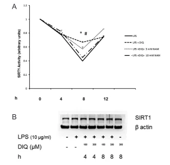
Although basal activation of PARP1 is needed for maintenance of cell function, overactivation of PARP1 consumes NAD+ and results in cell death due to depletion of intracellular NAD+stores. One class of HDACs that are affected by changes in intracellular levels of NAD+is the class III HDACs, also called as SIRT1. We next examined the effect of PARP1 inhibition on the nuclear SIRT1 activity in THP-1 cells pretreated with DIQ in the presence or absence of LPS by using a colorimetric assay. As shown in Figure 7A, nuclear SIRT1 activity was reduced to <40% 8 h after LPS treatment as compared with naïve cells. This reduction in SIRT1 activ-ity persisted at 12 h after exposure to LPS. DIQ pretreatment significantly abro-gates LPS-mediated reduction in SIRT1 activity and furthermore, maintains it at 66% as compared with untreated cells at 8 h (P≤ 0.05). At 12 h, the SIRT1 activity is comparable in DIQ untreated and treated cells that were exposed to LPS (P≥ 0.05)respectively. SIRT1 protein lev-els were unchanged in LPS-exposed cells treated with DIQ as compared with naïve and untreated cells (Figure 7B), data as shown for 4 and 8 h only. Consistent with this possibility, PARP1–/–cells demon-strated significantly higher SIRT1 activity as compared with WT cells (2.05 ± 0.1 versus 1 ± 0.09 arbitrary units (P≤ 0.05)) (Figure 7C).
To better understand the effect of PARP1 inhibition on SIRT1 activity and its dependence on NAD+concentration, we repeated the experiment in the presence of NAM. NAM is an end product of ADP-ribosylation reactions, a noncompetitive inhibitor of SIRT1, which blocks NAD+ hydrolysis by binding to a conserved pocket adjacent to NAD+binding site on SIRT1. Importantly, while DIQ pretreated cells exposed to LPS demonstrated an in-crease in SIRT1 activity, cells pretreated with DIQ and NAM (10 mmol/L) failed to exhibit an increase in SIRT1 activity (66% versus 44%; P> 0.05) (Figure 7A).
These findings underscore the importance of NAD+in mediating the increase in SIRT1 activity with PARP1 inhibition as the addition of NAM leads to an inhibi-tion of NAD+consumption, thereby blocking the increase in SIRT1 activity.
To further validate these findings, we examined the effect of SIRT1 activation on HMGB1 production. THP-1 cells treated with the SIRT1 activator, SRT1720 for 45 min and were subsequently ex-posed to 10 μg/mL LPS for 18 h. Super-natant HMGB1 levels were assessed by immunoblotting. As expected, cells incu-bated with LPS demonstrated increased HMGB1 secretion as compared with
naïve cells (Figure 7D). However, when the cells were pretreated with SRT1720, LPS-induced HMGB1 secretion was sig-nificantly attenuated (Figure 7D). Taken together, these findings indicate that PARP1 inhibition mitigates LPS-medi-ated HMGB1 acetylation and subsequent secretion by increasing the deacetylase activity of SIRT1, and along with the re-sults above, provide insight that this mechanism is NAD+dependent.
PARP1 and SIRT1 Interaction
To examine whether we could detect endogenous complexes between PARP1 and SIRT1 proteins in THP-1 cells in situ,
Figure 7.(A) Nuclear proteins were extracted from THP-1 cells treated with LPS in the pres-ence or abspres-ence of DIQ (300 μmol/L) and NAM (5 and 10 mmol/L). THP-1 cells were treated with LPS (10 μg/mL) for 4, 8, 12 h. SIRT1 activity was determined by colorimetric assay. (*Rep-resents P≤ 0.05 LPS exposed cells in the absence or presence of DIQ; #represents P
≤ 0.05 LPS exposed cells treated with DIQ or DIQ and NAM.) Assay shown is representative of three experiments with similar results. (B) Representative autoradiograph of Western blot analysis for nuclear SIRT1 concentration in THP-1 cells treated with LPS in the presence or absence of DIQ (100 and 300 μmol/L). THP-1 cells were treated with LPS (10 μg/mL) for 4 and 8 h. The gel is representative of three experiments with similar results.
Continued on next page
A
we performed proximity ligation assays (PLA) (63), whereby each red signal is in-dicative of one detected interaction event. As compared with naïve cells, a slight increase in the red signal, indicat-ing an increase in nuclear complexes of PARP1 and SIRT1 was observed with LPS exposure. Pretreatment with DIQ leads to a significant increase in the fluo-rescent signal, indicating a stronger inter-action between PARP1 and SIRT1. Speci-ficity of the assay was confirmed by testing this interaction in the absence of specific antibodies (Figure 8A).
To test whether SIRT1 is ribosylated, we immunoprecipitated nuclear lysates with antibodies against HMGB1, and these immunoprecipitated proteins were immunoblotted with anti-poly(ADP-ribose) antibody. Cells incu-bated with LPS demonstrated a slight in-crease in ribosylated SIRT1 as compared with naive cells (Figure 8B, panel 1, lane 2
versus lane 1). Cells treated with PARP1 inhibitor alone demonstrated a slight de-crease in the ribosylation of SIRT as com-pared with LPS treated cells (Figure 8B, panel 1, lane 3 versus lane 2). However, ribosylation of SIRT1 in nuclear extracts of cells that were pretreated with DIQ and subsequently exposed to LPS was similar to untreated cells (Figure 8B, panel 1, lane 4 versus lane 2). Although we did not find any modulation in SIRT1 ribosylation with DIQ pretreatment, the following two mechanisms underlie PARP1 inhibition of LPS- mediated HMGB1 secretion: (1) using an NAD+dependent mechanism, PARP1 inhibition increases the deacetylase activ-ity of SIRT1; (2) PARP1 interacts with SIRT1 as demonstrated by PLA.
DISCUSSION
Wang et al. first demonstrated the cytokine-like properties of HMGB1 and established HMGB1 as a prototype for
endogenous danger signals or the so-called “alarmins” (1–3). Alarmins are cel-lular proteins rapidly released from cells in response to infection or tissue damage that serve to activate the immune system (64). Active secretion of HMGB1 from monocytes/macrophages begins 8 to 12 h after exposure to the inflammatory stimulus and represents a delayed onset of release as compared with the early proinflammatory mediators. In a murine model of sepsis, circulating HMGB1 lev-els increase 18 h after induction of peri-tonitis and remain elevated for 3 d (65). The delayed kinetics of HMGB1 release parallels the onset of animal lethality in animal models of sepsis. Furthermore, treatment with neutralizing anti-HMGB1 antibodies can rescue mice from LPS- or sepsis-induced lethality (4), thereby, so-lidifying its role as a potential therapeu-tic target. Collectively, these in vivoand in vitrostudies support a paradigm in which HMGB1 plays a pathogenic role in sepsis, and is a late mediator of systemic inflammation. Therefore, fully under-standing the molecular mechanisms in-volved in HMGB1 release will expand our therapeutic armamentarium in sepsis.
In this study, we examine the role of acetylation in HMGB1 release after THP-1cells were treated with LPS. HMGB1 protein lacks a leader signal sequence; therefore, it cannot be secreted via the classical endoplasmic reticulum-Golgi se-cretory pathway. In resting cells, there is a continuous HMGB1 shuttle between the nucleus and cytoplasm. HMGB1 pro-tein undergoes several posttranslational modifications, for example, acetylation and phosphorylation prior to its nuclear export (66,67). Hassa et al. have previ-ously demonstrated that PARP1-depen-dent gene expression not only requires the enzymatic activity of p300/CBP but also that PARP1 itself is acetylated in vivo (68). Therefore, we hypothesized that the acetylation of HMGB1 may require the intrinsic HAT activity of CBP/p300 pro-teins, but we were unable to detect any evidence of alteration of HAT activity in LPS-treated. The coactivator activity of
Figure 7. Continued.(C) Nuclear proteins were extracted from WT and PARP1–/–cells and
SIRT1 activity was determined by colorimetric assay. (*Represents P≤ 0.05 versus WT cells.) (D) Representative autoradiograph of Western blot analysis for supernatant HMGB1 levels in THP-1 cells treated with LPS in the presence or absence of SRT1720 (0.01 and 0.1 μmol/L), a SIRT1 activator. THP-1 cells were treated with LPS (10 μg/mL) for 18 h. The gel is representa-tive of three experiments with similar results.
C
CBP/p300 for gene expression may be dependent on the stimuli and cell type, therefore, we were unable to detect any change in HAT activity.
HMGB1 has two main clusters of lysines that are crucial for acetylation and nuclear localization (69). Bonaldi et al. have previously demonstrated that when the balance between HATs and HDACs is tilted in favor of acetylation, nuclear im-port of HMGB1 is reduced and it accu-mulates in vesicles or secretory lyso-somes (69). Gardella et al. first noted that HMGB1 accumulates in the cytoplasm of activated monocytes, and is subsequently sequestered into lysosomes prior to its re-lease into the extracellular space (61). We provide evidence that LPS-treated cells demonstrate a significant reduction in nuclear HDAC activity as compared with naïve cells, and this reduction persists even 16 h after exposure to LPS, leading to an increase in HMGB1 secretion. DIQ pretreatment significantly abrogates LPS-mediated reduction in HDAC activity,
thereby tilting the balance to deacetyla-tion, consequently leading to reduced LPS-mediated HMGB1 secretion.
In addition to its influence on vital cel-lular processes, recent studies indicate PARP1 plays a role in gene-specific tran-scription (49,53,70,71). PARP1 regulates transcription by modifying associated proteins, and also functions as a cofactor for transcription factors, most notably, nuclear factor-κB (NF-κB) and activating protein (AP)-1 (46,72). PARP1 is frequently associated with transcrip-tionally active regions of chromatin sug-gesting a possible role of ribosyl)ation in the regulation of transcription (73,74). To this end, we found that pharmacological inhibition of PARP1 activity has no appreciable effect on HMGB1 gene expression as evidenced by steady-state HMGB1 mRNA levels.
It has been demonstrated previously that LPS stimulation induces PARP1 en-zymatic activity and the ADP-ribosyla-tion of histones in macrophages (75).
Fur-thermore, the targets of ADP-ribosylation in LPS-stimulated macrophages are PARP1, core histones (76), nucleosome-occupied promoters of IL-1β, MIP-2 and CSF2, facilitating NF-κB recruitment and transcription (76). Similarly, Davis et al., (55) have shown that HMGB1 release to the extracellular milieu requires not only directs protein–protein interaction with PARP1, but also PARP1 enzymatic activ-ity, which results in HMGB1 poly(ADP-ribosyl)ation. Therefore, LPS-mediated ADP-ribosylation of PARP1, HMGB1 and other inflammatory mediators is well published. However, we focus on another function of PARP1, its ability to cross-talk with another NAD+-dependent member of the Class III HDACs, SIRT1 (55–58). The effects of PARP1 inhibition on LPS-mediated HMGB1 secretion were associ-ated with the modulation of HDAC activ-ity, specifically SIRT1. SIRT1 has been implicated in transcriptional silencing, genetic control of aging, cell metabolism and calorie restriction-mediated longevity of the organism.
Both PARP1 and SIRT1 use NAD+for their activity and are capable of perform-ing several common functions, therefore, cross-talk between these proteins has been suggested (77). PARP and SIRT pro-teins operate as the “yin and yang” of the cellular response to stress that deter-mines cell survival or death (57,78). Using SIRT1 deficient mouse cardiomy-ocytes, Rajamohan et al. noted increased levels of PARP1 acetylation in response to mechanical stress (58). However, no similar posttranslational modification has been seen on SIRT1 by PARP1 in re-sponse to DNA damage (79). Our results are in agreement with this observation as we were unable to find any changes in ADP-ribosylation of SIRT1 after PARP1 inhibition in LPS-exposed THP-1 cells.
We believe that PARP1 modulates SIRT1 activity by two mechanisms: (a) Cellular NAD+levels: Since NAD+serves as a substrate for poly(ADP-ribosyl)ation and NAD+dependent deacetylases, cellu-lar NAD+concentrations form a frame-work for the basis of cross-talk between these proteins. The Km of PARP1 and
Figure 8.(A) Representative confocal images of proximity ligation assay (PLA). Nuclear pro-teins were extracted from THP-1 cells treated with LPS in the presence or absence of DIQ (300 μmol/L), a PARP1 inhibitor. THP-1 cells were treated with LPS (10 μg/mL) for 6 h. PLA ampli-fication corresponds with the interaction of PARP1 with SIRT1 and is visualized as red-pink spots localized mainly in the nucleus. (B) Coimmunoprecipitation analysis from THP-1 cell lysates. Panel 1: Samples were immunoprecipitated with SIRT1 and immunoblotted with anti-poly(ADP-ribose). The blot was then stripped and reprobed for SIRT1 (panel 2).
A
SIRT1 toward NAD+is 20–60 (80,81) and 100–200 μmol/L (82) respectively, indicat-ing that PARP1 has a 5- to 10-fold higher affinity for NAD+. Consequently, overac-tivation of PARP1 with a precipitous drop in cellular NAD+levels leads to re-duced SIRT1 activity (82). Therefore, while PARP1 will continue to consume NAD+, SIRT1 activity will decrease. Con-versely, PARP1 chemical or genetic inhi-bition will increase cellular SIRT1 activity. In our study, we demonstrate that DIQ pretreatment significantly abrogates LPS-mediated reduction in SIRT1 activity. However, cells pretreated with DIQ and NAM failed to show a significant in-crease in SIRT1 activity. These findings indicate that PARP1 inhibition mitigates LPS-mediated HMGB1 acetylation by in-creasing the deacetylase activity of SIRT1, and along with the results above, provide insight that this mechanism is NAD+ -de-pendent. (b) PARP1 and SIRT1 interac-tion: PARP1 and SIRT1 interact with each other in the presence of LPS stimulation; furthermore, this interaction is efficiently enhanced upon inhibition of PARP1.
Finally, there are remarkably few thera-peutic inhibitors of HMGB1 release dis-covered so far. One such inhibitor of HMGB1 release is ethyl pyruvate (EP). Mice subjected to cecal perforation can be rescued from lethal sepsis by treatment with anti-HMGB1 antibodies, HMG A box or EP, in a dose-dependent manner, even when the first dose is administered 24 h after the onset of sepsis (67,81). With such a limited number of clinically effica-cious inhibitors available, we wanted to focus on finding new targets. Further un-derstanding of these mechanisms will en-able us to design new drugs to neutralize HMGB1, a crucial late mediator of sepsis.
CONCLUSION
To summarize, two distinct mecha-nisms support our conclusion that PARP1 inhibition decreases LPS-mediated HMGB1 acetylation and its subsequent secretion. These include: (1) Using an NAD+-dependent mechanism, PARP1 in-hibition mitigates LPS-mediated HMGB1 acetylation by increasing the deacetylase
activity of SIRT1. (2) PARP1 and SIRT1 interact with each other in the presence of LPS stimulation and this interaction is ef-ficiently enhanced upon inhibition of PARP1. These novel findings further elu-cidate signaling pathways that govern cellular responses to sepsis and open up new avenues to develop targeted thera-pies to treat sepsis.
ACKNOWLEDGMENTS
This work was supported by the National Institutes of Health (grant R01GM098474 to RK Aneja).
DISCLOSURES
The authors declare they have no com-peting interests as defined by Molecular Medicine, or other interests that might be perceived to influence the results and discussion reported in this paper.
REFERENCES
1. Wang H, Yang H, Czura CJ, Sama AE, Tracey KJ. (2001) HMGB1 as a late mediator of lethal sys-temic inflammation. Am. J. Respir. Crit. Care Med. 164:1768–73.
2. Bianchi ME. (2007) DAMPs, PAMPs and alarmins: all we need to know about danger. J. Leukoc. Biol. 81:1–5.
3. Castiglioni A, Canti V, Rovere-Querini P, Man-fredi AA. High-mobility group box 1 (HMGB1) as a master regulator of innate immunity. Cell Tissue Res. 343:189–99.
4. Wang H, et al. (1999) HMG-1 as a late mediator of endotoxin lethality in mice. Science. 285:248–51. 5. Yang H, et al. (2004) Reversing established sepsis
with antagonists of endogenous high-mobility group box 1. Proc. Natl. Acad. U. S. A. 101:296–301. 6. Angus DC, et al. (2007) Circulating high-mobility
group box 1 (HMGB1) concentrations are elevated in both uncomplicated pneumonia and pneumo-nia with severe sepsis. Crit. Care Med. 35:1061–7. 7. Sunden-Cullberg J, et al. (2005) Persistent
eleva-tion of high mobility group box-1 protein (HMGB1) in patients with severe sepsis and sep-tic shock. Crit. Care Med. 33:564–73.
8. Yang R, et al. (2006) Anti-HMGB1 neutralizing antibody ameliorates gut barrier dysfunction and improves survival after hemorrhagic shock. Mol. Med. 12:105–14.
9. Liu S, et al. (2006) HMGB1 is secreted by im-munostimulated enterocytes and contributes to cy-tomix-induced hyperpermeability of Caco-2 mono-layers. Am. J. Physiol. Cell Physiol.290:C990–9. 10. Aneja RK, et al. (2008) Preconditioning with
high mobility group box 1 (HMGB1) induces lipopolysaccharide (LPS) tolerance. J. Leukoc. Biol. 84:1326–34.
11. Ame JC, Spenlehauer C, de Murcia G. (2004) The PARP superfamily. Bioessays. 26:882–93. 12. Bakondi E, et al. (2002) Detection of
poly(ADP-ribose) polymerase activation in oxidatively stressed cells and tissues using biotinylated NAD substrate. J. Histochem. Cytochem. 50:91–8. 13. Butler AJ, Ordahl CP. (1999) Poly(ADP-ribose)
polymerase binds with transcription enhancer factor 1 to MCAT1 elements to regulate muscle-specific transcription. Mol. Cell. Biol. 19:296–306. 14. Chiarugi A. (2002) Poly(ADP-ribose) polymerase:
killer or conspirator? The ‘suicide hypothesis’ re-visited. Trends Pharmacol. Sci. 23:122–9. 15. de Murcia G, Menissier de Murcia J. (1994)
Poly(ADP-ribose) polymerase: a molecular nick-sensor. Trends Biochem. Sci. 19:172–6.
16. de Murcia G, et al. (1994) Structure and function of poly(ADP-ribose) polymerase. Mol. Cell. Biochem. 138:15–24.
17. Desmarais Y, Menard L, Lagueux J, Poirier GG. (1991) Enzymological properties of poly(ADP-ribose)polymerase: characterization of automodi-fication sites and NADase activity. Biochim. Bio-phys. Acta. 1078:179–86.
18. Hassa PO, Hottiger MO. (2002) The functional role of poly(ADP-ribose)polymerase 1 as novel coactivator of NF-kappaB in inflammatory disor-ders. Cell Mol. Life. Sci. 59:1534–53.
19. Ogata N, Ueda K, Kawaichi M, Hayaishi O. (1981) Poly(ADP-ribose) synthetase, a main acceptor of poly(ADP-ribose) in isolated nuclei. J. Biol. Chem. 256:4135–7.
20. Oliver FJ, Menissier-de Murcia J, de Murcia G. (1999) Poly(ADP-ribose) polymerase in the cellu-lar response to DNA damage, apoptosis, and dis-ease. Am. J. Hum. Genet. 64:1282–8.
21. Zingarelli B, Szabo C, Salzman AL. (1999) Block-ade of poly(ADP-ribose) synthetase inhibits neu-trophil recruitment, oxidant generation, and mu-cosal injury in murine colitis. Gastroenterology. 116:335–45.
22. Zingarelli B, Salzman AL, Szabo C. (1998) Ge-netic disruption of poly (ADP-ribose) synthetase inhibits the expression of P-selectin and intercel-lular adhesion molecule-1 in myocardial ische-mia/reperfusion injury. Circ. Res. 83:85–94. 23. Zingarelli B, O’Connor M, Wong H, Salzman AL,
Szabo C. (1996) Peroxynitrite-mediated DNA strand breakage activates poly-adenosine diphos-phate ribosyl synthetase and causes cellular en-ergy depletion in macrophages stimulated with bacterial lipopolysaccharide. J. Immunol. 156:350–8. 24. Eliasson MJ, et al. (1997) Poly(ADP-ribose)
poly-merase gene disruption renders mice resistant to cerebral ischemia. Nat. Med. 3:1089–95. 25. Liaudet L, et al. (2000) Protection against
hemor-rhagic shock in mice genetically deficient in poly(ADP-ribose)polymerase. Proc. Natl. Acad. Sci. U. S. A. 97:10203–8.
27. Virag L, Szabo C. (2002) The therapeutic poten-tial of poly(ADP-ribose) polymerase inhibitors. Pharmacol. Rev. 54:375–429.
28. Mota RA, et al. (2008) Poly(ADP-ribose) poly-merase-1 inhibition increases expression of heat shock proteins and attenuates heat stroke-in-duced liver injury. Crit. Care Med. 36:526–34. 29. Jagtap P, et al. (2002) Novel phenanthridinone
in-hibitors of poly (adenosine 5′ ribose) synthetase: potent cytoprotective and an-tishock agents. Crit. Care Med. 30:1071–82. 30. Liaudet L, et al. (2001) Suppression of poly
(ADP-ribose) polymerase activation by 3-aminobenza-mide in a rat model of myocardial infarction: long-term morphological and functional conse-quences. Br. J. Pharmacol. 133:1424–30. 31. Oliver FJ, et al. (1999) Resistance to endotoxic
shock as a consequence of defective NF-kappaB activation in poly (ADP-ribose) polymerase-1 de-ficient mice. Embo. J. 18:4446–54.
32. Soriano FG, et al. (2002) Resistance to acute sep-tic peritonitis in poly(ADP-ribose) polymerase-1-deficient mice. Shock. 17:286–92.
33. Stern Y, Salzman A, Cotton RT, Zingarelli B. (1999) Protective effect of 3-aminobenzamide, an inhibitor of poly (ADP-ribose) synthetase, against laryngeal injury in rats. Am. J. Respir. Crit. Care Med. 160:1743–9.
34. Szabo C, et al. (1997) Inhibition of poly ribose) synthetase attenuates neutrophil recruit-ment and exerts antiinflammatory effects. J. Exp. Med. 186:1041–9.
35. Szabo E, et al. (2001) Peroxynitrite production, DNA breakage, and poly(ADP-ribose) poly-merase activation in a mouse model of oxa-zolone-induced contact hypersensitivity. J. Invest. Dermatol. 117:74–80.
36. Valenzuela MT, et al. (2002) PARP-1 modifies the effectiveness of p53-mediated DNA damage re-sponse. Oncogene. 21:1108–16.
37. Veres B, et al. (2003) Decrease of the inflamma-tory response and induction of the Akt/protein kinase B pathway by (ADP-ribose) poly-merase 1 inhibitor in endotoxin-induced septic shock. Biochem. Pharmacol. 65:1373–82. 38. Virag L, Salzman AL, Szabo C. (1998)
Poly(ADP-ribose) synthetase activation mediates mitochon-drial injury during oxidant-induced cell death. J. Immunol. 161:3753–9.
39. Virag L, Szabo C. (1999) Inhibition of poly(ADP-ribose) synthetase (PARS) and protection against peroxynitrite-induced cytotoxicity by zinc chela-tion. Br. J. Pharmacol. 126:769–77.
40. Virag L, Szabo C. (2000) BCL-2 protects peroxynitrite-treated thymocytes from poly(ADP-ribose) synthase (PARS)-independent apoptotic but not from PARS-mediated necrotic cell death. Free Radic. Biol. Med. 29:704–13.
41. Virag L, Szabo C. (2001) Purines inhibit poly(ADP-ribose) polymerase activation and modulate oxi-dant-induced cell death. FASEB J. 15:99–107. 42. Zingarelli B, Cuzzocrea S, Zsengeller Z, Salzman
AL, Szabo C. (1997) Protection against
myocar-dial ischemia and reperfusion injury by 3-aminobenzamide, an inhibitor of poly ribose) synthetase. Cardiovasc. Res. 36:205–15. 43. Zingarelli B, et al. (2004) Differential regulation of
activator protein-1 and heat shock factor-1 in my-ocardial ischemia and reperfusion injury: role of poly(ADP-ribose) polymerase-1. Am. J. Physiol. Heart Circ. Physiol. 286:H1408–15.
44. Zingarelli B, O’Connor M, Hake PW. (2003) In-hibitors of poly (ADP-ribose) polymerase modu-late signal transduction pathways in colitis. Eur. J. Pharmacol. 469:183–94.
45. D’Amours D, Desnoyers S, D’Silva I, Poirier GG. (1999) Poly(ADP-ribosyl)ation reactions in the regulation of nuclear functions. Biochem. J. 342 (Pt 2): 249–68.
46. Hassa PO, Covic M, Hasan S, Imhof R, Hottiger MO. (2001) The enzymatic and DNA binding ac-tivity of PARP-1 are not required for NF-kappa B coactivator function. J. Biol. Chem. 276:45588–97. 47. Oei SL, Griesenbeck J, Ziegler M, Schweiger M.
(1998) A novel function of poly(ADP-ribosyl)ation: silencing of RNA polymerase II-dependent tran-scription. Biochemistry. 37:1465–9.
48. Andreone TL, O’Connor M, Denenberg A, Hake PW, Zingarelli B. (2003) Poly(ADP-ribose) polymerase-1 regulates activation of activator protein-1 in murine fibroblasts. J. Immunol. 170:2113–20.
49. Cervellera MN, Sala A. (2000) Poly(ADP-ribose) Polymerase Is a B-MYB Coactivator. J. Biol. Chem. 275:10692–6.
50. Hassa PO, Buerki C, Lombardi C, Imhof R, Hot-tiger MO. (2003) Transcriptional coactivation of nuclear factor-kappaB-dependent gene expres-sion by p300 is regulated by poly(ADP)-ribose polymerase-1. J. Biol. Chem. 278:45145–53. 51. Hassa PO, Hottiger MO. (1999) A role of poly
(ADP-ribose) polymerase in NF-kappaB tran-scriptional activation. Biol. Chem. 380:953–9. 52. Kannan P, Yu Y, Wankhade S, Tainsky MA. (1999)
PolyADP-ribose polymerase is a coactivator for AP-2-mediated transcriptional activation. Nucleic Acids Res. 27:866–74.
53. Kraus WL, Lis JT. (2003) PARP goes transcrip-tion. Cell. 113:677–83.
54. Ditsworth D, Zong WX, Thompson CB. (2007) Activation of poly(ADP)-ribose polymerase (PARP-1) induces release of the pro-inflamma-tory mediator HMGB1 from the nucleus. J. Biol. Chem. 282:17845–54.
55. Davis K, et al. (2012) Poly(ADP-ribosyl)ation of high mobility group box 1 (HMGB1) protein en-hances inhibition of efferocytosis. Mol. Med. 18:359–69.
56. Bai P, Canto C. (2012) The role of PARP-1 and PARP-2 enzymes in metabolic regulation and disease. Cell Metab. 16:290–5.
57. Kolthur-Seetharam U, Dantzer F, McBurney MW, de Murcia G, Sassone-Corsi P. (2006) Control of AIF-mediated cell death by the functional inter-play of SIRT1 and PARP-1 in response to DNA damage. Cell Cycle. 5:873–7.
58. Rajamohan SB, et al. (2009) SIRT1 promotes cell survival under stress by deacetylation-dependent deactivation of poly(ADP-ribose) polymerase 1. Mol. Cell. Biol. 29:4116–29.
59. Wang ZQ, et al. (1995) Mice lacking ADPRT and poly(ADP-ribosyl)ation develop normally but are susceptible to skin disease. Genes. Dev. 9:509–20. 60. Robert SM, Sjodin H, Fink MP, Aneja RK. (2010) Preconditioning with high mobility group box 1 (HMGB1) induces lipoteichoic acid (LTA) toler-ance. J. Immunother. 33:663–71.
61. Gardella S, et al. (2002) The nuclear protein HMGB1 is secreted by monocytes via a non-classical, vesi-cle-mediated secretory pathway. EMBO Rep. 3:995–1001.
62. Evankovich J, et al. (2010) High mobility group box 1 release from hepatocytes during ischemia and reperfusion injury is mediated by decreased histone deacetylase activity. J. Biol. Chem. 285:39888–97. 63. Soderberg O, et al. (2006) Direct observation of
individual endogenous protein complexes in situ by proximity ligation. Nat. Methods. 3:995–1000. 64. Gallucci S, Matzinger P. (2001) Danger signals:
SOS to the immune system. Curr. Opin. Immunol. 13:114–9.
65. Yang H, et al. (2004) Reversing established sepsis with antagonists of endogenous high-mobility group box 1. Proc. Natl. Acad. Sci. U. S. A. 101:296–301.
66. Bonaldi T, et al. (2003) Monocytic cells hyper-acetylate chromatin protein HMGB1 to redirect it towards secretion. EMBO J. 22:5551–60. 67. Youn JH, Shin J-S. (2006) Nucleocytoplasmic
shuttling of HMGB1 is regulated by phosphory-lation that redirects it toward secretion. J. Im-munol. 177:7889–97.
68. Hassa PO, et al. (2005) Acetylation of poly(ADP-ribose) polymerase-1 by p300/CREB-binding pro-tein regulates coactivation of NF-kappaB- dependent transcription. J. Biol. Chem. 280:40450–64.
69. Bonaldi T, et al. (2003) Monocytic cells hyper-acetylate chromatin protein HMGB1 to redirect it towards secretion. EMBO J. 22:5551–60. 70. Anderson MG, Scoggin KE, Simbulan-Rosenthal
CM, Steadman JA. (2000) Identification of poly(ADP-ribose) polymerase as a transcrip-tional coactivator of the human T-cell leukemia virus type 1 Tax protein. J. Virol. 74:2169–77. 71. Pavri R, et al. (2005) PARP-1 determines
speci-ficity in a retinoid signaling pathway via direct modulation of mediator. Mol. Cell. 18:83–96. 72. Aguilar-Quesada R, et al. (2007) Modulation of
transcription by PARP-1: consequences in car-cinogenesis and inflammation. Curr. Med. Chem. 14:1179–87.
73. De Lucia F, Mennella MR, Quesada P, Farina B. (1996) Poly(ADPribosyl)ation system in tran-scriptionally active rat testis chromatin fractions. J. Cell Biochem. 63:334–41.
74. Rouleau M, Aubin RA, Poirier GG. (2004) Poly(ADP-ribosyl)ated chromatin domains: ac-cess granted. J. Cell Sci. 117:815–25.
ADP-ribosylation facilitates gene transcription by directly remodeling nucleosomes. Mol. Cell. Biol. 32:2490–502.
76. Petrilli V, et al. (2004) Noncleavable ribose) polymerase-1 regulates the inflammation response in mice. J. Clin. Invest. 114:1072–81. 77. Zhang J. (2003) Are poly(ADP-ribosyl)ation by
PARP-1 and deacetylation by Sir2 linked? Bioes-says. 25:808–14.
78. Pillai JB, Isbatan A, Imai S, Gupta MP. (2005) Poly(ADP-ribose) polymerase-1-dependent car-diac myocyte cell death during heart failure is mediated by NAD+ depletion and reduced Sir2alpha deacetylase activity. J. Biol. Chem. 280:43121–30.
79. Luna A, Aladjem MI, Kohn KW. (2013) SIRT1/ PARP1 crosstalk: connecting DNA damage and metabolism. Genome Integr. 4:6.
80. Ame JC, et al. (1999) PARP-2, A novel mam-malian DNA damage-dependent poly(ADP-ri-bose) polymerase. J. Biol. Chem. 274:17860–8. 81. Alvarez-Gonzalez R, Mendoza-Alvarez H. (1995)
Dissection of ADP-ribose polymer synthesis into individual steps of initiation, elongation, and branching. Biochimie. 77:403–7.
82. Houtkooper RH, Canto C, Wanders RJ, Auwerx J. (2010) The secret life of NAD+: an old