R E V I E W
Open Access
The nanosilica hazard: another variable entity
Dorota Napierska
1†, Leen CJ Thomassen
2†, Dominique Lison
3, Johan A Martens
2, Peter H Hoet
1*Abstract
Silica nanoparticles (SNPs) are produced on an industrial scale and are an addition to a growing number of com-mercial products. SNPs also have great potential for a variety of diagnostic and therapeutic applications in medi-cine. Contrary to the well-studied crystalline micron-sized silica, relatively little information exists on the toxicity of its amorphous and nano-size forms. Because nanoparticles possess novel properties, kinetics and unusual bioactiv-ity, their potential biological effects may differ greatly from those of micron-size bulk materials. In this review, we summarize the physico-chemical properties of the different nano-sized silica materials that can affect their interac-tion with biological systems, with a specific emphasis on inhalainterac-tion exposure. We discuss recentin vitroandin vivo investigations into the toxicity of nanosilica, both crystalline and amorphous. Most of thein vitrostudies of SNPs report results of cellular uptake, size- and dose-dependent cytotoxicity, increased reactive oxygen species levels and pro-inflammatory stimulation. Evidence from a limited number ofin vivostudies demonstrates largely reversi-ble lung inflammation, granuloma formation and focal emphysema, with no progressive lung fibrosis. Clearly, more research with standardized materials is needed to enable comparison of experimental data for the different forms of nanosilicas and to establish which physico-chemical properties are responsible for the observed toxicity of SNPs.
Introduction
Over the past decade, the definition of nanoparticles has been controversial. Nanoparticles are commonly defined as objects with a diameter less than 100 nm, but no clear size cut-off exists, and this usual boundary does not appear to have a solid scientific basis. Other defini-tions of nanoparticles have been proposed, and the most recent proposal [1] is based on surface area rather than size (a nanoparticle should have specific surface area > 60 m2/cm3), thus reflecting the critical importance of this parameter in governing the reactivity and toxicity of nanomaterials. Physico-chemical properties that may be important in understanding the toxic effects of nanoma-terials include primary particle size, agglomeration/ aggregation state, size distribution, shape, crystal struc-ture, chemical composition, surface chemistry, surface charge, and porosity. Aspects of these properties have been discussed in several reviews of nanotoxicology [2-4].
Silicais the common name for materials composed of silicon dioxide (SiO2) and occurs in crystalline and
amorphous forms. Crystalline silica exists in multiple forms. Quartz, and more specifically a-quartz is a wide-spread and well-known material. Upon heating,a-quartz is transformed intob-quartz, trydimite and cristobalite. Porosil is the family name for porous crystalline silica. Quartz exists in natural and synthetic forms, whereas all porosils are synthetic. Amorphous silica can be divided into natural specimens (e.g., diatomaceous earth, opal and silica glass) and human-made products.
The application of synthetic amorphous silica, espe-cially silica nanoparticles (SNPs), has received wide attention in a variety of industries. SNPs are produced on an industrial scale as additives to cosmetics, drugs, printer toners, varnishes, and food. In addition, nanosi-lica is being developed for a host of biomedical and biotechnological applications such as cancer therapy, DNA transfection, drug delivery, and enzyme immobili-zation [5-9]. Barik et al. [10] recently reviewed the impact of nanosilica on basic biology, medicine, and agro-nanoproducts. With the growing commercialization of nanotechnology products, human exposure to SNPs is increasing, and many aspects related to the size of these nanomaterials have raised concerns about safety [11]. Until recently, most research has focused on silica parti-cles 0.5 to 10μm, mainly in crystalline forms, but nano-silica may have different toxicological properties as * Correspondence: [email protected]
†Contributed equally 1
Unit of Lung Toxicology, Katholieke Universiteit Leuven, Herestraat 49, 3000 Leuven, Belgium
Full list of author information is available at the end of the article
compared with larger particles. The unique physico-chemical properties of nano-sized silica that make them attractive for industry may present potential hazards to human health, including an enhanced ability to pene-trate intracellular targets in the lung and systemic circulation.
Biocompatibility is a critical issue for the industrial development of nanoparticles [12,13]. Even though no acute cytotoxicity has been observed or reported, the uptake of the nanoparticles by cells may eventually lead to perturbation of intracellular mechanisms. The ability of silica-coated nanomaterials to penetrate the blood-brain barrier also strongly suggests that extensive studies are required to clarify the potential chronic toxicity of these materials [14].
A number of SNPs have recently been shown to cause adverse health effects in vitro andin vivo (discussed later in this review). However, most of the studies have used poorly characterized particles in terms of their composition and physico-chemical properties. The dis-tinct physico-chemical properties of nanoparticles indeed determine their interaction with the cell/within the cell, and even subtle differences in such properties can modulate the toxicity and modes of action. The results of toxicity studies then become difficult to inter-pret and compare, and, as a result, drawing appropriate conclusions is nearly impossible. Although SNPs could certainly provide benefits to society, their interaction with biological systems and potential toxic effects must be carefully addressed.
In this review, we discuss silica materials with a special attention to the physico-chemical properties that can affect their potential interaction with biologi-cal systems. We aim to provide an overview of the recentin vitro and in vivo investigations of the toxi-city of nanosilica, both in crystalline and amorphous forms, rather than review the toxicity of micron-sized silica and quartz. A summary of the present knowl-edge on the potential toxic effects of nano-sized silica particles is needed, because their toxicological pattern appears distinct from that of micron-sized silica particles.
Synthesis & Characterization of Silica Materials Classification of natural and synthetic silica materials “Silica”is the name given to materials with the chemical formula of silicon dioxide, SiO2. Silicas can be amor-phous or crystalline, porous or non-porous (dense), anhydrous or hydroxylated [15], regardless of their nat-ural or synthetic nature. In a silica material, the silicon atom is in tetrahedral coordination with 4 oxygen atoms. Theoretically, an infinite variety of 3-D-ordered structures can be built from oxygen-sharing silicate tet-rahedra. The number of known crystalline silica
materials is limited, which leaves much room for research and development. In amorphous silica, the tet-rahedra are randomly connected.
In nature, amorphous silica can have different origins. Silica can be condensed from vapors emitted in volcanic eruptions. Natural silica can also be deposited from supersaturated natural water or polymerized in living organisms (biogenic silica). These amorphous biogenic silicas can be found as isolated particles, skeletal struc-tures or surface elements in different living organisms. Many microcrystalline silica minerals such as flint, chert and chalcedony are derived from biogenic silica after crystallization by compaction. Kieselguhr (diatomaceous earth) occurs at various stages of transformation [15] and therefore often exhibits both crystalline and amor-phous silica constituents.
Physico-chemical characteristics of synthetic silica materials related to toxicity
The silica materials presenting a toxicological hazard to human health are mainly synthetic materials and natural quartz. The physico-chemical properties of silica materi-als largely depend on the synthetic procedures used for their preparation. Therefore, we will briefly discuss silica synthesis processes.
Silica synthesis
Silica is mainly synthesized from an aqueous solution, with dissociated monomeric silicic acid, Si(OH)4, or from a vapor of a silicon compound such as silicon tetrachloride.
Waterglass is a concentrated alkaline sodium silicate solution with anhydrous composition corresponding to Na2SiO3. It is the most common reagent for silica produc-tion in aqueous soluproduc-tion. Waterglass is a sodium salt of silicic acid that forms silicic acid upon acidification. When the concentration of Si(OH)4exceeds about 2.10-3M, con-densation to polysilicic acids (Figure 1) occurs, thus lead-ing to the formation of colloidal silica particles [15].
The polymerization and the formation of silica can be represented as follows:
[SinO2n-nx/2(OH)nx] + m Si(OH)4 ®
[Sin+mO2n-nx/2+2m(2-p)(OH)nx+4(m-p)] + 2 pm H2O Where:
n = number of silicon atoms in a polysilicic acid molecule or particle,
x = number of OH groups per silicon atom in the polymer (0≤×≤3),
m = number of monomeric silicic acid molecules added to the polymer, and
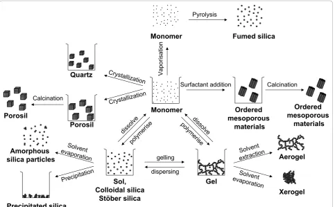
Amorphous silica particles are formed by polymeriza-tion of monomers in the aqueous solupolymeriza-tion supersatu-rated with silicic acid. Various silica materials are produced in liquid phase processes (Figure 2).
Colloidal silicaor silica sol is most often produced in a multi-step process in which the alkaline silicate
solution is partially neutralized with a mineral acid. Alternatively, this pH neutralization can be achieved by electrodialysis. The resulting silica suspension is stabi-lized by pH adjustment. Finally a solid concentration up to 50 wt% is reached by water evaporation. Silica sol nanoparticles show a perfect spherical shape and identi-cal size as a result of extensive Ostwald ripening [15].
Stöber silica sol is prepared by controlled hydrolysis and condensation of tetraethylorthosilicate (TEOS) in ethanol to which catalytic amounts of water and ammo-nia are added. The Stöber procedure can be used to obtainmonodisperse spherical amorphoussilica parti-cles with tunable size and porosity [16].
Silica gel is obtained by destabilizing silica sol. Silica gel is an open 3-D network of aggregated sol particles. The pore size is related to the size of the original silica sol particles composing the gel.
Precipitated silica is formed when a sol is destabi-lized and precipitated.
Ordered mesoporous silicais obtained by a supra-molecular assembly of silica around surfactant micelles. Typical surfactant molecules are amphiphilic polymers such as tribloc copolymers or quaternary alkylammo-nium compounds. These organic supramolecular tem-plates are evacuated from the mesopores, typically via a Si
OH
O H
OH OH
Si O H
O H
OH
Si
OH O
OH
OH Si OH
O H
OH OH
- H
2
O
Figure 1Polymerization of silicic acid molecules through formation of siloxane bond and water.
Monomer
Sol, Colloidal silica
Stöber silica
Gel
gelling dispersing
polyme
rise diss
olve
diss olve
polyme rise
Xerogel Aerogel
Solvent extraction
Solvent evaporation
Surfactant addition
Ordered mesoporous
materials
Precipitation
Solvent evaporation
Precipitated silica Amorphous silica particles
Crystallization
Quartz
Porosil
Crystallizat ion
Calcination
Vaporisation
Monomer
Pyrolysis
Fumed silica
Calcination
Porosil
Ordered mesoporous
materials
calcination step. Calcination is a controlled combustion process leading to oxidation and decomposition of the template molecules into small volatile products such as NOx, CO2 and H2O, which can leave the pores. The dia-meter of the mesopores (2-50 nm) is determined by the type of surfactant applied [17,18].
A completely different synthesis route of amorphous silica starts from SiCl4in the vapor phase. Silicon tetra-chloride is oxidized in a hydrogen flame at temperatures exceeding 1000°C and polymerized into amorphous non-porous SNPs. This nanopowder has very low bulk density and high specific surface area, typically 200 to 300 m2/g. This material is calledpyrogenic or fumed silica, referring to the special synthesis conditions [15].
The synthesis of dense crystalline silica such asquartz
from aqueous solution is a slow process requiring heat-ing the solution to accelerate the formation process in a so-called hydrothermal synthesis [15]. Alternatively, under high pressure, amorphous silica can be trans-formed to crystalline material by microcrystallization. The appearance of quartz ranges from macroscopic crystals to microcrystalline powders. Large crystals are grown at high temperature and pressure in industry. Smaller quartz crystals are conveniently obtained by grinding large crystals. Alpha-quartz is formed under moderate temperature and pressure conditions and is the most abundant form of quartz. At temperatures exceeding 573°C, a-quartz can transform intob-quartz [19]. At atmospheric pressure and temperatures higher than 870°C, quartz is transformed into tridymite and at temperatures more than 1470°C into cristobalite [15,20]. These high-temperature polymorphs of quartz have the same elemental composition but a different crystal structure and can persist metastably at lower temperatures.
Dense and porous crystalline materials can be distin-guished by framework density. The framework density is conveniently defined as the number of tetrahedrally coordinated atoms (T-atoms) per nm3. For dense struc-tures, such as quartz, tridymite and cristobalite, values of 22 to 29 T-atoms/nm3 are common, whereas for por-osils belonging to the zeolite material family, as few as 12.1 T-atoms/nm3 are present [21]. The framework structure of a porosil is denoted with a 3-letter code. Descriptions are available in theAtlas of Zeolite
Frame-work Types [22].
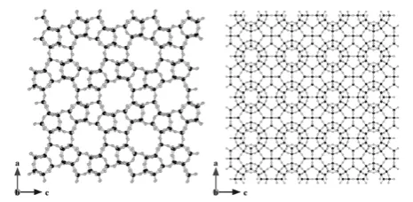
Porosils are crystallized in aqueous media in the pre-sence of organic molecules that act as porogens or tem-plate molecules defining the size and shape of the pores. Their evacuation is typically achieved through calcina-tion. Among the porosils are clathrasils and zeosils [23,24].Zeosilshave cages with windows or channels of a sufficiently free dimension to allow molecules to diffuse in and out, a property known as molecular sieving [25].
Clathrasilshave cages with windows that are delineated with a 6-membered ring of SiO units, thus presenting a free aperture of barely 0.28 nm. Even a molecule as small as oxygen has no access to the cavities of a clathrasil. The organic template molecules engaged in the crystallization of a clathrasil cannot be removed easily from the pores [23,24].
When heated above 1700°C, any type of silica (amor-phous or crystalline) melts. During cooling, the disor-dered structure is solidified, and a dense amorphous silica glass orvitreous silicais formed [15].
Physico-chemical properties
The properties of silica materials considered essential for their potential toxicity are crystallinity, particle size and morphology, porosity, chemical purity, surface chemistry and solubility [26]. An overview of the prop-erties of silica materials involved in silica toxicity is pro-vided in Table 1.
Crystallinity
In crystalline structures such as quartz and porosils, the arrangement of atoms is ordered in all dimensions. According to the International Union of Pure and Applied Chemistry (IUPAC), the atoms must be arranged periodically with long-range order (at least 10 repeats in all directions) and produce sharp maxima in a diffraction experiment to observe x-ray diffraction (XRD) crystallinity [27]. The threshold for observing crystallinity depends on the unit cell size (size of the repeated unit in a crystal). For materials with large unit cells, such as porosils, the minimum particle size required is about 10 nanometers to observe a distinct, sharp XRD pattern. Amorphous silica may present some short-range order but lacks long-range order in 3 dimensions and does not exhibit a sharp XRD pattern. Of note, the surface of a crystal represents a discontinu-ity that can be seen as a defect. With the presence of a less-structured or even partially amorphous rim, crystals may behave like amorphous particles. Thus, particles with an ordering at limited-length scales or with amor-phous regions may be classified as amoramor-phous.
Particle size and morphology
Table 1 Overview of silica materials and relevant properties
Material Nature of product
Crystallinity Particle size Porosity Polarity Purity Applications Ref
Colloidal silica
Sol Amorphous 1-1000 nm Dense Hydrophillic Very
high
Binders, ink [15]
Stober silica Sol Amorphous 10-1000 nm Tunable porosity Hydrophillic Very high
Research [16]
Precipitated silica
Powder Amorphous 5-6 nm primary particles precipitated to 500 nm - 50μm aggregates
Tunable porosity Hydrophillic Very high
Filler and performance additive
[15]
Silica gel Powder Amorphous 0.5–5 nm primary particles gelled to networks and milled to 500μm -6 mm aggregates
Tunable, void spaces between primary particles
Hydrophillic Very high
Dessicant, filler and performance additive
[15]
Mesoporous silica
Powder Amorphous 50–1000 nm, aggregated because of calcinations
Mesoporous Hydrophobic Very high
Drug delivery, catalysis, imaging
[8]
Pyrogenic silica (fumed silica)
Powder Amorphous 2-50 nm primary particles fused to 1-250μm aggregates
Void spaces between primary particles
Hydrophobic Very high
Tickner, performance additive
[15]
Vitreous silica (fused silica glass)
Powder Amorphous 50-2000μm Dense Hydrophobic
(grinded: hydrophilic)
Variable Glass [15,19]
Quartz Powder Crystalline 50 nm- severalμm Dense Hydrophobic/ (grinded: hydrophilic)
Variable Geologic mineral, Piezoelectricity
[19,20]
Cristobalite Powder Crystalline 1μm - several cm Dense Hydrophobic Variable Geologic mineral
[20]
Zeosils (porosil)
Powder Crystalline 0.05-5000μm Porous Pore diameter: 0.4-1.2 nm
Hydrophillic/ hydrophobic
Very high
Adsorbent [25]
Clathrasils (porosil)
Powder Crystalline 0.5-5000μm Porous
Pore diameter: 0.2-0.3 nm
Hydrophillic/ hydrophobic
Very high
Gas separation [24]
Diatomeus earth, kieselguhr
Powder Amorphous, partially crystalline
5-120μm Dense Hydrophillic/
hydrophobic Low (90%)
Filter, filling material
[15]
silica. The obtained products generally have a broad size distribution.
Crystalline particles exhibit crystal planes at the sur-face, and the morphology of the crystalline nanoparticles depends on the crystal class such as cubic, hexagonal, tetragonal, and orthorhombic (Figure 3 right). For all nanomaterials, in aqueous environment, the primary nano-sized silica particles tend to form aggregates.
Porosity
According to IUPAC [28], pores are classified according to their diameter into micropores (< 2 nm), mesopores (2-50 nm) and macropores (> 50 nm). Amorphous sol particles can be microporous or non-porous (dense). The porosity of Stöber silica can be tuned by adapting the synthesis parameters: decreasing the ratio of water to TEOS promotes particle growth by aggregating smal-ler sub-particles, thus leading to rough particle surfaces with micropores. In contrast, smooth particle surfaces are obtained with conditions of high ratio of water to TEOS [29]. Silica gel is a powder with particle size in the micrometer range or larger and is, typically, mesoporous.
Zeosils and clathrasils have characteristic pores and cages in the micropore size range, depending on frame-work topology. Examples of porosil frameframe-works are shown in Figure 4 [22].
When the silica is presented as a nanopowder, poros-ity can be an intrinsic and extrinsic characteristic: sta-pling of the elementary nanoparticles gives rise to an interparticle porosity, which often is difficult to distin-guish from the intrinsic intraparticle porosity, especially when dealing with mesoporosity.
Hydrophilic-hydrophobic properties
The hydrophilicity of a silica material increases with the number of silanols, or silicon-bonded hydroxyl groups, capable of forming hydrogen bonds with physical water molecules. The chemical formula of silica is represented
as SiO2.xH2O, in which water represents chemical water contained in silanol groups present on the surface of the silica material. These water molecules are not to be con-fused with crystal water, such as that present in many inorganic salt crystals. The surface chemistry of silica is depicted in Figure 5. Vicinal hydroxyl groups (one hydroxyl group per tetrahedron) located at mutual dis-tances smaller than 3 nm are engaged in hydrogen bonding. Geminal hydroxyls (2 hydroxyl groups per tet-rahedron) are considered to occur in minor concentra-tions. Isolated silanols are positioned too far apart to be engaged in hydrogen bonding. Because of the differing chemistry of these 3 types of silanol groups, they are not all equivalent in their adsorption behavior or chemi-cal reactivity. Vicinal hydroxyls interact strongly with water molecules and are responsible for the excellent water adsorption properties of silica, which are exploited in industrial gas drying operations, for example.
The reported concentration of hydroxyl groups per square nanometer on the surface of amorphous silica ranges from 4 to 5 OH/nm2 [12]. As compared with amorphous silica, the crystalline forms of silica generally contain a lower concentration of surface hydroxyl groups [15]. Hydrogen-bonded water molecules are removed when silica is heated at 170°C under atmo-spheric pressure or at room temperature under vacuum.
Colloidal silica, precipitated silica and ordered meso-porous silica and silica gel are hydrophilic because of their high concentration of silanols. Silicagel, for exam-ple, can adsorb water in quantities up to 100% of its proper weight.
Porosils typically are hydrophobic because they lack silanols in the pores of their framework. Silica produced at high temperature, such as pyrogenic and vitreous Figure 4 Atomic representation of (left) a zeosil with
microporous channels (MFI type) and (right) clathrasil with a denser framework (SOD type). Black and gray circles represent silicon and oxygen atoms, respectively. Figure made with Vesta 2.0.3 [178] with unit cell coordinates from [22].
Si Si O H
Si Si
O H
Si O H
Si Si
O H
Si O H
Si O
H
H
Isolated Vicinal,
anhydrous
Vicinal,
hydrated
Si Si O O
H H
Si
Geminal
Si O
Si Si
Siloxane, dehydrated
Surface
Hydrophilic surface
Hydrophobic surface
silica, or calcined at temperatures exceeding 800°C, is almost entirely dehydroxylated. In a dehydroxylation reaction, neighboring silanol groups are condensed into siloxane bonds (Figure 5 bottom) and water molecules. Some isolated silanol groups may persist on the surface [15]. Because hydrogen bonding on siloxanes is unfavor-able, dehydroxylated silica is hydrophobic. Grinding of hydrophobic bulk materials such as quartz and vitreous silica induces silicon and oxygen radicals and surface charges. These charges increase the hydrophilic surface [19,30].
Solubility
The dissolution and precipitation of silica in water che-mically involves hydrolysis and condensation reactions, respectively, catalyzed by OH-ions (Figure 1).
For micrometer-sized nonporous amorphous silica, the equilibrium concentrations of Si(OH)4 at 25°C in water corresponds to 70 ppm at pH 7. The silica solubility depends on the surface curvature of the (nano)particles. SNPs and nanoporous silica show enhanced equilibrium solubility, of 100-130 ppm [12]. According to Vogelsber-ger et al. [31], the solubilization of amorphous SNPs in physiological buffer at 25°C is accelerated because of the large surface area exposed. The solubility equilibrium is reached only after 24 to 48 h. Crystalline silica such as quartz has a much lower equilibrium solubility, of 6 ppm [15].
In summary, when dealing with silica, the physico-chemical properties such as amorphous versus crystal-line nature, porosity, particle size and degree of hydro-xylation must be specified. An overview of silica materials described in the scientific literature and in the research and development environment is provided in Table 1.
Toxicity Of Silica Background
Health effects of silica and epidemiological studies
Until recently, toxicological research into silica particles focused mainly on“natural”crystalline silica particles of 0.5 to 10μm (coarse or fine particles). This research was/is fed by the clear association of occupational inha-lation exposure and severe health effects, mainly on the respiratory system. The typical lung reaction induced by chronic inhalation of crystalline silica is silicosis, a gen-erally progressive fibrotic lung disease (pneumoconiosis), exemplified by the development of silicotic nodules composed of silica particles surrounded by whorled col-lagen in concentric layers, with macrophages, lympho-cytes, and fibroblasts in the periphery. Epidemiologic studies have found that silicosis may develop or progress even after occupational exposure has ended; therefore, above a given lung burden of particles, silicosis was sug-gested to progress without further exposure [32-34].
Calvert et al. [35] recently reported an association of crystalline silica (mainly quartz) exposure and silicosis, as well as lung cancer, chronic obstructive pulmonary disease (COPD), and pulmonary tuberculosis. The carci-nogenicity of quartz and cristobalite has been shown in several epidemiological studies [36-38]. In 1997, the International Agency for Research on Cancer (IARC) classified some crystalline silica polymorphs (quartz and cristobalite) in group 1 (sufficient evidence for the carci-nogenicity to experimental animals and to humans), whereas amorphous silica (silicon dioxide without crys-talline structure) was classified in group 3 (inadequate evidence for carcinogenicity) [39]. This classification has recently been confirmed [40]. Checkoway and Franzblau [41] reviewed occupational epidemiologic literature on the interrelations among silica exposure, silicosis and lung cancer and concluded that the appearance of silico-sis is not necessarily required for the development of silica-associated lung cancer. Hnizdo and Vallyathan [42] suggested that chronic exposure to levels of crystal-line silica dust, which does not cause disabling silicosis, may cause chronic bronchitis, emphysema, and/or small airway disease leading to airflow obstruction, even in the absence of radiological evidence of silicosis. Evidence has linked silica exposure to various autoimmune dis-eases (systemic sclerosis, rheumatoid arthritis, lupus, chronic renal disease), as reviewed by Steenland and Goldsmith [43]. A study by Haustein et al. [44] reported on silica-induced (silica dust) scleroderma.
partially reversible inflammation [50,51], granuloma for-mation and emphysema, but no progressive fibrosis of the lungs [52,53]. However, high doses of amorphous silica may result in acute pulmonary inflammatory responses, which could conceivably trigger long-term effects, despite a low biopersistence of the particles [54]. The debate on the health effects of micron-sized crystal-line or amorphous silica is beyond the scope of this article. Readers are referred to other publications [35-38,41,55-57].
Mechanisms of toxic action
As mentioned, most of the toxicological research into silica has focused on crystalline silica particles of 0.5 to 10 μm (coarse or fine particles). Despite the relatively large amount of available studies, the mechanisms of crystalline silica toxicity at the cellular and molecular levels are still unclear, and whether any single mechan-ism underlies all the above-mentioned diseases induced by these particles is uncertain [43]. However, severe inflammation following exposure to silica particles appears to be a common initiating step [58,59].
The crucial role of reactive oxygen species (ROS) in the inflammatory, fibrogenic and carcinogenic activity of quartz is well established [60,61]. Oxidative membrane and DNA damage are considered the most important mechanisms involved in the health effects of micron-sized crystalline silica. A few of the numerous reports clearly demonstrate these findings: ROS generated by the silica surface can induce cell membrane damage via lipid peroxidation that may subsequently lead to increased cellular permeability [62], perturbation of intracellular calcium homeostasis [63] and alterations in signaling pathways. Schins et al. and Fanizza et al. [64,65] demonstrated that respirable quartz particles induce oxidative DNA damage in human lung epithelial cells. Li et al. [66,67] demonstrated that micron-sized quartz particles induce.OH generation through an iron-dependent mechanism. A close association of .OH and iron ion concentration has been reported for amorphous silica particles [66,67]. The study of Ghiazza et al. [30] indicates that crystallinity might not be a necessary pre-requisite to make a silica particle toxic; both quartz and vitreous silica showed stable surface radicals and sus-tained release of HO. radicals. When tested on macro-phages, vitreous silica and pure quartz showed a remarkable potency in cytotoxicity, release of nitrite and tumor necrosis factora(TNF-a) production, suggesting a common behavior in inducing of oxidative stress [30]. Ding et al. [68] discuss the molecular mechanisms of silica-induced lung injuries with a focus on NF-kB acti-vation, generation of cyclooxygenase II and tumor necrosis factor a (TNF-a). The review of Castranova [69] summarizes evidence that in vitro and in vivo
exposure to crystalline silica results in activation of NF-kB and AP-1 signaling pathways.In vitroand in vivo animal studies, as well as investigations in humans, strongly support the role of macrophage products in the development and progression of silicosis [70]. Such pro-ducts include a large panel of cytokines [71], with TNF-a seeming to determine the development of silica-induced pulmonary fibrosis [72]. In addition, recent evi-dence implicates interleukin 1b(IL-1b) and its activation by the NALP-3 inflammasome [73].
A large body of experimental work in the past 20 years has shown that 2 main factors seem to govern the hazardous nature of crystalline silica: particle surface reactivity and the form of silica [74]. Fenoglio et al. [75] evaluated these factors systematically, studying synthetic quartz samples differing only in size and shape. Cyto-toxicity appeared to be primarily governed by the form of the particles and the extent of the exposed surface. Several studies indicate that the surface silanol groups are directly involved both in membranolysis [76-78] and in toxicity to alveolar cells [79,80]. Therefore, the distri-bution and abundance of silanols determines the degree of hydrophilicity (see “Physico-chemical properties of synthetic silica materials related to toxicity” described above) and seems to modulate cell toxicity [80,81]. Experimental work with respirable silica particles and the survey of published data by Bagchi [82] suggest that the toxicity of these particles is caused by the large amount of positive charges they carry. Ghiazza et al. [83] reported that formation of a vitreous phase at the surface of some commercial diatomaceous earth pre-vents the onset of oxidative stress effects. Donaldson and Borm [84] emphasized that the ability of quartz to generate ROS could be modified by a range of sub-stances that affect the quartz surface, such as subsub-stances originating from other minerals. The authors concluded that the toxicity of quartz is not a constant entity and may vary greatly depending on the origin/constitution of the sample.
dissolution rate was reported as being more rapid than the reverse precipitation rate [86]. Solubility has been defined as a key driver in the clearance mechanisms involved in amorphous silica removal from lung [87]. Warheit [45] reviewed pulmonary responses to different forms of silica and reported that cristobalite produced the greatest lung injury, quartz produced intermediate effects, and amorphous silica produced minimal effects. In terms of analytical technique, small differences in dis-solution exist among these different forms of silica, and dissolution, in turn, influences pulmonary effects through the concept of persistence. In addition, compo-nents from the biological system may react with the surface of the particle. A systematic investigation of iron-containing SNPs as used in industrial fine-chemical synthesis demonstrated the presence of catalytic activity that could strongly alter the toxic action of nanoparti-cles [88].
On the whole, considering the great variety of silica forms, degree of crystallinity, surface state and the pre-sence of contaminants, there is a critical need for care-fully characterized standard silica samples to unravel the relationships between physico-chemical factors and toxi-city, both micron- and nano-sized. The main goal of this review is to focus on the toxicity of nanosilica, which has never been properly reviewed. Moreover, nanosilica occurs mainly in amorphous forms, and the potential hazard posed by these nanomaterials cannot be simply related to, as has already been reviewed many times, studies of micron-sized crystalline materials.
Silica nanoparticles
The growing abundance and industrial applications of nanotechnology has resulted in a recent shift of toxico-logical research towards nanoparticles [89-94]. Ultrafine particles (< 0.1 μm) have been demonstrated to cause greater inflammatory responses and particle-mediated lung diseases than have fine particles (< 2.5 μm) per given mass [95-97]. Also, experiments involving silica have shown that nanoparticles, both ultrafine colloidal silica [98,99] and crystalline silica [99], have a greater ability to cause lung injury as compared with fine parti-cles. Thus, the unique properties (i.e., small size and corresponding large specific surface area; cell penetrat-ing ability) of nano-sized SiO2are likely to impose bio-logical effects that differ greatly from micron-scale counterparts.
In vitrostudies of nanosilica toxicity
A structured summary ofin vitrostudies of the toxicity of SNPs can be found in Table 2.
Chen and von Mikecz [100] investigated the effects of nanoparticles on structure, function, and proteasomal proteolysis in the cell nucleus by incubating different
cell lines with unlabeled and fluorescently labeled amor-phous silicaparticles of different sizes [100]. SiO2 parti-cles between 40 nm and 5μm were applied to epithelial cells in culture and observed on confocal laser scanning microscopy with differential interference contrast. Parti-cles of all tested sizes penetrated the cytoplasm; how-ever, nuclear localization was observed exclusively in cells treated with SiO2 nanoparticles between 40 and 70 nm. Fine and coarse SiO2 particles (0.2-5μm) were exclusively located in the cytoplasm and accumulated around the nucleus, forming nuclear indentations. The uptake of SNPs in the nucleus induced aberrant clusters of topoisomerase I and protein aggregates in the nucleo-plasm–the former inhibiting replication, transcription, and cell proliferation –without altering cell viability. Cells treated with fine (0.5μm) or coarse (5 μm) SiO2 particles had the same replication and transcription activity as that of untreated control cells [100].
Jin et al. [101] investigated the potential toxicity of luminescentamorphous SNPs (50 nm) in freshly iso-lated rat alveolar macrophage cells and human lung epithelial cells (A549 cells). The SNPs penetrated the cells but were not detected in the nuclear region and did not cause significant toxic effects at the molecular and cellular levels below a concentration of 0.1 mg/ml.
Table 2 In vitro studies on nanosilica particles (SNPs) toxicity
Silica form Size (primary) Material characterization Cells used Test Biological endpoints and findings
Ref
Amorphous 40 nm- 5μm Not specified A549
HEp-2 RPMI 2650 RLE-6TN N2a
•Replication and transcription assays
•Cell proliferation and cell viability assay
•Proteasome activity assay
•
Immunofluorescence and microscopy
•Uptake of all particles into the cytoplasm and nuclear localization of nanoparticles between 40 and 70 nm
•The uptake of NSPs in the nucleus induced aberrant clusters of topoisomerase I and protein aggregates in the nucleoplasm
[100]
Amorphous (luminescent)
50 nm •Synthesis (ref. to literature) A549 rat alveolar macrophages
•laser scanning confocal microscope
•Comet Assay
•Pulse Field Gel Electrophoresis (PFGE)
•Western Blot Analysis of DNA Adducts/DNA Agarose Gel
•DNA Repair Enzyme Activity Assay
•Cell Proliferation Assay
•Vybrant Apoptosis Assay
•Uptake not detected in the nuclear region
•As compared to the A549 cells, the nanoparticle penetration rate was much faster in the rat alveolar macrophages
•No significant toxic effects observed at the molecular and cellular levels below a concentration of 0.1 mg/ml
[101]
Amorphous (colloidal)
15 and 46 nm •Particle sizes and distribution
•Surface area (268 and 52.5 m2/g for 15 and 46 nm particle, respectively), crystalline structure, major trace metal impurities
•Hydrodynamic particle size in water suspension
A549 •SRB
(sulforhodamine B) and LDH assays
•Reduced glutathione (GSH) level
•DCFH assay (ROS generation)
•Malondialdehyde (MDA) assay
•Cytotoxicity was dose- and time-dependent
•Reduced glutathione (GSH) levels and elevated MDA production after exposure to 15 nm SNPs
[102]
Amorphous 60 and
100 nm •
Size distribution analysis
•Endotoxin concentration
A549 THP-1 Mono Mac 6; co-cultures
•LDH assay
•Cytokine expression (TNF-a, IL-6, IL-8)
•Light and transmission electron microscopy (TEM)
•Cytotoxicity differed among the cell lines and was dose-and size-dependent (smaller particles were more toxic)
•co-cultures showed an increased sensitivity to particles concerning the cytokine release in comparison to the mono-cultures of each cell type
[103]
Amorphous ~14 nm •Size distribution A549
L-132 HeLa MNNG/ HOS
•MTT and WST-1 assays
•Trypan blue exclusion and LDH assay
•Annexin V-PI assay (fluorescence microscopy)
•DCFH assay
•IL-8 expression (ELISA)
•Little cytotoxic effects in 4 cell lines tested at the
concentration below 250μg/ml within 48 h
•Exposing cancer cells to high concentrations (250-500μg/ml) for 72 h resulted in an inflammatory response with oxidative stress and membrane damage, which varied with cell type (A549>HOS > HeLa)
•SNPs triggered an
inflammation response without causing considerable cell death for both cancer cells and normal cells
Table 2 In vitro studies on nanosilica particles (SNPs) toxicity(Continued)
Amorphous 10 and 80 nm o Provided by producer for the primary particles (surface area: 640 and 440 m2/g for 10 and 80 nm particle, respectively) o Hydrodynamic particle size (in cell culture medium)
A549 •MTT and LDH assays
•DCFH assay
•Intracellular glutathione (GSH) concentration
•Membrane lipid peroxidation (LPO)
•Assay of glutathione reductase and glutathione peoxidase
•Cytotoxicity was dose-dependent
•SNPs induced reactive oxygen species and membrane lipid peroxidation in dose-dependent manner
•Both sizes of SNPs had little effect on GSH level and the activities of glutathione metabolizing enzymes
[105]
Amorphous 7 and 5-15 nm o Surface area (350 and 644 m2/g for 7 and 5-15 nm particle, respectively) o Size distribution (in the test medium)
Beas-2B •Incorporation of SNPs into the cells (confocal LSM)
•MTT assay
•PI staining (flow cytometry)
•Apoptosis
•DCFH assay
•Oxidative stress responding transcription factors (Western blotting)
•SNPs were incorporated into the cells and distributed around the nucleus area
•SNPs induced oxidative stress via ROS formation and induction of of antioxidant enzymes (SOD and HO-1)
•Induction of Nrf-2-ERK MAP kinase signaling pathway was observed
•Overall, cells exposed to 5-15 nm SNPs (porous) showed a more sensitive response than those exposed to 7 nm SNPs (fumed)
[106]
Amorphous 10-20 nm o Provided by manufacturer (surface area: 140-180 m2/g) o Primary particle size o Endotoxin content (LPS)
A549 •MTT and LDH assays
•DCFH assay
•SOD activity determination
•Nitrate/nitrite determination
•DNA oxidative damage assay
•Cytotoxicity was dose- and time-dependent
•SNPs stimulated the ROS generation, GSH depletion and lower expression of SOD activity in a dose-dependent manner
•No NO production and significant DNA oxidative damage was observed after treatment of cells with SNPs
•Co-treatment of LPS with SNPs enhanced observed cytoxicity and generation of oxidative stress
[107]
Amorphous 30, 48, 118 and
535 nm •
Synthesis method
•Hydrodynamic particle size (in water and cell culture medium)
HEL-30 •MTT and LDH assays
•Reduced glutathione (GSH) and DCFH assay
•Transmission electron microscopy (TEM)
•Cytotoxicity was dose- and size-dependent (smaller particles were more toxic)
•Uptake of all particles into the cytoplasm (nuclear uptake not studied)
•GSH level reduced significantly of after exposure to 30 nm nanoparticles
•No significant Reactive Oxygen Species (ROS) formation
[108]
Amorphous 70, 300 and 1000 nm
Not specified XS52 •TEM analysis of cells
•LDH assay
•Proliferation ([3 H]-Thymidine incorporation assay)
•SNPs of 300 and 1000 nm were incorporated into the cells and located in cytoplasm only; nanoparticles of 70 nm were located in nucleus as well as cytoplasm
•Cell proliferation was inhibited by treatment with SNPs of all sizes in dose-dependent manner
•The growth of the cells was more strongly inhibited by smaller-sized SNPs
Table 2 In vitro studies on nanosilica particles (SNPs) toxicity(Continued)
Amorphous 15, 30 and 365 nm •Size distribution
•Zeta potential
•Amorphous structure
HaCaT •CCK assay
•Cell cycle assay
•Annexin V-PI assay (Flow cytometry)
•2D-DIGE and, IEF and SDS_PAGE (protein expression)
•Western blot
•Cytotoxicity was dose- and size-dependent (smaller particles were more toxic)
•Apoptosis was dose- and size-dependent (smaller particles induced higher apoptosis frequency)
•Up-regulated proteins were classified as oxidative stress-associated proteins; cytoskeleton-associated proteins; molecular chaperones; energy metabolism-associated proteins; apoptosis and tumor-associated proteins
[110]
Amorphous 15 nm •Size distribution
•Zeta potential
•Amorphous structure
HaCaT •Flow cytometric analysis of methylated DNA
•Real-time PCR
•Western blot
•Treatment with SNPs induced Global DNA hypomethylation
[111]
Amorphous 21 and 80 nm •Particle preparation and dispersion
•Size, morphology and chemical states of elements
•Hydrodynamic particle size (dispersed in water)
WS1 CCD-966sk MRC-5 A549 MKN-28 HT-29
•MTT and LDH assays
•Toxicity was seen at concentrations exceeding 138 μg/ml
•Susceptibility to NSPs differed among tested cell lines
[113]
Amorphous 20 nm Only provided by producer (surface area: 640 ± 50 m2/g)
RAW264.7 •Membrane fluidity measurements (FRAP technique by LSCM)
•DCFH assay
•Intracellular free calcium content
•Exposure to SNPs increased ROS generation and decrease of the membrane fluidity
•Perturbation of Intracellular free calcium homeostasis was responsible for observed cytotoxicity
[114]
Amorphous 14 nm Only provided by producer
(surface area: 200 m2/g) Caco-2 •assayLDH and WST-1
•Fpg-modified comet assay
•Total GSH content
•Cytotoxicity observed
•Oxidative DNA damage
•Significant depletion of intracellular GSH
[115]
Amorphous 21, 48 and 86 nm •Size distribution analysis
•Surface area (225, 106 and 39 m2/g for 21, 48 and 86 nm particle, respectively)
•structure
L-02 •MTT and LDH assays
•TEM assay
•DCFH, MDA and GSH assay
•Annexin V-PI assay (flow cytometry)
•DNA ladder assay
•Western blot
•Cytotoxicity was dose time -and size-dependent (smaller particles were more toxic)
•21 nm SNPs induced ROS generation, lipid peroxidation and GSH depletion in a dose-dependent manner
•21 nm SNPs induced apoptosis in a dose-dependent manner
[116]
Amorphous 4-40 nm (mean size: 14)
Not specified HDMEC •MTS assay
•transmission electron microscopy (TEM)
•Ki67 expression and IL-8 release
•The particles were internalized but they did not exert cytotoxic effects
•Reduction of the proliferative activity and a pro-inflammatory stimulation were observed
[117]
Amorphous (monodisperse)
14, 15, 16, 19, 60, 104, 335 nm
•Particle preparation and stability
•shape and size distribution
•surface area (196, 179, 183, 145, 33, 28 and 7.7 m2/g for 14, 15, 16, 19, 60, 104 and 335 nm particle, respectively)
•micropore volume
•Hydrodynamic particle size (in water and cell culture medium)
EAHY926 •MTT and LDH assays
•Annexin V-PI assay
•Cytotoxicity was dose- and size-dependent (smaller particles were more toxic and affected the exposed cells faster)
•Cell death predominantly caused by necrosis
Table 2 In vitro studies on nanosilica particles (SNPs) toxicity(Continued)
Amorphous 21 and 48 nm •Size distribution analysis
•Surface area (225 and 106 m2/g for 21 and 48 nm particle, respectively)
•structure
H9c2(2-1) •MTT and LDH assays
•Hematoxylin and eosin staining
•DCFH, intracellular MDA and GSH assays
•Flow cytometry (cell cycle)
•Western blot
•Cytotoxicity was dose time -and size-dependent (smaller particles were more toxic)
•ROS generation in a dose-dependent manner; increased level of MDA and decreased concentration of GSH indicated oxidative stress
•Cell cycle arrest in G1 phase
•Dose-dependent expression of p53 and p21 for 21 nm SNP
[119]
Amorphous From 20 nm to below 400 nm •
the dispersion characteristics (size, size distribution, size evolution)
•zeta potential
3T3-L1 •comet assay •No detectable genotoxicity (the results were independently validated in two separate laboratories)
[120]
Amorphous (monodisperse)
16, 60 and 104 nm •Particle preparation and stability
•shape and size distribution
•surface area (183, 33 and 28 m2/g for 16, 60 and 104 nm particle, respectively)
•micropore volume
•Hydrodynamic particle size (in water and cell culture medium)
A549 •MTT assay
•cytochalasin-B micronucleus assay (CBMN) alone or in combination with FISH-centromeric staining
•Alkaline Comet assay
•Measurements of cell-associated silica (ICP-MS)
•Results suggest that non-cytotoxic doses of SNPs may be capable of inducing slight chromosome breakage, loss and mitotic slippage, and at higher concentration possibly mitotic arrest.
[122]
Amorphous (monodisperse)
from 2 up to 335
nm •
Particle preparation and stability
•shape and size distribution
•surface area (from 232 to 7.7 m2/g)
•micropore volume
•Hydrodynamic particle size (in water and cell culture medium)
•Zeta potential
J774 EAHY926 3T3 Human erythrocytes
•MTT and WST-1 assays
•RBC hemolysis
•in murine macrophages, the cytotoxic response, after treatment with SNPs of 17 different sizes, increased with external surface area and decreased with micopore volume
•in human endothelial cells and mouse embryo fibroblast the cytotoxicity increased with surface roughness and decrease in diameter
•the hemolytic activity of SNPs in human erythrocytes increased with the diameter of SNPs
[141]
Amorphous 30 nm •Provided by producer for primary partilcles (surface area: 165 m2/g)
•Hydrodynamic particle size (in PBS and cell culture medium)
•Adsorption of proteins from the test media in the absence of cells
3T3 hT RAW264.7
•MTS assay
•Uptake (flow cytometry)
•DCFH assay
•Lysosomal membrane integrity
•Mitochodrial membrane potential
•Apoptosis (caspase-3, and caspase-7 activation; Annexin V-PI assay)
•SNPs depleted serum proteins from cell culture media
•SNPs cytotoxicity was dose-, time- and cell line dependent-dependent
•SNPs induced significant ROS generation in all cell lines
•No detectable destabilization of lysosomal membranes was observed
•Incubation with SNPs decreased mitochodrial membrane potential in hT and RAW cells
•SNPs triggered different extent of cell apoptosis depending on the cell line tested
[140]
Amorphous (mesoporous)
110 nm (pore diameter of ~2.5 nm)
•Structure
•surface area (910 m2/g)
•pore volume
•stability in aqueous solution 3T3-L1 MCF-7 K562
•Confocal microscope
•TEM
•Flow cytometry
•Particles were internalized into cells and accumulated in cytoplasm
•No apparent cytotoxicity
Table 2 In vitro studies on nanosilica particles (SNPs) toxicity(Continued) Amorphous (mesoporous) Not specified (MCM-41 particle type)
•Synthesis and
functionalization of particles
•Zeta potential
•Cylindrical pores with a diameter around 5 nm
HeLa •MTT, WST-1 and LDH assays
•Flow cytometry for PI
•TEM observations
•No cytotoxicity was observed up to 50μg/ml
•Particles interfered with MTT assay
[126]
Amorphous (mesoporous)
108, 110, 111 and 115 nm
•Synthesis (ref to the previous study) and surface
modification
•Zeta potential
•Surface area (780, 980, 930 and 1050 m2/g for 108, 110, 111 and 115 nm particle, respectively)
•pore volume and pore size distribution (2.6-2.0 nm)
hMSCs 3T3-L1
•MTT assay
•Flow cytometry for the uptake
•Cellular differentiation and cytochemical assay
•The modulation of surface charge and its threshold affects the uptake and is specific to cell type
•Positive correlation of positive surface charge and the uptake by the cells
•Uptake was through clathrin and actin-dependent endocytosis
•Uptake did not affect cells viability, proliferation and differentiation [125] Amorphous (mesoporous silica nanorods capped with iron oxide NPs)
200 × 80 nm (pore diameter of ~3 nm)
•Preparation and functionalization
HeLa •Confocal fluorescence microscopy
•Particles were endocytosed by the cells and biocompatible (concentration used: 0.2 mg/ mL)
[127]
Amorphous (mesoporous)
30, 50, 110, 170 and 280 nm
•Synthesis, suspension stability (no interparticle aggregation), hydrodynamic diamaters, zeta potential
HeLa •MTT
•onfocal laser scanning microscopy
•ICP-MS
•Cellular uptake is highly particle size-dependent (with the optimum size of 50 nm); little cytotoxicity up to 100 mg/ ml [128] Amorphous (mesoporous) loaded with anticancer drugs)
<130 nm (pore diameter of ~2 nm)
•Preparation, shape, aggregation/stability in aqueous solution PANC-1 AsPC-1 Capan-1 MKN45 SW480
•Fluorescence and confocal microscopy •
The particles offer the possibility of controlled release of anticancer drugs (non-loaded particles did not caused cytotoxicity)
[129]
Amorphous (mesoporous)
150 nm (pore diameter of ~2.4 nm)
•Synthesis, functionalization, surface area (850 m2/g), zeta potential
HeLa •Flow cytometry
•Fluorescence microscopy
•Uptake of particles can be regulated by different surface functionalization
•More negatively charged particles were able to escape from endosomes [130] Amorphous (mesoporous) Commercially available amorphous silica material
100 - 300 nm (pore diameter of ~3 nm)
-•Synthesis (ref. to the previous study), funcionalization, surface area (1138 m2/g), pore volumes, number of silanol group
•Funcionalization
Rabbit RBCs •Hemolysis assay
•UV/Vis spectroscopy
•Flow cytometry
•The hemolytic activity of silica nanoparticles depends only on the concentration of negatively charged silanol groups
•Mesoporous particles exhibit a high compatibility towards RBCs as most of the silanols are located in the interior of the particles that are not accessible by the RBCs membranes
[131]
Amorphous (mesoporous)
300-650 nm (pore diameter of 31Å) and SBA-15 type (>hundreds of nm, pore diameter of 55 Å)
•Synthesis,
•Order of mesostructures, surface area (821 and 506 m2/ g), wall thickness, composition
HL-60 Jurkat
•Oxygen consumption assay
•ATP formation assay
•Cellular GSH assay
•Particles with larger size and larger pores caused concentration- and time dependent inhibition of cellular respiration
•Both nanoparticles were toxic to the isolated mitochondria
•No significant changes in cellular glutathione level was observed
Table 2 In vitro studies on nanosilica particles (SNPs) toxicity(Continued)
Amorphous (mesoporous and silica nanospheres)
250 nm; 166x320 nm (pore diameter = 3.5 nm)
•Synthesis and functionalization
•Number of particles per gram, surface area (4.1 and 0.2 m2/particle for mesoporous and spherical particle, respectively)
SK-N-SH •Staining with trypan blue and
determination of viable cells using a hemacytometer
•The cytotoxicity of particles was related to the adsorptive surface area of the particle (the most toxic malodorous silica are those with the largest BET surface areas)
•Dependency of cytotoxicity on the nature of the attached functional groups cannot be ruled out
[133]
Amorphous (mesoporous)
270 ± 50 nm (pore diameter of 3.9 nm) and 2.5μm ± 500 nm (pore diameter of 2.8 nm)
•Synthesis
•The structural and textural characterizations
•Surface area(520 and 547 m2/ g for 270 nm and 2.5μm particle, respectively)
•LPS concentration analysis
Human monocyte-derived dendritic cells
•Apoptosis/necrosis (Annexin V/PI assay)
•production of cytokines10 and IL-12p70,IL-12, IL-10
•confocal microscopy, TEM
•Viability, uptake and immune regulatory markers were affected with increasing size and dose
[134]
Amorphous (mesoporous)
190, 420 and 1220
nm •
Synthesis and functionalization
•Size distribution
•Dispersity and porosity
•Surface area (220-650 m2/g)
•Zeta potential
MDA-MB-468
COS-7 •
MTT assay
•The biodegradation experiments
•Intracellular localization of particles
•The cytotoxicity of particles was highly correlated with particle sizes ((smaller particles were more toxic)
•The biodegradation products of spherical E-MS particles showed no toxicity
•The residual surfactant bound to the particles has a much smaller contribution to the cytotoxicity than the free one
•The smaller particles were more easily endocytosed and consequently located within lysosomes
[135]
Amorphous 100 and 200 nm •rod-shaped and spherical particles (Stöber), not-coated and coated with fibronectin or polyethylene glycol (PEG),
•Primary and aggregate size, surface area (9.2 and 4.6 m2/g for silica rods and 27.3 and 14.2 m2/g for silica spheres), crystallinity, impurities, zeta potential
MET-5A •LDH assay
•Expression of IL-8
•Simulated stretch imposed on the cells
•Dosimetric comparison of acicular and isotropic particulate materials is not straightforward
•In the absence of simulated lung function (stretch), cells showed no significant enhancement of cytotoxicity or inflammation release
•PEG surface treatment tended to reduce the cytotoxicity and IL-8 release from particle exposures suggesting the significance of adhesive interactions e.g. for membrane binding/signal transduction
[136]
Amorphous 130 nm and 155 nm; iron oxide particle with silica shell (80 nm)
•Size distribution
•Reference given for the description in detail
Hmy2 Jurkat U937 PC3; human peripheral blood cells
•MTT assay and Trypan Blue exclusion
•Scanning electron microscopy
•DCFH assay
•The cytotoxicity of particles depended on the cell type tested
•No direct correlation between ROS production and cell toxicity.
•PEG-ylation of SNP protected the particles from protein adsorption on the external surface of the NPs and consequently no agglomeration in culture medium was observed.
•The availability of the particles to be internalized by the cells depended on the size and morphology of the aggregates.
bronchial epithelial cells (BEAS-2B) and observed the formation of ROS and induction of antioxidant enzymes. Shi et al. [107] exposed A549 cells to amorphous
SNPs (10-20 nm) at concentrations up to 200 μg/ml and observed low cytotoxicity as measured by MTT and LDH assays. However, co-treatment with the same nanoparticles and lipopolysaccharide, a bacterial product that may contaminate (nano)materials, significantly enhanced the cytotoxicity.
Yu et al. [108] examined the cytotoxic activity (by MTT and LDH assay) of well-dispersed amorphous silicaparticles (30-535 nm) in mouse keratinocytes. All sizes of particles were taken up into the cell cytoplasm; nuclear uptake was not studied. The toxicity was dose and size dependent, with 30- and 48-nm particles being more cytotoxic than 118- and 535-nm particles. The reduced GSH level significantly decreased only after exposure to 30-nm nanoparticles [108]. Nabeshi et al. [109] showed the size-dependent cytotoxic effects of
amorphous silica particles (70, 300 and 1000 nm) on mouse epidermal Langerhans cells. The smallest parti-cles induced greater cytotoxicity (by LDH assay) and inhibited cellular proliferation (by [3H]-thymidine incor-poration). The observed effects were associated with the quantity of particle uptake into the cells.
Yang et al. [110] evaluated the effects ofamorphous
SNPs (15 and 30 nm) and micron-sized silica particles on cellular viability, cell cycle, apoptosis and protein expression in the human epidermal keratinocyte cell line HaCaT. Microscopy examination revealed morphological changes after 24-h exposure; cell growth also appeared to be significantly inhibited. The cellular viability of HaCaT cells was significantly decreased, and the amount of apoptotic cells was increased in a dose-dependent manner after treatment with nano- and micron-sized SiO2 particles. Furthermore, smaller silica particles were more cytotoxic and induced a higher apoptotic rate. Proteomic analysis revealed differential induction of expression of 16 proteins by SiO2 exposure; proteins were classified into 5 categories according to their func-tions: oxidative stress-associated proteins, cytoskeleton-associated proteins, molecular chaperones, energy meta-bolism-associated proteins, and apoptosis and
tumor-associated proteins. The expression levels of the differ-entially expressed proteins were associated with particle size [110]. In a recently published study [111], the same research group used these SNPs to study the global DNA methylation profiles in HaCaT cells; the authors reported that nanosilica treatment can induce epigenetic changes.
Cousins et al. [112] exposed murine fibroblasts to smallamorphous (colloidal) silicaparticles (7, 14 and 21 nm) over a long incubation period (1, 3 and 7 days and up to 7 weeks) and observed a distinctive cellular response affecting the morphologic features, adhesion and proliferation of the fibroblasts but not cell viability. Chang et al. [113] exposed selected human fibroblast and cancer cell lines for 48 h to amorphous SNPs and assessed cellular viability by MTT and LDH assays. Cytotoxicity was seen at concentrations > 138 μg/ml and depended on the metabolic activity of the cell line. However, the average primary size of tested silica parti-cles was 21 and 80 nm, but their average hydrodynamic particle size was 188 and 236 nm, respectively, so in media, aggregates/agglomerates were formed.
In the study of Yang et al. [114], cell membrane injury induced by 20-nm amorphous silica nanoparticles in mouse macrophages was closely associated with increased intracellular oxidative stress, decreased mem-brane fluidity, and perturbation of intracellular calcium homeostasis.
Besides inhalation, ingestion is considered a major uptake route of nanoparticles into the human body [3]; however, the possible harmful effects of engineered nanoparticles in the gastrointestinal tract are still largely unknown. Recently, Gerloff et al. [115] investigated the cytotoxic and DNA damaging properties ofamorphous
fumed SiO2 nanoparticles (14 nm) in the human colon epithelial cell-line Caco-2. Exposure to SNPs for up to 24 h caused cell mortality, significant DNA damage and total glutathione depletion. The results of an in vivo study of mice fed nanosized silica are discussed in sec-tion 3.2.2.
Ye et al. [116] reported on induced apoptosis in a human hepatic cell line after exposure to amorphous (colloidal) SNPs (21, 48 and 86 nm). The viability of Table 2 In vitro studies on nanosilica particles (SNPs) toxicity(Continued)
Crystalline Particle sizes not uniform (7.21, 9.08 and 123.21 nm)
•Size and concentration WIL2-NS •MTT assay
•Population Growth Assay
•Apoptosis Assay by Flow Cytometry
•Cytokinesis Block Micronucleus Assay
•Comet Assay
•HPRT Mutation Assay
•Significant dose-dependent decrease in viability
•with increasing dose of particles
•Fourfold increase in micronucleated binucleated cells frequency was detected, while no significant difference was measured by the Comet assay
Table 3 In vivo studies on nanosilica particles (SNPs) toxicity
Silica form
Size (primary)
Material characterization
Exposure model Test Biological endpoints and
findings
Ref
Quartz 10-20 nm (average size: 12), 30-65 (average size: 50), 300 nm -2μm
•Synthesis
•Surface area
•Crystallinity
•Metal impurities
Rats instilled intratracheally with various particle types (1 or 5 mg/kg), sacrificed at 24 h, 1 week, 1 month, and 3 months post-exposure
•Bronchoalveolar lavage (BAL) fluid analysis: cell counts, differentials, and pulmonary biomarkers (Lactate
dehydrogenase (LDH), alkaline phosphatase (ALP), and lavage fluid protein)
•Cell proliferation
•Morphological/
Histopathology examination
•Hemolytic Potential of particles
Exposures to the various quartz particles produced differential degrees of pulmonary inflammation and cytotoxicity, which were not consistent with particle size but correlated with surface activity, particularly hemolytic potential.
[148]
Silica dust 10 ± 5 nm; and 0.5-10 μm (80% of the particles 1-5μm)
•Composition uknown
•Surface area
Rats instilled intratracheally (20 mg), sacrificed 1 and 2 months after dosing
•The changes of lung/body coefficient and
hydroxyproline content
•Pathologic examination
•Immunohistochemical staining for IL-4 and TGF-beta1
One month after instillation cellular nodules (Stage I silicosis) were found in the nanosized SiO2group, while in microsized SiO2group Stage II, II+ of silicotic nodules were observed. Two months after instillation, still only Stage I silicotic nodules in nanosilica group were found, while in the micro-silica group the disease progressed and Stage II+, and III silicotic nodules were found.
The experiment revealed that in rats the effect of fibrogenesis of nano-SiO2 might be milder than that of micro-SiO2.
[147]
Ludox colloidal silica
- •Mass median aerodynamic diameter (2.9, 3.3 and 3.7μm)
•Chamber Ludox concentration
Rats Inhalation (nose-only) for 2 or 4 weeks at concentrations 10, 50 and 150 mg/m3.
Additional groups of rats exposed for 4 weeks were given a 3-month recovery period
•Lung silica analysis
•BAL analysis: cell differential counts and biochemical assay (LDH, ALP, lavage fluid protein)
•Pulmonary macrophage cell culture and phagocytosis assay
•SEM ananlysis
•Additional groups of animals were processed for cell labeling studies or lung deposition studies.
The inflammatory responses, mainly seen as increased numbers of neutrophils in BALF, following the 2 and/or 4 weeks of exposure was evident at 50 mg/m3(or higher) group. Three months after exposure most biochemical parameters returned to control values. Results showed that exposures to 150 mg/m3 Ludox for 2 or 4 weeks produced pulmonary inflammation along with increases in BAL protein, LDH, and alkaline phosphatase values (p less than 0.05) and reduced macrophage phagocytosis.
Autoradiographic studies demonstrated that the labeling indices of terminal bronchiolar and lung parenchymal cells were generally increased in the 50 and 150 mg/m3 groups after 2 and 4 weeks of exposure but, with one exception, returned to normal levels following a 3-month postexposure period.
Table 3 In vivo studies on nanosilica particles (SNPs) toxicity(Continued)
Aerosol containing colloidal silica
Average size:
22 nm •
Mass median aerodynamic diameter (2.9, 3.3 and 3.7μm)
•Chamber Ludox concentration
Rats inhalation (from 10 to 150 mg/m3), 6 h/day, 5 days/week for 4 weeks; 3 months postexposure
•Lung silica determination
•Body weights and clinical observations
•Clinical pathology (urine and blood samples)
•Histopathology
No effects after exposure to the lowest concentration Lung weights were increased significantly after 4 exposure to 50 and 150 mg/m3. A dose dependent alveolar macrophage response, polymorphonuclear leukocytic infiltration, and Type II pneumocyte hyperplasia in alveolar duct regions was reported.
Lung-deposited nanosilica cleared rapidly from the lungs with half-times of
approximately 40 and 50 days for the 50 and 150 mg/m3 groups, respectively. The lungs did not show fibrotic scar tissue formation or alveolar bronchiolarization.
[52]
Colloidal silica (UFCSs, average size of 14 nm) fine colloidal silica particles (FCSs; average size of 213 nm)
•Size distribution
•Surface area
•Metal composition
Mice instilled intratracheally (3 mg) and sacrificed 0.5, 2, 6,12 and 24 h after dosing
•Histopathology
•Immunohistochemistry
•Electron microscopy
Histopathological examination revealed for both sizes bronchiolar degeneration, necrosis, neutrophilic inflammation, alveolar type II cell swelling and alveolar macrophage accumulation. UFCs induced extensive alveolar hemorrhage, a more severe bronchiolar epithelial cell necrosis and neutrophil influx in alveoli compared to FCSs.
Electron microscopy demonstrated UFCSs and FCSs on bronchiolar and alveolar wall surface as well as in the cytoplasm of alveolar epithelial cells, alveolar macrophages and neutrophils.
The findings suggest that UFCSs (possibly linked to larger surface area) have greater ability to induce lung inflammation and tissue damages than FCSs.
Table 3 In vivo studies on nanosilica particles (SNPs) toxicity(Continued)
Colloidal silica average size:
14 nm •
Size distribution
•Surface area
•Metal composition
Mice instilled intratracheally (0.3,3,10,30 or 100μg) and sacrificed 3 days after dosing; 1 to 30 days postexposure
•BAL analysis: cells quantification, viability and differentiation, total protein concentration
•Histopathology
•Immunohistochemistry
•Apoptosis (TUNEL assay)
Exposure up to 100μg of UFCSs produced moderate to severe pulmonary
inflammation and tissue injury 3 days post exposure. Mice instilled with 30μg of UFCSs and sacrificed at intervals from 1 to 30 days post-exposure showed moderate pulmonary inflammation and injury on BALF indices at acute period; however, these changes gradually regressed with time. Histopathological and immunohistochemical examination correlated to BALF data.
A significant increase of the apoptotic index (TUNEL) in lung parenchyma at all observation times was reported.
The findings suggest that instillation of a small dose of UFCSs caused an acute, but transient, lung inflammation and tissue damage in which oxidative stress and apoptosis may be involved.
[146]
Amorphous silica
14 nm •Endotoxins content
Mice instilled intratracheally (2,10 and 50 mg/kg) and sacrificed 24 h, 1,4 and 14 weeks after dosing
•BAL analysis: total protein and endotoxin concentration, cell differential counts
•Histopathology
•Real-time PCR
•Immunohistochemistry
Significantly increased lung weights, total BAL cells and proteins were observed until 1 week after treatment. Particles induced acute inflammation (with neutrophils) at an early stage and chronic granulomatous inflammation at the later stage.
The significant up-regulation of cytokines (IL-1b, IL-6, IL-8, and TNF-a) and chemokines (MCP-1 and MIP-2) was observed during the early stages, but there were no changes after week 1. In conclusion, Instillation of nanoparticles induced transient but very severe lung inflammation.
Table 3 In vivo studies on nanosilica particles (SNPs) toxicity(Continued)
Amorphous silica
37.9 ± 3.3
nm •
Size distribution
•Surface area
•Particle number
Rats inhalation (24.1 mg/ m3, 40 min/day, 4 weeks The age factor involved 3 levels (young/
adult/old)
•Electrocardiography
•BAL analysis
•Hemorheological analysis
•Serum biomarker assay
•Pathology
Inhalation of SNP under identical conditions caused the strongest pulmonary and cardi ovascular alterations in old rats, yet less change in young and adult rats. Observed changes included pulmonary inflammation, myocardial ischemic damage, atrio-ventricular blockage, and increase in fibrinogen concentration and blood viscosity.
Old individuals were more sensitive to nanoparticle exposure
than the young and adult rats. The risk of causing pulmonary
damages was: old > young > adult. The risk of
cardiovascular disorder was observed only in old age.
[145]
Amorphous silica
37 nm and 83 nm
•The generation of nanosilica aerosol
•Size distribution
Rats inhalation (3.7 × 10⁷or 1.8 × 10⁸ particles/cm3), 6h/day, for 1- or 3-days
several post-exposure time points (up to 2 months)
•Bal analysis: cell counts, differentials, enzymatic activity of LDH,and ALP
•Genotoxicity endpoints (micronuclei induction)
One- or three-day aerosol exposure produced no significant pulmonary inflammatory, genotoxic, or adverse lung
histopathological effects in rats exposed to very high particle numbers
corresponding to a range of mass concentrations (1.8 or 86 mg/m3).
[149]
Amorphous silica
14 nm o Daily mean mass median aerodynamic diameter (2.1 ± 0.1μm)
Rats inhalation
(head/nose only; 26.9 ± 3 mg/m3), 6h/day during 6 days);
Challenging the animals by inhalation to a minimally irritating concentration of allergen trimellitic anhydride (TMA)
•Breathing parameters
•Cellular and biochemical changes in BAL
•Histopathological airway changes
Exposure to SNPs alone resulted in transient changes in breathing parameters during exposure, and in nasal and alveolar inflammation with neutrophils and macrophages.
Exposure to particles before a single TMA challenge resulted in only a slightly irregular breathing pattern during TMA challenge. Pre-exposure to particles also diminished the effect of TMA on tidal volume, laryngeal ulceration, laryngeal inflammation, and the number of BAL eosinophils in most animals. When the additional group of animals was exposed to nanosilica before a second challenge to TMA, the pulmonary eosinophilic infiltrate and edema induced by a second TMA challenge in control animals was diminished by the preceding silica exposure, but the number of lymphocytes in BAL was increased.