Available online
www.ijpras.com
International Journal of Pharmaceutical Research & Allied Sciences, 2018, 7(4):77-85
Research Article
CODEN(USA) : IJPRPM
ISSN : 2277-3657
Osteogenic Differentiation of Stem Cells Treated with Fast Set NeoMTA Plus
Sawsan T. Abu Zeid1,2*, Najlaa M. Alamoudi3, Hazem M. Atta4,5, Hadeel Edrees1, Abeer
Mokeem Saleh1
1Endodontic Department, Faculty of dentistry, King Abdulaziz University, Jeddah, Saudi Arabia. 2Endodontic Department, Faculty of Dentistry, Cairo University, Cairo, Egypt.
3Department of Pediatric Dentistry, Faculty of Dentistry, King Abdulaziz University, Jeddah, Saudi Arabia. 4Clinical Biochemistry Department, Faculty of Medicine-Rabigh, King Abdulaziz University, Jeddah, Saudi
Arabia.
5Medical Biochemistry and Molecular Biology Department, Faculty of Medicine, Cairo University, Cairo,
Egypt.
*Email: sawsanabuzeid55 @ hotmail.com
ABSTRACT
Aim: To evaluate the osteogenic activity of NeoMTA Plus using rat mesenchymal stem cells (MSCs). Materials & Methods: Pre-seeded rat MSCs were cocultured with either NeoMTA Plus or ProRoot-MTA and incubated for 1, 3 and 7 days (6 well for each) .The cell growth, viability and proliferation were evaluated with light and scanning electron microscope. The specimens were also subjected to quantitative real time-polymerase chain reaction for osteogenic gene expression. Results: After the treatment with either material, cell viability gradually increased by time, and the cells showed signs of differentiation into osteoblasts. ProRoot-MTA exhibited a significant higher mean value than NeoMTA plus after 7 days. At the third and seventh days, the cells treated with both materials exhibited deep alkaline phosphatase staining more than control untreated cells. Upregulation of osteogenic gene expression including bone morphogenetic-2, alkaline phosphatase, bone sialoprotein, osteopontin and osteocalcin was observed with both materials. Conclusion: Both NeoMTA Plus and ProRoot-MTA had osteogenic activity when used as pulp and endodontic repair material.
Key words: Mesenchymal Stem Cells, Root Repair Materials, Alkaline Phosphatase, Mineral Trioxide Aggregates,
Osteogenic Gene Expression, Calcium Silicate Cement, Fast-Set Neomta
INTRODUCTION
Mineral trioxide aggregates (MTA) is the standard material used in endodontic treatment and pediatric dentistry as pulp capping, repair for root perforation, retrograde filling restoration, apexification and/or revascularization. Due to its handling difficulty and prolonged setting, an accelerator has been added for its improvement, although it had a negative impact on its biocompatible properties [1]. Accordingly, new fast setting NeoMTA Plus has been advocated in the market. It is a powder/gel system of tricalcium silicate-based bioactive cement. Like conventional MTA, it has nearly similar composition with varying amount of aluminum, sulfate and zirconium oxide with addition of tantalum oxide [2].
78
Germany). The expected null hypothesis had no difference between both materials when applied on Mesenchymal stem cells (MSCs) to induce gene expression related to osteogenic activity of stem cells.
MATERIALS AND METHODS
The institutional ethical approval was obtained from King Abdulaziz university ethical committee (# 057-15). The osteogenic potential of NeoMTA Plus was analyzed as compared with white ProRoot-MTA.
Materials procedure
Under sterile condition, both NeoMTA Plus and ProRoot-MTA were mixed, powder to liquid in ratio 3:1 according to manufacture recommendation until homogenous mix and backed into polyethylene ring (4 mm radius and 2 mm height) to prepare small discs of each material. For 24 hours at 370 C, the prepared discs were
covered with moistened gauze, and incubated until complete setting and then sterilized by UV light for 1 hour.
Culture of Mesenchymal stem cells
In King Fahd Research center, King Abdulaziz University, the procedure of stem cells culture was performed. MSCs were derived from bone marrow of 6-8 weeks old, 120 gram weight albino rat femurs and tibias. The cells were then suspended in DMEM supplemented with 10% FBS and antibiotic of penicillin/streptomycin, incubated for 24 hours at 37oC in 100% humidified 5% carbon dioxide (CO
2) incubator [3]. Using Flow
cytometry (FACSAria III cell sorter, BD Bioscience, Belgium), the isolated cells were evaluated to be MSCs by labeling their monoclonal antibodies for CD29+/CD45-[4].
The living stem cells were pre-plated in 96-well culture plate containing osteogenic culture DMEM and 10% FBS supplemented with pencilline/streptomycin antibiotic at a density of 1x 10⁵ cell/ well for cell proliferation
assay [5], while seeded in 6-well culture plate at a density of 3x 10⁵ cell per well for gene expression
procedures. The culture plates were incubated for 10 days at 37o C in 100% humidified 5% carbon dioxide
(CO2) incubator with regular changes of culture medium every 2-3 days until the appropriate cell/well density
[5, 6].
Cell proliferation Assay
The discs of each material were applied on the pre-seeded cells, and incubated for 1, 3 and 7 days (6 well for each) at 37oC in 100% humidified 5% carbon dioxide (CO
2) incubator [7]. Further 6 wells of culture cell were
incubated without treatment (negative control). After each incubation period, 10 µl of cell proliferation reagent (WST-1 cell proliferation assay kit reagent, Sigma-Aldrich, Inc. Germany) was added for 4 hours, then the cells were dissolved by sodium Dodecyl sulfate for 10-15 minutes. Using ELISA absorbance reader (ELx 808, Bio Tek Instrument, Inc., USA), cell viability, growth and proliferation were then evaluated at 450 nm absorbance [8]. The procedures were repeated three times to confirm the results. The cell cultured wells exposed to the investigated materials as well as the untreated cells of different incubation periods, were also evaluated by scanning electron microscope (Field Emission SEM, 450 FEI, Amsterdam, Netherlands)
Staining procedure for Alkaline phosphatase
After each incubation period, the MSCs were extracted from culture media using 70% ethanol followed by deionized water rinsing 3 times. For alkaline phosphatase staining, the cells were treated with 300µL/well live stain (1-Step NBT/BCIP solution; Thermo Fisher Scientific Inc, Rockford IL. USA) for 15 minutes, then scanned and photographed under light microscope.
Quantitative Real-time Polymerase Chain Reaction (qPCR):
Pre-seeded MSCs, as previously described, were further incubated after applying the investigated materials for 1, 3 and 7 days (6 wells for each). 6 wells of untreated cells were used as control. After each incubation periods, RNA was extracted from co-cultured cells, and then complementary deoxyribonucleic acid (cDNA) was synthesized using cDNA reverse transcription kit (ImPromIITM reverse Transcription System cat #A3800,
Promega, Madison, USA). The reaction was performed at 25 oC for 5 minutes, 42oC for 120 minutes and 70oC
for 15 minutes respectively. Then qPCR was done using QuantiFast® SYBR® Green PCR Kit Master Cat No
204054 (QIAGEN GmbH, QIAGEN Strasse, Hilden, Germany), in StepOne Plus Real-Time PCR system (AB Applied Biosystem, Life technology, Foster City, CA) by initial denaturation at 95oC for 10 minutes followed
by 34 cycles at 950C for 5 seconds, 650C for 10 seconds, and 720C for 15 seconds [9]. Different gene expression
79
AACACCGTGCTCAGCTTCCA-3'AGT-3'), Osteocalcin (rOC-2 reverse:5'-CATAGATGCGCTTGTAGG-3', forward: 5’-TACCAGGGAGGTGTGTGA-3’), Alkaline phosphatase (rAlp-1 reverse: 5' - ACTGGTCAGAGTCACCTG-3', forward: 5'-GCTCATTTCCAACATCATGGTC-3) bone sialoprotein (rBsp-1reverse: 5'-TTACCCCTGAGAGTATGG-3', forward: 5'-GATAGTTCGGAGGAGGAG-3',)osteopontin (rOP reverse 5’-CAGGATTCCATACCCAAGAAGG-3', Forwad 5’-GAGAAGATCTGGCACACCT-3'and GAPDH (rGapdh-2 reverse: 5'-GCAGGGATGATGTTCTGG-3', forward: 5'-AGTCCATGCCATCATTGC-3') as endogenous control gene [10].The reactions were triplicated.
Statistical analysis
The statistical analysis for cell growth results at different incubation periods were performed by ANOVA and Post-Hoch tests using SPSS software (Munich, Germany) at significance level of 0.05.
For qPCR, the data of five target genes and reference gene (GAPDH) results were statistically analyzed by Livak method (∆∆CT) and comparative cycle threshold (∆CT) [11]. After normalization of ∆CT of the target
gene to the ∆CT of the reference gene, ΔΔCT was determined and statistically analyzed by StepOne software
version 2.3 (Applied Biosystems). The Quantification of control target was normalized to be 1.
RESULTS
Cell viability
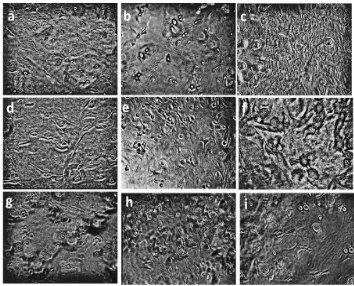
Figure 1 shows the variation in viability of MSCs during different experimental periods according to the effect of the material used. There was no significant difference in viable cell account among the materials and control group in the first and 3th days (F = 3.765 at P =.047 and F = .079 at P = .925 respectively). The significant difference among groups was only detected at the 7th day experimental period (F =9.044 at P = .003)
The ProRoot-MTA exhibited gradual increases in the MSCs account by time (0.281 ± 0.06, 0.222 ± 0.05 and 0.364 ± 0.05 at the 1st, 3rd and 7th day respectively) with statistically significant difference (F = 10.994at P =
0.001). The cell count in ProRoot-MTA group was greater than control and NeoMTA Plus groups in the first and seventh days (table1).
Figure 1: Photomicrographs of viable control untreated mesenchymal stem cells (a-c) and cells treated with either NeoMTA Plus (d-f) or ProRoot-MTA (g-i) and incubated for 1, 3 and 7 days respectively. Magnification
80
Table 1: MSCs viability after using the investigated root repair materials (Means ± standard deviation) Duration
Materials One day 3 days 7 days One Way ANOVA
Control 0.212 ± 0.08 0.243 ± 0.09 0.217 ± 0.06 F = 0.268 at P = 0.768 NeoMTA Plus 0.193 ± 0.02 0.234 ± 0.12 0.296 ± 0.07 F = 2.474 at P = 0.118 ProRoot-MTA 0.281 ± 0.06 0.222 ± 0.05 * 0.364 ± 0.05 F = 10.994at P = 0.001
F = 3.765 at P =.047 F = .079 at P = .925 F = 9.044 at P = .003
Scanning Electron Microscopy (SEM)
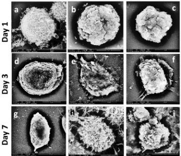
Upon scanning electron microscopic examination, there was no marked difference in the appearance of control untreated MSCs than that cells treated with either NeoMTA Plus and ProRoot-MTA (figure 2 a, b and c respectively) at the first day. Some of MSCs treated with ProRoot-MTA, showed an evidence of cell mitosis starting from day 1.
At day three, no changes were observed in control untreated MSCs except little cell elongation of some of them (figure 2 d). The cells treated with either NeoMTA Plus or ProRoot-MTA showed differentiation to osteoblasts- and fibroblasts-like cells (figure 2 e and f) isolated or collected in group (clump). The cells appeared ready for the proliferation stage.
At day seven, some of control untreated MSCs underwent apoptotic changes (figure 2 g). MSCs exposed to NeoMTA Plus or ProRoot-MTA become more elongated with evidence of mitotic activity (figure 2 hand i).
Figure 2: SEM photographs of control MSCs (a) and cells treated with either NeoMTA Plus (b) and ProRoot-MTA (c) showed no marked difference at day one. At the third and seventh days, compared with the control untreated MSCs (d and g), the cells treated with NeoMTA Plus (e and h) or ProRoot-MTA (f and i) showed
differentiation into osteoblast-like cells with an active process.
Alkaline phosphatase
81
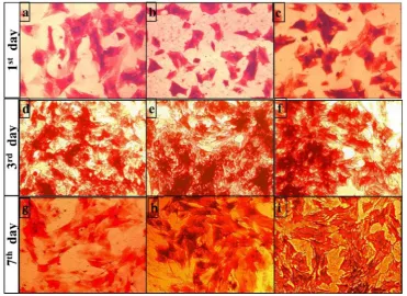
Figure 3: The Alkaline phosphatase staining of control MSCs at day 1 (a) appeared nearly similar to that treated with NeoMTA Plus (b) and ProRoot-MTA (c). At day 3 and 7, the control untreated MSCs (d and g) showed lighter staining density than that treated with NeoMTA Plus (e and h) and ProRoot-MTA (f and i) ; respectively.
Reverse-transcription Polymerase Chain Reaction and Quantitative Real-time Polymerase Chain Reaction
Bone morphogenetic protein-2
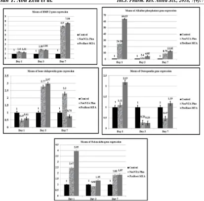
Both NeoMTA PlusNeo and ProRoot-MTA exhibited the upregulation of BMP-2 gene expression that was increased by time up to the seventh day. The greatest expression values were obtained by ProRoot-MTA all over the experimental periods (figure 4).
Alkaline phosphatase
Both investigated materials exhibited upregulation of alkaline phosphatase gene expression at day one with the greatest mean CT value obtained by ProRoot-MTA (64.19) compared with NeoMTA Plus (24.28) and control untreated MSCs. The expression markedly decreased at the third day, then re-increased at the seventh day (figure 4).
Bone sialoprotein
At day one, both investigated root repair materials exhibited downregulation of bone sialoprotein gene expression. However, at the third day, they showed marked upregulation of gene expression, This expression was maintained by NeoMTA Plus (mean CT value= 2.3) while down-regulation was obtained by ProRoot-MTA (mean CT value= 0.75) (figure 4).
Osteopontin
At day one, ProRoot_MTA exhibited upregulation of osteopontin gene expression (2.19) more than that obtained by NeoMTA Plus (1.11) as compared with control untreated MSCs. Whereas, both materials exhibited down regulation of this gene expression at the third day. At the seventh day, ProRoot-MTA was the only one to induce upregulation (1.19) (figure 4).
Osteocalcin
82
Figure 4: Gene expression of bone and osteogenesis genes (including BMP-2, alkaline phosphatase, bone sialoprotein, osteopontin and osteocalcin) induced by NeoMTA Plus and ProRoot-MTA compared with control
untreated MSCs.
DISCUSSION
Based on the prolonged setting time and handling difficulty of conventional MTA (ProRoot-MTA), fast setting NeoMTA Plus was developed to offer its bioactive and osteogenic potential. It was reported that there was some variation of their constituents as NeoMTA Plus contained higher aluminum and sulfur than conventional MTA that might interfere with its biological behavior [2]. There has been scarcity of studies evaluating their biocompatibility and bioactivity. In a previous in-vitro study, the evidence of bioactivity of NeoMTA Plus was reported as the calcium phosphate crystals precipitated on the material surface [2]. The current study was designed to evaluate their bioactivity on stem cells, and their ability to induce osteogenic differentiation when used as a root repair material.
Because MSCs of mice are multipotent cells, they are capable of in vitro differentiation into various non mesenchymal lineages such as calcified-forming cells including osteoblasts-, cementoblasts- and/or odontoblasts-like cells [9, 12]. This is why they were used in the current in vitro study. MSCs were proved to have high rate of proliferation with possible growth regulation [13], and to effectively evaluate the osteogenic potential of dental repair materials [14].
83
In the present study, ProRoot-MTA exhibited higher cell viability/proliferation along during the whole experiment than that obtained by NeoMTA Plus with significant difference at day seven. This finding might be related to higher calcium hydroxide (CH) content of the material. CH has been the main byproduct of both NeoMTA Plus and ProRoot-MTA, with extra amount within the original composition of ProRoot-MTA [2]. As CH dissociated to calcium and hydroxyl ions in the culture medium, the extracellular calcium might stimulate the cell viability [6].
Bone formation or bone repair has been a complex process based on the differentiation of MSCs present in pulp, periapical and/or periodontal tissues into osteoblast-like cells in association with the expression of different staged osteogenic-related factors and several genes [15] under suitable environment and/or therapeutic effect of repair material to generate calcified deposits, and repair the root defects [9, 16, 17].
In the current study, both investigated repair materials enhanced the gene expression of BMP-2 that gradually increased by time up to the seventh day, so the null hypothesis of this point was accepted. The calcium silicate-based material including MTA was reported to induce significant upregulation of BMP-2 gene expression in the rat dental pulp cells [18] and human periodontal cells [9]. It was suggested that BMP-2 is the most osteo-inductive factor [19], regulating cell differentiation and proliferation [10]. This fact has been supported by the scanning electron microscopic finding as the cell proliferation and differentiation increased gradually synchronized with the upregulation of BMP-2 gene expression. Maeda et al 2010 determined that cocultures of MTA with human periodontal ligament cells induced early upregulation of BMP-2 gene expression through calcium sensing receptor stimulation. This stimulation might attribute to increase the extracellular calcium ion that released from MTA into culture media [9].
The differentiation process can be enhanced not only by BMP-2 but also by increasing the expression of various pro-mineralization genes like alkaline phosphatase concentration and non-collagenous matrix proteins like bone sialoprotein, osteopontin and osteocalcin [16] that are responsible for bone deposition and maturation process [20]. Each gene has a specific role in different stages during bone repair. Alkaline phosphatase, osteopontin and bone sialoprotein genes accompany osteoblast differentiation [15, 20, 21].
The present result showed the upregulation of alkaline phosphatase by both repair materials during the whole experiment with the significant increase after the first day. ProRoot-MTA induced higher expression than NeoMTA Plus. Its expression was decreased at day three, then re-increased at day seven. This finding might be attributed to the role of alkaline phosphatase in cell differentiation at the first day of using material [20]. On the other hand, the increase of alkaline phosphatase production at day seven might be attributed to its ability to initiate mineralization by supplying phosphate during cyto-differentiation stage [10] as it has been encoded by differentiated osteoblasts and enhanced bone turnover [17].
The present finding reported the higher expression of alkaline phosphatase, osteopontin and osteocalcin by both investigated materials in day one than that of third and seventh days. The same finding was previously reported that might be attributed to pH-changes during setting reaction [17]. Furthermore, the release of calcium ions from MTA upregulated the biologic marker including BMP-2, alkaline phosphatase, bone sialoprotein and octeocalcin [9, 17].
Bone sialoprotein, or osteopontin and osteocalcin have been mineralized tissue- proteins having important roles in regulation and differentiation of mineral-cells like osteoblasts, odontoblasts and cementoblasts [22]. In the current study, bone sialoprotein (BSP) was highly expressed by both investigated materials compared with control at day 3, whereas, at day 7, its expression was down-regulated by ProRoot-MTA, and up-regulated by NeoMTA Plus. Bone sialoprotein has been a noncollagenous protein, essential for the organization of collagen matrix of new bone and nucleation as well as the growth of hydroxyapatite crystals [21]. It has been expressed by fully differentiated osteoblasts during the mineralization phase responsible for nucleation and growth of hydroxyapatite crystals to form highly specific mineralized tissues in the presence of high affinity of calcium ions [17, 21] enhancing the mineral (hydroxyapatite) crystal formation [13, 20]. Therefore, it has been expected to be expressed at later stages after the differentiation and maturation. The null hypothesis related to the expression of alkaline phosphatase and bone sialoprotein was rejected as both investigated materials had different behavior during the incubation periods.
84
investigated materials. While osteocalcin was formed by the mature osteoblasts during the bone mineralization [20].
Osteocalcin is an osteoblast-specific gene encoding a secreted protein essential for regulating the bone mineralization process. This fact was confirmed in the current study as the investigated materials showed their therapeutic effect on the undifferentiated mesenchymal stem cells that differentiated to osteoblast-like cells by encoding the bone morphogenetic protein-2 [21, 23]. Osteocalcin is the terminal differentiation of osteoblastic gene [21].
CONCLUSION
The relevance of the present result was that the upregulation of several bone-related genes expression including bone morphogenetic protein-2, alkaline phosphatase, bone sialoprotein, osteopontin and osteocalcin by MSCs exposed to either NeoMTA Plus or MTA indicated their ability to induce osteogenic activity. ProRoot-MTA had a greater osteogenic potential as it induced higher gene expression than that obtained by NeoProRoot-MTA Plus. Furthermore, rat bone marrow derived MSCs were effectively used to evaluate the osteogenic gene expression of dental materials.
REFERENCES
1. Roberts HW, Toth JM, Berzins DW, Charlton DG. Mineral trioxide aggregate material use in endodontic treatment: a review of the literature. Dental Materials 2008; 24: 149-164.
2. Abu Zeid ST, Alamoudi NM, Khafagi MG, Abou Neel EA. Chemistry and Bioactivity of NeoMTA Plus™ versus MTA Angelus® Root Repair Materials. Journal of Spectroscopy 2017; Article ID 8736428, 9 pages https://doi.org/10.1155/2017/8736428
3. Braga JM, Oliveira RR, Martins RC, Ribeiro Sobrinho AP. The effects of a mineral trioxide aggregate-based sealer on the production of reactive oxygen species, nitrogen species and cytokines by two macrophage subtypes. International Endodontic Journal 2014; 47: 909-919.
4. Faidah M, Noorwali A, Atta H, Ahmed N, Habib H, Damiati L, et al. Mesenchymal stem cell therapy of hepatocellular carcinoma in rats: Detection of cell homing and tumor mass by magnetic resonance imaging using iron oxide nanoparticles. Advances in clinical and experimental medicine: official organ Wroclaw Medical University 2017; 26: 1171-1178.
5. Sun Y, Luo T, Shen Y, Haapasalo M, Zou L, Liu J. Effect of iRoot Fast Set root repair material on the proliferation, migration and differentiation of human dental pulp stem cells in vitro. PloS one 2017; 12: e0186848.
6. Castro-Raucci LMSd, Oliveira IRd, Teixeira LN, Rosa AL, Oliveira PTd, Jacobovitz M. Effects of a novel calcium aluminate cement on the early events of the progression of osteogenic cell cultures. Brazilian Dental Journal 2011; 22: 99-104.
7. Chen I, Salhab I, Setzer FC, Kim S, Nah H-D. A new calcium silicate-based bioceramic material promotes human osteo-and odontogenic stem cell proliferation and survival via the extracellular signal-regulated kinase signaling pathway. Journal of Endodontics 2016; 42: 480-486.
8. Ding S-J, Shie M-Y, Hoshiba T, Kawazoe N, Chen G, Chang H-C. Osteogenic differentiation and immune response of human bone-marrow-derived mesenchymal stem cells on injectable calcium-silicate-based bone grafts. Tissue Engineering Part A 2010; 16: 2343-2354.
9. Maeda H, Nakano T, Tomokiyo A, Fujii S, Wada N, Monnouchi S,et al. Mineral trioxide aggregate induces bone morphogenetic protein-2 expression and calcification in human periodontal ligament cells. Journal of Endodontics 2010; 36: 647-652.
10. Asgary S, Nazarian H, Khojasteh A, Shokouhinejad N. Gene expression and cytokine release during odontogenic differentiation of human dental pulp stem cells induced by 2 endodontic biomaterials. Journal of Endodontics 2014; 40: 387-392.
85
12. Somoza R, Juri C, Baes M, Wyneken U, Rubio FJ. Intranigral transplantation of epigenetically induced BDNF-secreting human mesenchymal stem cells: implications for cell-based therapies in Parkinson's disease. Biology of Blood and Marrow Transplantation 2010; 16: 1530-1540.
13. Kirkham GR, Cartmell SH. Genes and proteins involved in the regulation of osteogenesis. Ashammakhi N, Reis R, Chiellini E, editores. Topics in Tissue Engineering 2007; 3., pp.22
14. Donzelli E, Salvade A, Mimo P,Vigano M, Morrone M, Papagna R,et al. Mesenchymal stem cells cultured on a collagen scaffold: In vitro osteogenic differentiation. Archives of Oral Biology 2007; 52: 64-73.
15. Shen J, Hovhannisyan H, Lian JB,Montecino MA, Stein GS, Stein JL, et al. Transcriptional induction of the osteocalcin gene during osteoblast differentiation involves acetylation of histones h3 and h4. Molecular Endocrinology 2003; 17: 743-756.
16. Duncan HF, Smith AJ, Fleming GJP, Cooper PR. HDACi: cellular effects, opportunities for restorative dentistry. Journal of Dental Research 2011; 90: 1377-1388.
17. Lee B-N, Lee K-N, Koh J-T, Min K-S, Chang H-S, Hwang I-N, et al. Effects of 3 Endodontic Bioactive Cements on Osteogenic Differentiation in Mesenchymal Stem Cells. Journal of Endodontics 2014; 40: 1217-1222.
18. Yasuda Y, Ogawa M, Arakawa T, Kadowaki T, Saito T. The effect of mineral trioxide aggregate on the mineralization ability of rat dental pulp cells: an in vitro study. Journal of Endodontics 2008; 34: 1057-1060.
19. Seo M-S, Hwang K-G, Lee J, Kim H, Baek S-H. The effect of mineral trioxide aggregate on odontogenic differentiation in dental pulp stem cells. Journal of Endodontics 2013; 39: 242-248.
20. Wang J-j, Ye F, Cheng L-j, Shi Y-j, Bao J, Sun H-q, et al. Osteogenic differentiation of mesenchymal stem cells promoted by overexpression of connective tissue growth factor. Journal of Zhejiang University SCIENCE B 2009; 10(5): 355–367.
21. Hsu Y-L, Chang J-K, Tsai C-H, Chien T-TC, Kuo P-L. Myricetin induces human osteoblast differentiation through bone morphogenetic protein-2/p38 mitogen-activated protein kinase pathway. Biochemical Pharmacology 2007; 73: 504-514.
22. Harris NL, Rattray KR, Tye CE, Underhill TM, Somerman MJ, D’errico JA, et al. Functional analysis of bone sialoprotein: identification of the hydroxyapatite-nucleating and cell-binding domains by recombinant peptide expression and site-directed mutagenesis. Bone 2000; 27: 795-802.