Frontiers in Dentistry
Evaluation of the Shear Bond Strength and Adhesive
Remnant Index in Debonding of Stainless Steel
Brackets Assisted with Nd:YAG Laser Irradiation
Maryam Nasiri
1, Amir Hossein Mirhashemi
1, Ardavan Etemadi
2, Mohammad
Javad Kharazifard
3, Elahe Soltanmohamadi Borujeni
4, Maryam Javaheri
Mahd
1, Pegah Khazaei
5, Mohammad Sadegh Ahmad Akhoundi
1,2,3*1. Department of Orthodontics, School of Dentistry, Tehran University of Medical Sciences, Tehran, Iran
2. Laser Research Center of Dentistry, Dentistry Research Institute, Tehran University of Medical Sciences, Tehran, Iran 3. Dental Research Center, Dentistry Research Institute, Tehran University of Medical Sciences, Tehran, Iran
4. Department of Orthodontics, School of Dentistry, Qom University of Medical Sciences, Qom, Iran
5. Research Center of Caries Prevention, Dentistry Research Institute, Tehran University of Medical Sciences, Tehran, Iran
Article Info A B S T R A C T
Article type:
Original Article Objectives: and adhesive remnant index (ARI) in debonding of stainless steelThe purpose was to compare shear bond strength (SBS), pulp temperature, brackets from enamel surface using neodymium-doped yttrium aluminum garnet (Nd:YAG) laser versus the conventional debonding method.
Materials and Methods: Forty-eight extracted premolars were bonded to stainless steel brackets. The samples were divided into three experimental groups and one control group. In the first three groups, Nd:YAG laser was used for debonding with the power of 1, 1.5, and 2 W, respectively, for 10 seconds. The SBS and ARI of the samples were assessed. Pulp temperature was recorded before and after irradiation. Two samples from each group were used for determining enamel morphology after debonding using scanning electron microscopy (SEM).
Results: The mean SBS in the groups was 33.05, 28.69, 24.37, and 31.53 MPa, respectively, with no statistically significant differences (P=0.205). Significant differences in post-irradiation temperature were noted among the lased groups (P=0.000). Debonding mainly occurred at the adhesive-enamel interface in the 1-W laser and control groups and at the bracket-adhesive interface in the 1.5-W and 2-W laser groups. Enamel structure was amorphous and irregular following laser irradiation.
Conclusion: Based on the results of this study, the use of Nd:YAG laser could not significantly affect the SBS. Therefore, this laser would not be suitable for debonding of metal brackets. The use of a 2-W laser could significantly raise the pulpal temperature. Nd:YAG laser renders a more heterogeneous enamel morphology compared to conventional debonding methods.
Keywords: Nd:YAG Laser; Dental Debonding; Orthodontic Brackets
Article History:
Received: 8 May 2018 Accepted: 11 August 2018 Published: 20 January 2019
* Corresponding author:
Laser Research Center of Dentistry, Dentistry Research Institute, Tehran University of Medical Sciences, Tehran, Iran; Dental Research Center, Dentistry Research Institute, Tehran University of Medical Sciences, Tehran, Iran
Email: ahmadakh@tums.ac.ir
Cite this article as: Nasiri M, Mirhashemi AH, Etemadi A, Kharazi Fard MJ, Soltanmohamadi Borujeni E, Javaheri Mahd M, et al. Evaluation of the Shear Bond Strength and Adhesive Remnant Index in Debonding of Stainless Steel
INTRODUCTION
Proper bonding of brackets to enamel can significantly facilitate orthodontic treatments. Since the 1960s to the present, various changes have been made to this process, including new techniques and equipment for bonding,
rebonding, and debonding [1-4]. One of the most brilliant methods for improving the quality of the bonding process is to use different lasers in various stages of treatment from the beginning to the time of removal of resin
38
Front Dent, Vol. 16, No. 1, Jan-Feb 2019
of the treatment [5].
Lasers can be used for enamel preparation prior to bonding instead of conventional acid-etching. Although the results may be controversial, many researchers have concluded that the shear bond strength (SBS) of stainless steel brackets to enamel decreases following enamel preparation using lasers [3-5].
Several articles have been published on the application of lasers for bonding of brackets to enamel, but few studies have focused on using lasers in debonding procedures. Since ceramic brackets are popular among adult patients, different methods have been introduced for debonding them from enamel with the lowest frequency of enamel fractures and cracks, such as ultrasonic methods, use of special pliers, and laser therapy [6-11]. Carbon dioxide (CO2), neodymium-doped yttrium aluminum garnet (Nd:YAG), and erbium-doped yttrium aluminium garnet (Er:YAG) lasers are the most popular lasers used for this goal as they can cause degradation in the adhesive layer, thereby facilitating the debonding process [12,13].
Tocchio et al [14] believe that the laser energy can degrade the adhesive layer through thermal softening, thermal ablation, and photoablation. It is postulated that the heat penetrates the tooth structure and damages the dental pulp. Although some authors have stated that an increase in the pulp temperature by 5.5°C might cause pulpal necrosis [15], some reports showed that appropriate laser irradiation can decrease the bond strength of brackets without significant increase of pulp temperature [16-19].
Feldon et al [20] concluded that diode lasers decrease the force required for debonding of monocrystalline ceramic brackets without significant increase of pulp temperature. Oztoprak et al [21] showed that the force required to remove polycrystalline ceramic brackets can be reduced by using Er:YAG laser. Nevertheless, the laser type, the technique, and the characteristics of brackets have to be fully considered to prevent undesirable results [22-26].
Nd:YAG laser is one of the lasers used for debonding purposes, especially for ceramic brackets [6]. Since the use of this laser for debonding of metal brackets has been evaluated only in one article [27], it is rational to perform a study to assess the effects of this laser in
debonding of metal brackets. Therefore, the present study was performed to compare the debonding of stainless steel brackets from enamel surface using Nd:YAG laser and the conventional debonding method in terms of the SBS, pulp temperature, and adhesive remnant index (ARI).
MATERIALS AND METHODS
1. Teeth were irradiated by a 1-W Nd:YAG laser (Fotona, LightWalker AT-S, M021-5AF/1S, Slovenia) for 10 seconds at the enamel-bracket interface for composite softening (frequency=20 Hz, pulse duration=0.2 milliseconds).
2. Teeth were irradiated by a 1.5-W Nd:YAG laser.
3. Teeth were irradiated by a 2-W Nd:YAG laser.
4. Teeth were debonded conventionally using the cutting blade of a universal testing machine (Zwick/Roell, Ulm, Germany). An access cavity was prepared on the occlusal surface of each sample of the laser-treated groups to measure and compare the pre- and post-irradiation pulp temperatures using a thermocouple (K-type; Delta Electronics Inc., Mashhad, Iran).
In order to reach the maximum effect of laser irradiation, the debonding process should be done as soon as possible. Therefore, immediately after laser irradiation, the samples in the first three groups were placed in the universal testing machine with a 1-kN load cell to measure the SBS. The blade had an occlusogingival direction and moved downward towards the tooth-bracket interface at a crosshead speed of 0.5 mm/minute. The loads were recorded in Newton (N) and were converted to Megapascal (MPa) automatically using a computer according to the bracket's base area which was precisely calculated using an electronic gauge:
SBS = debonding force (N)/surface area of the bracket (mm2)
After debonding, the samples were stored in distilled water. The ARI was assessed using the stereomicroscope at 10× magnification according to the study by Oliver (1986) [28], with scores from 1 to 5:
Score 1 = 100% adhesive remnant left on the tooth.
Score 2 = more than 90% adhesive remnant left on the tooth.
Score 3 = 10-90% adhesive remnant left on the tooth.
Score 4 = less than 10% adhesive remnant left on the tooth.
Score 5 = no adhesive remnant left on the tooth.
As the last step, two samples from each group were assessed under a scanning electron microscope (SEM; TESCAN VEGA, Czech
Republic) to evaluate the enamel morphology. Data were analyzed in SPSS 22 software (IBM Co., Chicago, IL, USA). One-way analysis of variance (ANOVA) was used to compare the SBS of the four groups. The level of significance was set at P<0.05.
RESULTS 1. SBS:
According to Table 1, there were no significant differences among the four groups in terms of the SBS.
Table 1. Mean, standard deviation (SD), minimum and
maximum shear bond strength (SBS) values (MPa)
P value Max
Min SD
Mean Groups
0.205 51.94
17.97 10.94
33.05
1-W laser
48.95 11.96
11.91 28.69
1.5-W laser
39.51 13.87 6.86
24.37
2-W laser
49.37 12.67 11.38
31.53
Control
SD: Standard Deviation
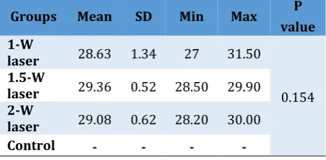
2. Pre-irradiation pulp temperature:
No significant differences were found among the irradiated groups in terms of the pre-irradiation pulp temperature (Table 2).
3. Post-irradiation pulp temperature:
According to Table 3, the lased groups showed statistically significant differences with regard to post-irradiation pulp temperatures, and the maximum post-irradiation temperature was noted in the 2-W laser group.
Table 2. Pre-irradiation pulp temperature (°C) of the
three laser-treated groups.
P value Max
Min SD
Mean Groups
0.154 31.50
27 1.34 28.63
1-W laser
29.90 28.50
0.52 29.36
1.5-W laser
30.00 28.20
0.62 29.08
2-W laser
- -
- -
Control
40
Front Dent, Vol. 16, No. 1, Jan-Feb 2019
Table 3. Post-irradiation pulp temperature (°C) of
the three laser-treated groups.
P value Max Min SD Mean Groups <0.001 34.50 29.10 1.65 31.88 1-W laser 34.90 32.50 0.83 33.59 1.5-W laser 37.00 33.50 1.05 34.82 2-W laser - - - - Control
SD: Standard Deviation
4. Temperature difference (ΔT):
There was a significant difference in ΔT among
the studied groups (Table 4).
Table 4. Difference between pre- and
post-irradiation pulp temperatures (°C)
P value Max Min SD Mean Groups <0.001 6.50 0.20 1.57 3.25 1-W laser 5.30 2.90 0.75 4.22 1.5-W laser 7.00 4.70 0.68 5.74 2-W laser - - - - Control
SD: Standard Deviation
5. ARI:
According to Table 5, it can be concluded that in the 1-W laser and control groups, most of the composite was removed from enamel surface during bracket debonding, i.e. debonding mainly occurred at the adhesive-enamel interface, increasing the risk of enamel fractures and cracks.
Table 5. Adhesive remnant index (ARI) scores of the
four studied groups
ARI N(%)
Groups 1 2 3 4 5
2 (16.7) 4 (33.3) 4 (33.3) 2 (16.7) 0 1-W laser 0 2 (16.7) 7 (58.3) 3 (25) 0 1.5-W laser 0 1 (8.3) 6 (50) 3 (25) 2 (16.7) 2-W laser 2 (16.7) 4 (33.3) 5 (41.7) 1 (8.3) 0 Control
In contrast, in the 1.5-W and 2-W laser groups, a higher tendency existed towards debonding at the bracket-adhesive interface with a lower risk of enamel damage (Fig. 1).
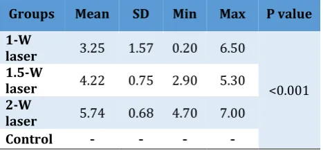
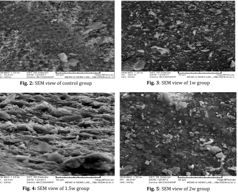
6. SEM analysis:
Enamel morphology after acid-etching is categorized into three major types:
1. Type 1: honeycomb appearance due to the preferential removal of the core material, leaving prisms peripherally intact.
2. Type 2: preferential dissolution of peripheral regions of the prisms, leaving the prism cores intact.
3. Type 3: both types 1 and 2 can be seen in SEM analysis.
The honeycomb appearance (type 1) was clearly evident in the control group after debonding. In spite of heavy forces used for debonding the brackets, no obvious enamel crack was found in these samples (Fig. 2). In contrast to this distinct morphology in the control group, the lased samples showed an irregular and coarse structure with no obvious patterns in any region (Fig. 3-5). In the samples of group 2, some enamel cracks were found, which can be a sign of intense damage to enamel. Nevertheless, the depths of the cracks could not be estimated because SEM images were two-dimensional (2D). In one slide, the difference between the etched enamel under the bracket base and the enamel at the bonding interface was obvious; the enamel under the bracket showed a honeycomb appearance, while the interfacial enamel was amorphous and irregular.
There was no difference between the three laser-treated groups with regard to enamel morphology. One can conclude that Nd:YAG laser can damage enamel and create irreversible cracks and micro-fractures.
DISCUSSION
Fig. 1: ARI index of all groups after debonding
Fig. 2: SEM view of control group Fig. 3: SEM view of 1w group
Fig. 4: SEM view of 1.5w group Fig. 5: SEM view of 2w group
There is a lack of literature on the application of lasers in debonding of stainless steel brackets since the majority of the related studies have focused on the effect of laser debonding on ceramic brackets [6-10,12,14-16,20,21,25,26]. In a comprehensive review on this topic, Ghazanfari et al [25] concluded that irradiation of Nd:YAG laser can be considered as an efficient
and safe way to reduce the SBS and debonding time of ceramic brackets with minimal impacts on the intrapulpal temperature and enamel surface.
1. SBS:
application of a high-peak-power Nd:YAG laser at 2.0 J or more is useful for debonding of ceramic brackets. Lasers with energies lower than 2.0 J did not show any significant reduction in the SBS [6]. Iijima et al [12] investigated the effects of CO2 laser debonding of ceramic brackets on the mechanical properties of enamel. They found that the SBS decreased under all laser irradiation conditions, irrespective of the output power of the laser; this phenomenon can facilitate bracket debonding. Similar results have been reported by other researchers after using various lasers including Er:YAG laser (Oztoprak et al [22]), diode laser (Feldon et al [20] and Nalbantgil et al [16]), and CO2 laser (Saito et al [26]).
2. Pulp temperature:
Zach and Cohen [29] stated that the maximum safe temperature increase for the dental pulp is lower than 5.5°C. In the present study, significant differences were noted among the three laser-treated groups with regard to the post-irradiation pulp temperature. The maximum temperature increase was detected in the 2-W laser group (5.74°C), which was a little higher than the critical temperature increase threshold (5.5°C). Lai et al [27] found that using Nd:YAG laser for 5 minutes at a high energy can cause irreversible damage to pulpal tissue. They assessed the effect of using Nd:YAG laser for metal bracket debonding instead of conventional methods. The authors found a significant difference in pulpal temperature change among groups with different laser energies [27]. This finding is in line with the results of the present study as the highest temperature change was related to the samples under 2-W laser emission.
Hayakawa [6] concluded that when using Nd:YAG for ceramic bracket debonding, the maximum temperature rise measured on the pulpal walls at the lasing points was 5.1°C. Iijima et al [12] stated that lasers with higher output powers are more harmful to the dental pulp due to more temperature increase (200°C with 5-W and 6-W outputs versus 100°C to 150°C for 3-W and 4-W outputs). The authors mentioned that the results of temperature change should be interpreted with caution due to some differences between the vital pulp and the samples investigated at room temperature; as the pulpal cavity is located far from the tooth surface, the propagation behavior of heat might be different from the experimental situation
[12]. Nevertheless, some studies have reported a mean temperature increase of lower than 5.5°C [30-34]. Also, Dostalova et al [23] stated that temperature increase after using Er:YAG laser for debonding of metal brackets is trivial.
3. ARI:
In the 1-W laser and control groups of the present study, most of the brackets were debonded at the adhesive-enamel interface with an increased risk of enamel damage because of the higher adhesive bond strength. In contrast, the samples in the 1.5-W and 2-W laser groups had lower adhesive bond strengths; therefore, more composite remnants were left on the tooth surface with a lower risk of enamel injury. Nalbantgil et al [16] found a reverse correlation between the ARI and SBS. Tehranchi et al [13] stated that conventional mechanical debonding methods can be more dangerous due to more bond failures at the adhesive-enamel interface. Yassaei et al [30] performed a study on ceramic bracket debonding using a diode laser and reported no significant difference between the lased samples and the samples debonded conventionally with regard to the ARI. In contrast, Anand et al [32] stated that using a diode laser for debonding purposes can increase the ARI, thereby facilitating the debonding and reducing enamel fractures.
4. Enamel morphology:
We found an amorphous and irregular enamel structure in lased areas, irrespective of the power of the laser, while the samples in the control group and the areas of enamel not affected by laser irradiation in the experimental groups showed the typical honeycomb appearance without any irregularity or crack. In some areas under the emission of the 1.5-W laser, deep cracks were obvious, which can be a sign of intense injuries to enamel.
After using Er:YAG laser instead of the conventional acid-etching technique in bonding and rebonding procedures, Ahmad Akhoundi et al [17] stated that laser emission makes enamel structure more heterogeneous and irregular compared to the homogenous enamel morphology following conventional tooth surface preparation.
groups. The 60mJ-laser-treated group showed vertical scratches on the enamel surface. In the 120J-laser-treated group, the enamel surface was covered by craters and cracks, while the 160mJ-laser-treated group showed a completely altered enamel structure with columns separated by voids and with a glass-like surface. The authors reported that laser treatment at low energy levels (<60 mJ) produces a protective glass-like surface without loss of integrity, while higher energy levels lead to the formation of craters and cracks [35]. Nevertheless, Ahrari et al [15], Dostalova et al [23], Mundethu et al [34], and Keller and Hibst [19] found opposite results and stated that laser irradiation and conventional debonding methods are similar in terms of enamel damage. It can be inferred from the above-mentioned studies that our research has the same direction as many other relevant studies regarding SBS, ARI, enamel morphology, and pulpal temperature increase. Nd:YAG laser might have various advantages in debonding of stainless steel brackets; however, its probable long-term effects on the enamel morphology and pulp temperature should not be ignored. Further research seems to be useful for more thorough evaluations of the potential benefits and side effects of this brilliant and novel modality for bracket debonding.
CONCLUSION
Based on the results of the present study, the use of Nd:YAG laser could not significantly affect the SBS. Therefore, this laser would not be suitable for debonding of metal brackets. The use of a 2-W laser could significantly raise the pulpal temperature. This laser can adversely affect the enamel morphology, making the tooth structure more heterogeneous compared to the use of conventional debonding methods. The long-term effects of this phenomenon are still unknown.R
REFERENCES
1. Hashim NT, Gasmalla BG, Sabahelkheir AH, Awooda AM. Effect of the clinical application of the diode laser (810 nm) in the treatment of dentine hypersensitivity. BMC Res Notes. 2014 Jan 13; 7:31.
2. Maenosono RM, Bim Junior O, Duarte MA, Palma-Dibb RG, Wang L, Ishikiriama SK. Diode laser irradiation increases microtensile bond strength of dentin. Braz Oral Res. 2015;
29:1-5.
3. Van Waveren Hogervorst WL, Feilzer AJ, Prahl-Andersen B. The air-abrasion technique versus the conventional acid-etching technique: A quantification of surface enamel loss and a comparison of shear bond strength. Am J Orthod Dentofacial Orthop. 2000 Jan; 117(1):20-6. 4. Van Meerbeek B, De Munck J, Yoshida Y, Inoue S, Vargas M, Vijay P, et al. Buonocore memorial lecture. Adhesion to enamel and dentin: current status and future challenges. Oper Dent. 2003 May-Jun; 28(3):215-35. 5. Marimoto AK, Cunha LA, Yui KC, Huhtala MF, Barcellos DC, Prakki A, et al. Influence of Nd:YAG laser on the bond strength of self-etching and conventional adhesive systems to dental hard tissues. Oper Dent. 2013 Jul-Aug; 38(4):447-55.
6. Hayakawa K. Nd:YAG laser for debonding ceramic orthodontic brackets. Am J Orthod Dentofacial Orthop. 2005 Nov; 128(5):638-47.
7. Swartz ML. Ceramic brackets. J Clin Orthod. 1988 Feb; 22(2):82-8.
8. Bishara SE, Forrseca JM, Fehr DE, Boyer DB. Debonding forces applied to ceramic brackets simulating clinical conditions. Angle Orthod. 1994; 64(4):277-82.
9. Bishara SE, Fehr DE, Jakobsen JR. A comparative study of the debonding strengths
of different
ceramic brackets, enamel conditioners, and adhesives. Am J Orthod Dentofacial Orthop. 1993 Aug; 104(2):170-9.
10. Jou GL, Leung RL, White SN, Zernik JH. Bonding ceramic brackets with light-cured glass ionomer cements. J Clin Orthod. 1995 Mar; 29(3):184-7.
11. Krejci I, Simunovic K, Lutz F. [Substance ablation with a superpulsed CO2 laser]. [Article in German]. Schweiz Monatsschr Zahnmed. 1992; 102(6):693-9.
12. Iijima M, Yasuda Y, Muguruma T, Mizoguchi I. Effects of CO2 laser debonding of a ceramic bracket on the mechanical properties of enamel. Angle Orthod. 2010 Nov; 80(6):1029-35.
13. Tehranchi A, Fekrazad R, Zafar M, Eslami B, Kalhori KA, Gutknecht N. Evaluation of the effects of CO2 laser on debonding of orthodontics porcelain brackets vs. the conventional method. Lasers Med Sci. 2011 Sep; 26(5):563-7.
Standing KG. Laser debonding of ceramic orthodontic brackets. Am J Orthod Dentofacial Orthop. 1993 Feb; 103(2):155-62.
15. Ahrari F, Heravi F, Fekrazad R, Farzanegan F, Nakhaei S. Does ultra-pulse CO(2) laser reduce the risk of enamel damage during debonding of ceramic brackets? Lasers Med Sci. 2012 May; 27(3):567-74.
16. Nalbantgil D, Tozlu M, Oztoprak MO. Pulpal Thermal Changes following Er-YAG Laser Debonding of Ceramic Brackets. Sci World J. 2014; 2014:912429.
17. Ahmad Akhoundi MS, Etemadi A, Nasiri M, Soltanmohamadi Borujeni E. Comparison of enamel morphologic characteristics after conditioning with various combinations of acid etchant and Er:YAG laser in bonding and rebonding procedures: a SEM analysis. J Dent (Tehran). 2017 May; 14(3):144-152.
18. Sawan MN, Hussain N, Alkurdi MM. Etching of enamel by laser energy for direct bonding of orthodontic appliance and evaluation of shear bond strength. Energy Procedia. 2015 Aug; 74: 1452-58.
19. Keller U, Hibst R. Experimental studies of the application of the Er:YAG laser on dental hard substances: II. Light microscopic and SEM investigations. Lasers Surg Med. 1989; 9(4):345-51.
20. Feldon PJ, Murray PE, Burch JG, Meister M, Freedman MA. Diode laser debonding of ceramic brackets. Am J Orthod Dentofacial Orthop. 2010 Oct; 138(4):458-62.
21. Oztoprak MO, Nalbantgil D, Erdem AS, Tozlu M, Arun T. Debonding of ceramic brackets by a new scanning laser method. Am J Orthod Dentofacial Orthop. 2010 Aug; 138(2):195-200.
22. Verdonschot EH, Bronkhorst EM, Wenzel A. Approximal caries diagnosis using fiber-optic transillumination: a mathematical adjustment to improve validity. Community Dent Oral Epidemiol. 1991 Dec; 19(6):329-32.
23. Dostalova T, Jelinkova H, Remes M, Šulc
J, Němec M. The Use of the Er:YAG Laser for
Bracket Debonding and Its Effect on Enamel Damage. Photomed Laser Surg. 2016 Sep; 34(9):394-9.
24. Zulkifli N, Suhaimi FM, Razab AAM, Jaafar MS, Mokhtar N. The Use of Nd:YAG Laser for Ablation of Dental Material. 5th
International Conference on Biomedical Engineering and Technology. 2015;81(8):40-7. 25. Ghazanfari R, Nokhbatolfoghahaei H, Alikhasi M. Laser-aided ceramic bracket debonding: a comprehensive review. J Lasers Med Sci. 2016 Winter; 7(1):2-11.
26. Saito A, Namura Y, Isokawa K, Shimizu N. CO2 laser debonding of a ceramic bracket bonded with orthodontic adhesive containing thermal expansion microcapsules. Lasers Med Sci. 2015 Feb; 30(2):869-74.
27. Lai RF, Wang HY, Chen T, Liu XN. [Pulsed Nd:YAG laser-aided debonding for removing the metal brackets]. [Article in Chinese]. Zhonghua Kou Qiang Yi Xue Za Zhi. 2010 Jul; 45(7):407-10.
28. Oliver RG. Bond strength of orthodontic attachments to enamel from unerupted and erupted young permanent teeth. Eur J Orthod. 1986 May; 8(2):123-6.
29. Zach L, Cohen G. Pulp response to externally applied heat. Oral Surg Oral Med Oral Pathol. 1965 Apr; 19:515-30.
30. Yassaei S, Soleimanian A, Nik ZE. Effects of Diode Laser Debonding of Ceramic Brackets on Enamel Surface and Pulpal Temperature. J Contemp Dent Pract. 2015 Apr 1; 16(4):270-4. 31. Macri RT, de Lima FA, Bachmann L, Galo R, Romano FL, Borsatto MC, et al. CO2 laser as auxiliary in the debonding of ceramic brackets. Lasers Med Sci. 2015 Sep; 30(7):1835-41. 32. Anand P, Anand PB, Prabhakar R, Rajvikram N, Rajakumar P, Atali VR, et al. Immediate and delayed effects of diode laser on debonding of ceramic brackets: an in vitro study. J Contemp Dent Pract. 2016 Apr 1; 17(4):275-81.
33. Yilanci H, Yildirim ZB, Ramoglu SI. Intrapulpal Temperature Increase During Er:YAG Laser-Aided Debonding of Ceramic Brackets. Photomed Laser Surg. 2017 Apr; 35(4):217-222.
34. Mundethu AR, Gutknecht N, Franzen R. Rapid debonding of polycrystalline ceramic orthodontic brackets with an Er:YAG laser: an in vitro study. Lasers Med Sci. 2014 Sep; 29(5):1551-6.