Effect of Vitamin E and Sodium Selenite on the Expression
of
Bax
and
Bcl2
Genes and Renal Histopathology in the
Electromagnetic Field-Exposed Mice
Introduction
Electromagnetic waves are a form of energy that are emitted by charged particles and create magnetic fields through passing a wave from a path in space. Electromagnetic fields (EMFs) are produced following the development of various devices in the world (1). More than 3 billion people are exposed to the mentioned EMF every day. Owing to the overuse of devices that generate electrical waves, exposure to EMF is inevitable. EMF and its harmful effects on the human body, therefore, are important issues that have recently been considered in the medical and engineering areas (2).
Today, there are controversies about the biological adverse effects of EMF on the human body. In recent studies, the adverse effects of EMF on reproductive system, hormonal cycle, induction of cancer, and effects on other organs of the body have clearly been observed (3). The effects of EMF on the kidneys have been investigated from a variety of dimensions, of which two dimensions of morphologic aspects and changes in the activation processes of antioxidant enzymes are the most notable. In
the first case, studies have shown that EMF leads to the formation of small and large cysts in different regions of the kidney, inflammation of epithelial cells and glomeruli, necrosis of these regions, and reduction of the number of tubules, which ultimately results in pathological changes in the organ (4,5). Regarding the antioxidant enzymes, depending on the length of exposure of the kidneys to EMF, decreased superoxide dismutase (SOD), glutathione peroxidase (GPx), and total antioxidant capacity (TAC), as well as increased malondialdehyde (MDA) have been observed (6).
Penetration of magnetic and electrical fields into the cell membranes increases the risk of DNA damage and tissue damage by prolonging free radical stability, altering membrane potential, reducing the activity of antioxidant enzymes, and subsequently altering the ROS metabolism (7,8). Previous studies have confirmed the destructive effects of oxidative stress occurred due to the EMF exposure (5,9). These studies have also shown that the administration of antioxidants can reduce the adverse effects of EMF.
Abstract
Objectives:Electromagnetic field (EMF) causes damages to the kidney through the increase of oxidative stress, and the use of antioxidants can reduce the effects caused by EMF. The present study investigated the effects of vitamin E and sodium selenite on the expression levels of Bax and Bcl2 genes and renal histopathology in the EMF-exposed mice.
Materials and Methods: In this study, 48 mice with the approximate weight of 24-28 g were used. The mice were randomly assigned to six groups (n=8): 1) control; 2) EMF; 3) EMF + vitamin E; 4) EMF + sodium selenite; 5) EMF + vitamin E + sodium selenite; and 6) vitamin E + sodium selenite. Mice were exposed to the EMF for 4 hours a day for 2 months. After this duration, the mice were sacrificed by cervical dislocation and the right kidney was taken out for the evaluation of the histopathological changes and the left kidney was used to determine the expression of Bax and Bcl2 genes by real-time polymerase chain reaction (PCR).
Results: Statistical analyses showed that EMF significantly decreased the kidney corpuscle diameter and bcl2 gene expression. Moreover, increased urinary space, basal membrane thickness, and increased expression of Bax gene were observed compared to the control group (P < 0.05). However, the treatment of mice exposed to EMF with vitamin E and sodium selenite significantly decreased the effects of EMF on these parameters (P < 0.05).
Conclusions: The results of this study showed that vitamin E and sodium selenite could significantly inhibit the destructive effects of EMF on the expression of Bax and bcl2 genes and prevent the histopathological changes in the kidneys of mice.
Keywords:EMF, Kidney, Vitamin E, Sodium selenite, Bax, Bcl2
Mohammad Eyvazi1, Hamid Tayefi1, Ali Abedelahi1, Ramin Salimnejad1, Abbas Majdi1*
Open Access Original Article
Crescent Journal of Medical and Biological Sciences
Received 17 May 2018, Accepted 26 November 2018, Available online 20 December 2018 eISSN 2148-9696
Vitamin E is a lipid-soluble vitamin found in the cell membrane, which protects the cell membrane against oxidative damage. It consists of four tocopherols and four tocotrienols. Moreover, vitamin E supplementation influences the cellular reply to oxidative stress through the inflection of signal-transduction pathway. In fact, this vitamin has been well known as an antioxidant that plays an important role in neutralizing the toxic effects of reactive
oxygen species (ROS) (10). Angiotensin converting
enzyme activity, blood pressure, and GFR remained nearly at normal level under vitamin E supplementation (11).
Sodium selenite is also an essential element in human health. There are much evidence conforming that this mineral plays an important role in normal growth and reproductive ability of animals and humans (12). Sodium selenite is a necessary co-Factor for glutathione peroxidase. This substance, in combination with protein, generates selenoprotein, which is a critical antioxidant enzyme protecting the cell against free radical damage (11).
Given that increased oxidative stress and cellular damage is one of the mechanisms of kidney tissue damage caused by EMF exposure, as well as considering that there are contradictory results about the effects of EMF and inhibitory and antioxidant impacts of vitamin E and sodium selenite on urinary system, the purpose of this study was to investigate the effects of vitamin E and sodium selenite on the expression of Bax and Bcl2 genes and renal histopathology in the EMF-exposed mice.
Materials and Methods
Study Design and Experimental Groups
In this experimental study, 48 mice with approximate weight of 24-28 g were used. The mice were prepared from the animals’ house of Tabriz University of Medical Sciences and kept in a standard condition (temperature 24 ± 2°C, with a light/dark cycle of 12 hours). Having the mice adapted to the conditions of the animal house, they were divided randomly into 6 groups, each including 8 animals, and treated as follows: 1) control 2); EMF; 3) EMF + vitamin E; 4) EMF + sodium selenite; 5) EMF + vitamin E + sodium selenite; and 6) vitamin E + sodium selenite.
Electromagnetic radiation of 3 mT and 50 Hz was radiated 4 hours daily for 2 months. Vitamin E (1.35 mg/kg body weight) and sodium selenite (0.1 mg/kg body weight) were administrated by a gavage daily for 2 months (4). In the end, the mice were sacrificed by cervical dislocation and their kidneys were removed. The
left kidney was used for the assessment of Bax and Bcl-2 gene expression by real-time PCR. The right kidney was also paraffin embedded after the tissue processing, and finally, 5 µm sections were prepared and evaluated by the hematoxylin and eosin (H&E) staining (for examination of tissue changes) and periodic acid–Schiff (PAS) (for measuring the thickness of the basement membrane).
RNA Extraction and DNA Synthesis
In order to evaluate the expression of Bax and Bcl-2 genes, the kidney tissue was crushed using liquid nitrogen and mortar, and then homogenized using TRIZOL solution according to the manufacturer’s instructions. Next, the total RNA was extracted and its purity was determined via a NanoDrop. Then, cDNA was synthesized using the oligo primers and Revert AID TM first strand cDNA synthesis kit (manufactured by Fermentas Company).
Real-Time PCR
The NCBI website was used to design the primers. Table 1 shows the primers of Bax and Bcl2 genes and also the GAPDH gene as a reference gene. According to the kit instructions, the multiplication reaction was carried out as triple repetition in 20 μL volume, containing 10 μL SYBR Green, 0.5 μL forward primer, 0.5 μL reverse primer, 2 μL synthesized cDNA, and 7μL distilled water. The real-time PCR reaction was set up in three steps: 95°C for 15 seconds, 60°C for 30 seconds, and 72°C for 30 seconds, in 40 consecutive cycles. The drawing temperature of the melting curve was between 60°C to 95°C. Real-time data were calculated by the ratio formula (ratio= 2−∆∆Ct), where ∆∆Ct = ∆CT (control sample) - ∆CT (target sample).
Histological Studies
To evaluate the histological changes after tissue processing, 5 μm sections were prepared and stained with H&E and PAS. Tissue changes including renal corpuscle diameters, glomeruli and urinary space were examined by H&E staining, and the changes of the basement membrane were evaluated by PAS staining. In order to measure the diameter of the renal corpuscle, 16 slides were primarily prepared from each group and 5 renal corpuscles were examined in every slide, in such a way that two large and small diameters were measured, then their average was calculated. Finally, in order to track changes in the urinary space, the glomerular diameter was calculated by mentioned method and the result was considered as the differences between renal corpuscle diameter and
Table 1. Sequences of the Designed Primers Used for Real-time PCR
Gene Forward Primer (5'–3') Reverse primer (5'–3') Product Length
Bax GACTCCCCCCGAGAGGTCTT ACAGGGCCTTGAGCACCAGTT 121
Bcl2 GAGCGTCAACAGGGAGATGTC TGCCGGTTCAGGTACTCAGTC 72
glomeruli as urinary space (12). Moreover, changes in the basement membrane were determined semi-quantitatively using a four-point scale: -none; +weak; ++moderate; +++ strong (13, 14).
Periodic Acid-Schiff Staining
Cells were seeded in 24-well plates and induced for 10 days, then fixed with 4% paraformaldehyde for 15 minutes. After washing with PBS, cells were incubated with 0.5% periodic acid solution for 5 minutes, and then stained with Schiff reagent for 15 minutes, followed by counterstaining with hematoxylin solution for 3 minutes. All steps were taken at room temperature, and cells were rinsed with tap water after each step.
Results
Histopathological Observations
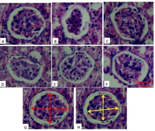
Table 2 presents histopathological changes in the renal tissues of mice in different groups. Statistical analysis revealed that EMF significantly decreased the renal corpuscle diameter, and increased the urinary space (Figure 1) and the thickness of the basement membrane (Figure 2) compared to the control group (P < 0.05) (Table
2, Figure 3). On the other hand, the treatment of the mice by vitamin E and sodium selenite in related groups showed that the administration of these antioxidants would significantly reduce adverse effects of EMF on the mentioned parameters during EMF exposure (P < 0.05). Moreover, in the group that received only sodium selenite and vitamin E, there were no significant changes in comparison to the control group. Similarly, histopathological changes in the group receiving vitamin E and sodium selenite were not significant compared to the groups receiving EMF plus vitamin E and EMF plus sodium selenite (Figures 1 and 2).
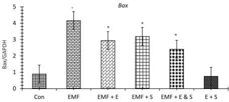
Molecular Aspects of Bax and bcl2 Genes Expression The real-time PCR results indicated that EMF significantly increased the expression of Bax (Figure 4) gene (as a pro-apoptotic gene) and decreased the expression of bcl2 (Figure 5) gene (as an anti-apoptotic gene) in the kidneys of mice in comparison with the control group (P < 0.05). Prescribing vitamin E and sodium selenite as antioxidants prevented the harmful effects of EMF on the expression of mentioned genes in the treated groups (P < 0.05). In the group receiving only vitamin E and sodium selenite,
Table 2. Effects of Vitamin E and Sodium Selenite on Corpuscle Diameter and Urinary Space in Mice Treated With EMF
Groups Control EMF EMF+E EMF+S EMF+E+S E+S
Corpuscle diameter 79.38 ± 1.87 57.31 ± 1.23b 71.74 ± 1.74a 68.66 ± 2.00a 69.46 ± 1.37a 76.19 ± 2.11
Urinary space (µm) 9.45 ± 0.48 12.90 ± 1.01b 10.78 ± 0.92a 10.84 ± 1.03a 10.71 ± 0.82a 10.19 ± 1.00
Data are mean ± SE. aP< 0.05 compared with EMF group; bP< 0.05 compared with control group. Figure 1. Histomorphometry of Renal Corpuscle of Mice in Different Groups.
the gene expression was not significant compared to the control group.
Discussion
Electromagnetic waves are a form of energy emitted by charged particles and dispersed in the space through passing a waveform path (15). According to the World Health Organization statistics, the EMFs play an important role in the development of numerous diseases, such as ALS, childhood leukemia, and brain tumor (16).
A number of studies have shown that some emaciated glomeruli, leukocytes infiltration between the kidney tubules, and the vacuolation of some tubules are other problems that EM waves cause in the kidneys (5). The renal hypoperfusion may result from an imbalance between vasodilators and vasoconstrictors. It is well established that free radicals play a major role in the induction of the cellular responses upon EM exposure (17).
The EMF effects on body tissues are generally evaluated under two dimensions of histopathological changes and molecular alteration, both of which were considered in this study (2). The results of this study showed that EMF causes histopathological changes, as well as altering the expression of Bax and Bcl2 genes in the kidneys of mice.
In the context of histopathologic changes, in this study, it was observed that EMF could make various changes in kidneys such as atrophy and shrinkage of glomeruli, tubular degeneration, increased urinary space, and increased thickness of the basement membrane. In this regard, Odacı et al realized that during the prenatal development, EMF can cause edema in the renal tissue, as well as visible small and large cysts in the medullary and cortico-medullary regions of the kidney (18).
Previous studies have attributed the destructive effects of EMF on tissues to oxidative stress (4, 17). The magnetic and electrical fields present in the environment, after
Figure 2. Photomicrograph of the Renal Corpuscle of Mice in Different Groups.
Note. EMF significantly increased the thickness of the basement membrane. The treatment of mice with vitamin E and sodium selenite decreased the effects of EMF on the thickness of the basement membrane (A: Control, B: EMF, C: EMF+E, D: EMF+S, E: EMF+E+S, F: E+S) (PAS staining) (PAS×100).
Figure 3. Effect of Vitamin E and Sodium Selenite on the Thickness of Basement Membrane of the Renal Corpuscle in Mice Treated With EMF.
Note. Data are mean ± SE. +P < 0.05 compared with control (con) group; *
P < 0.05 compared with EMF group.
+
*
*
0 0.5 1 1.5 2 2.5 3 3.5 4
Control Emf Emf + E Emf + S Emf + E+ S E+S
Basement membrane
Figure 4. Effects of Vitamin E and Sodium Selenite on the Relative Expression of Bax Gene to GAPDH Gene in Mice Treated With EMF.
Note. Data are mean ± SE. +P < 0.05 compared with control (con) group; *
P < 0.05 compared with EMF group.
0 1 2 3 4 5
Con EMF EMF + E EMF + S EMF + E & S E + S
Bax
Ba
x/GAPDH
+
* *
*
0 0.5 1 1.5 2 2.5
Con EMF EMF + E EMF + S EMF + E & S E + S
Bcl2
Bcl2/GAPDH
Figure 5. Effects of Vitamin E and Sodium Selenite on the Relative Expression of Bcl2 Gene to GAPDH Gene in Mice Treated With EMF.
Note. Data are mean ± SE. +P < 0.05 compared with control (con) group; *
P < 0.05 compared with EMF group. +
* *
passing the cell membrane, increase damages to DNA and cause the oxidative stress by prolonging free radical stability, changing the membrane potential, reducing the activity of the antioxidant enzymes, and the subsequent changes in the ROS metabolism (19). In this respect, Aydin et al found that in the presence of EMF, the activity of antioxidant enzymes decreases, while the production of ROS increases. They also showed that long-term EMF radiation leads to the production of more free radicals and carcinogens. Similarly, in the present study, EMF caused adverse effects on the kidney tissue, probably through the oxidative stress (20).
Regarding molecular changes, in this study, it was observed that EMF radiation led to the increased Bax gene expression (as a pro-apoptotic gene) and decreased Bcl2 gene expression (as an anti-apoptotic gene) in the kidney as compared to the control group. Previous studies have indicated that oxidative stress causes changes in the expression of Bax and Bcl2 genes (21, 22). Therefore, considering that EMF increases the oxidative stress, and since oxidative stress is a stimulant for the Bax gene expression and an inhibitor of Bcl2 gene expression, in the present study, EM waves affected the expression of Bax and Bcl2 genes likewise. This effect certifies the role of EMF in the initiation of the apoptosis process.
Previous studies have suggested that the use of antioxidants can reduce the adverse effects of oxidative stress (23). For instance, the study of Al-Damegh demonstrated that the use of vitamins C and E can reduce the harmful effects of oxidative stress on testicular tissue in mice by reducing the lipid peroxidation in mitochondria and microsomes (24). In addition, various studies have shown that antioxidants such as melatonin, caffeine, and phenyl ester acid inhibit oxidative stress and apoptosis formed by EMF (24). Furthermore, a number of previous studies have suggested that the use of vitamin E and selenium can reduce the effects of EMF on ovarian tissue (4).
In the current study, it was observed that in groups treated with vitamin E and sodium selenite, the pathological changes due to EMF, including tubular degeneration and glomerular atrophy, were slightly reduced. Moreover, in the group receiving vitamin E and sodium selenite plus EMF, the mentioned histopathologic changes were significantly improved. The results of a study conducted by Naziroglu et al on the sodium selenite and vitamin E effects on liver and kidney in the mice treated with cisplatin showed that usage of sodium selenite and vitamin E significantly increased the antioxidant enzymes. These findings are in line with the results of our study (25).
Furthermore, evaluating the expression of Bax and Bcl2 genes in groups treated with vitamin E and sodium selenite revealed that the use of these antioxidants reduced the expression of Bax gene and increased the expression of Bcl2 gene. Similarly, previous evidence suggests that antioxidants such as vitamins C and A reduce side effects
of oxidative stress and decrease the expression of Bax gene, while increasing the expression of Bcl2 gene (25,26). Given that vitamin E and sodium selenite have antioxidant properties, in this study, it was also assumed that these two antioxidants reduce the expression of Bax gene and increase the expression of Bcl2 in this way; thereby leading to the reduction of kidney histopathologic changes (27).
There is, however, some contradictory data about the effects of sodium selenite on the kidney tissue. Some studies have indicated that sodium selenite has a damaging effect on the kidney, which is inconsistent with our results (28). Such difference in results can be attributed to the dosage of sodium selenite and the exposure duration (28).
Vitamin E is a fat-soluble vitamin and a well-known antioxidant that plays an important role in neutralizing the toxic effects of ROS. This vitamin is able to prevent cellular damages by preserving the sulfhydryl group of membrane-bound proteins and eliminating free radicals (10). Sodium selenite is also a mineral that is found in large quantities in marine products, liver, and cereals. Sodium selenite has been known as an antioxidant in the selenoprotein formulation (25). Some earlier studies have displayed the synergistic effects of sodium selenite and vitamin E (4, 11). Sodium selenite integrates with vitamin E in the intracellular antioxidant system as the co-factor of the peroxidase glutathione enzyme. In the present study, vitamin E and sodium selenite likely prevented the adverse effects of EMF on the kidney via reducing oxidative stress.
Conclusions
As found in this study, EMF exerted adverse effects on the kidney and caused alterations in the expression of Bax and bcl2 genes through oxidative stress. Treatment with vitamin E and sodium selenite, however, could prevent the destructive effects of EMF owing to the antioxidant characteristics of these two substances.
Conflict of Interests
Authors have no conflict of interests.
Ethical Issues
All the procedures was in accordance with the guidelines for care and use of laboratory animals, adopted by the Ethics Committee of Tabriz University of Medical Sciences.
Financial Support
This work was financially supported by the Stem Cells Research Center, Anatomy Department, Tabriz University of Medical Sciences, Tabriz, Iran.
References
1. Ongel K, Gumral N, Ozguner F. The potential effects of electromagnetic field: a review. Cell Membranes and Free Radical Research. 2009;1(3):85-89.
Verdugo-Díaz L. Effect of acute extremely low frequency electromagnetic field exposure on the antioxidant status and lipid levels in rat brain. Arch Med Res. 2012;43(3):183-189. doi:10.1016/j.arcmed.2012.04.003
3. Yalçın S, Erdem G. Biological effects of electromagnetic fields. Afr J Biotechnol. 2012;11(17):3933-3941. doi:10.5897/AJB11.3308
4. Razavi SM, Majdi Seghinsara A, Abedelahi A, Salimnejad R, Tayefi H. Effect of Vitamin E and Selenium on Oxidative Stress and Tissue Damages Induced by Electromagnetic Fields in Immature Mice Ovarian. Crescent Journal of Medical and Biological Sciences. 2017;4(3):120-125. 5. Martínez-Sámano J, Torres-Durán PV, Juárez-Oropeza
MA, Elías-Viñas D, Verdugo-Díaz L. Effects of acute electromagnetic field exposure and movement restraint on antioxidant system in liver, heart, kidney and plasma of Wistar rats: a preliminary report. Int J Radiat Biol. 2010;86(12):1088-1094. doi:10.3109/09553002.2010.50184 1
6. Ulubay M, Yahyazadeh A, Deniz ÖG, et al. Effects of prenatal 900 MHz electromagnetic field exposures on the histology of rat kidney. Int J Radiat Biol. 2015;91(1):35-41. doi:10.3109/09553002.2014.950436
7. Ivancsits S1, Diem E, Jahn O, Rüdiger HW. Intermittent extremely low frequency electromagnetic fields cause DNA damage in a dose-dependent way. Int Arch Occup Environ Health. 2003;76(6):431-436. doi:10.1007/s00420-003-0446-5
8. Roushangar L, Rad JS. Ultrastructural alterations and occurrence of apoptosis in developing follicles exposed to low frequency electromagnetic field in rat ovary. Pak J Biol Sci. 2007;10(24):4413-4419.
9. Sies H. Oxidative stress: oxidants and antioxidants. Exp Physiol. 1997;82(2):291-295.
10. Aslanturk A, Uzunhisarcikli M, Kalender S, Demir F. Sodium selenite and vitamin E in preventing mercuric chloride induced renal toxicity in rats. Food Chem Toxicol. 2014;70:185-190. doi:10.1016/j.fct.2014.05.010
11. Kiafar E, Tayefi Nasrabadi H, Abedelahi A, Shoorei H, Majdi Seghinsara A. Protective Effects of Vitamin E and Selenium on Liver Tissue Damages Induced by Electromagnetic Field: An Ultrastructural Study. Crescent Journal of Medical and Biological Sciences.2018;5(4):338-344.
12. Kahya MC, Nazıroğlu M, Çiğ B. Selenium reduces mobile phone (900 MHz)-induced oxidative stress, mitochondrial function, and apoptosis in breast cancer cells. Biol Trace Elem Res. 2014;160(2):285-293. doi:10.1007/s12011-014-0032-6
13. Zdravković M, Kostov M, Stojanović M. Identification of postmortem autolytic changes on the kidney tissue using PAS stained method. Med Biol. 2006;13(3):181-184. 14. Banaei S, Ahmadiasl N, Alihemmati A. Comparison of
the protective effects of erythropoietin and melatonin on renal ischemia-reperfusion injury. Trauma Mon. 2016;21(3):e23005. doi:10.5812/traumamon.23005
15. Kiray A, Tayefi H, Kiray M, et al. The effects of exposure to electromagnetic field on rat myocardium. Toxicol Ind Health. 2013;29(5):418-425. doi:10.1177/0748233711434957 16. Cecconi S, Gualtieri G, Di Bartolomeo A, Troiani G, Cifone
MG, Canipari R. Evaluation of the effects of extremely low frequency electromagnetic fields on mammalian follicle development. Hum Reprod. 2000;15(11):2319-2325. doi:10.1093/humrep/15.11.2319
17. Amara S, Abdelmelek H, Garrel C, et al. Zinc supplementation ameliorates static magnetic field-induced oxidative stress in rat tissues. Environ Toxicol Pharmacol. 2007;23(2):193-197. doi:10.1016/j.etap.2006.09.001 18. Odacı E, Ünal D, Mercantepe T, et al. Pathological effects
of prenatal exposure to a 900 MHz electromagnetic field on the 21-day-old male rat kidney. Biotech Histochem. 2015;90(2):93-101. doi:10.3109/10520295.2014.947322 19. Wolf FI, Torsello A, Tedesco B, et al. 50-Hz extremely low
frequency electromagnetic fields enhance cell proliferation and DNA damage: possible involvement of a redox mechanism. Biochim Biophys Acta. 2005;1743(1-2):120-129. doi:10.1016/j.bbamcr.2004.09.005
20. Aydin B, Akar A. Effects of a 900-MHz electromagnetic field on oxidative stress parameters in rat lymphoid organs, polymorphonuclear leukocytes and plasma. Arch Med Res. 2011;42(4):261-267. doi:10.1016/j.arcmed.2011.06.001 21. Korsmeyer SJ, Yin XM, Oltvai ZN, Veis-Novack DJ, Linette
GP. Reactive oxygen species and the regulation of cell death by the Bcl-2 gene family. Biochim Biophys Acta. 1995;1271(1):63-66. doi:10.1016/0925-4439(95)00011-r 22. Xu J, Lian LJ, Wu C, Wang XF, Fu WY, Xu LH. Lead induces
oxidative stress, DNA damage and alteration of p53, Bax and Bcl-2 expressions in mice. Food Chem Toxicol. 2008;46(5):1488-1494. doi:10.1016/j.fct.2007.12.016 23. Salimnejad R, Soleimani Rad J, Mohammad Nejad D,
Roshangar L. Effect of ghrelin on total antioxidant capacity, lipid peroxidation, sperm parameters and fertility in mice against oxidative damage caused by cyclophosphamide. Andrologia. 2018;50(2). doi:10.1111/and.12883
24. Al-Damegh MA. Rat testicular impairment induced by electromagnetic radiation from a conventional cellular telephone and the protective effects of the antioxidants vitamins C and E. Clinics (Sao Paulo). 2012;67(7):785-792. doi:10.6061/clinics/2012(07)14
25. Naziroglu M, Karaoğlu A, Aksoy AO. Selenium and high dose vitamin E administration protects cisplatin-induced oxidative damage to renal, liver and lens tissues in rats. Toxicology. 2004;195(2-3):221-230. doi:10.1016/j. tox.2003.10.012
26. Reagan-Shaw S, Nihal M, Ahsan H, Mukhtar H, Ahmad N. Combination of vitamin E and selenium causes an induction of apoptosis of human prostate cancer cells by enhancing Bax/Bcl-2 ratio. Prostate. 2008;68(15):1624-1634. doi:10.1002/pros.20824
27. Haendeler J, Zeiher AM, Dimmeler S. Vitamin C and E prevent lipopolysaccharide-induced apoptosis in human endothelial cells by modulation of Bcl-2 and Bax. Eur J Pharmacol. 1996;317(2-3):407-411. doi:10.1016/s0014-2999(96)00759-5
28. Hussein AM, Saleh HA, H NM. Effect of sodium selenite and vitamin E on the renal cortex in rats: an ultrastructure study. Tissue Cell. 2014;46(3):170-177. doi:10.1016/j. tice.2014.03.002
Copyright © 2019 The Author(s); This is an open-access article distributed under the terms of the Creative Commons Attribution