Open Access
Research
Evolution of DNA ligases of Nucleo-Cytoplasmic Large DNA viruses
of eukaryotes: a case of hidden complexity
Natalya Yutin and Eugene V Koonin*
Address: National Center for Biotechnology Information, National Library of Medicine, National Institutes of Health, Bethesda, MD 20894, USA
Email: Natalya Yutin - [email protected]; Eugene V Koonin* - [email protected] * Corresponding author
Abstract
Background: Eukaryotic Nucleo-Cytoplasmic Large DNA Viruses (NCLDV) encode most if not all of the enzymes involved in their DNA replication. It has been inferred that genes for these enzymes were already present in the last common ancestor of the NCLDV. However, the details of the evolution of these genes that bear on the complexity of the putative ancestral NCLDV and on the evolutionary relationships between viruses and their hosts are not well understood.
Results: Phylogenetic analysis of the ATP-dependent and NAD-dependent DNA ligases encoded by the NCLDV reveals an unexpectedly complex evolutionary history. The NAD-dependent ligases are encoded only by a minority of NCLDV (including mimiviruses, some iridoviruses and entomopoxviruses) but phylogenetic analysis clearly indicated that all viral NAD-dependent ligases are monophyletic. Combined with the topology of the NCLDV tree derived by consensus of trees for universally conserved genes suggests that this enzyme was represented in the ancestral NCLDV. Phylogenetic analysis of ATP-dependent ligases that are encoded by chordopoxviruses, most of the phycodnaviruses and Marseillevirus failed to demonstrate monophyly and instead revealed an unexpectedly complex evolutionary trajectory. The ligases of the majority of phycodnaviruses and Marseillevirus seem to have evolved from bacteriophage or bacterial homologs; the ligase of one phycodnavirus, Emiliana huxlei virus, belongs to the eukaryotic DNA ligase I branch; and ligases of chordopoxviruses unequivocally cluster with eukaryotic DNA ligase III.
Conclusions: Examination of phyletic patterns and phylogenetic analysis of DNA ligases of the NCLDV suggest that the common ancestor of the extant NCLDV encoded an NAD-dependent ligase that most likely was acquired from a bacteriophage at the early stages of evolution of eukaryotes. By contrast, ATP-dependent ligases from different prokaryotic and eukaryotic sources displaced the ancestral NAD-dependent ligase at different stages of subsequent evolution. These findings emphasize complex routes of viral evolution that become apparent through detailed phylogenomic analysis but not necessarily in reconstructions based on phyletic patterns of genes.
Reviewers: This article was reviewed by: Patrick Forterre, George V. Shpakovski, and Igor B. Zhulin.
Published: 18 December 2009
Biology Direct 2009, 4:51 doi:10.1186/1745-6150-4-51
Received: 17 December 2009 Accepted: 18 December 2009
This article is available from: http://www.biology-direct.com/content/4/1/51
© 2009 Yutin and Koonin; licensee BioMed Central Ltd.
Background
Viruses are ubiquitous parasites of all cellular life forms. In recent years, extensive genome sequencing and com-parative analysis of both viral and host genomes yielded unprecedented insights into the evolution of viruses. In particular, it has been shown that 4 diverse families of large DNA viruses of eukaryotes (NCLDV), namely, Pox-viridae, AsfarPox-viridae, IridoPox-viridae, and PhycodnaPox-viridae, share a set of conserved genes with functions implicated in replication, transcription and virion morphogenesis, suggesting an origin from a single ancestral virus. This apparently monophyletic class of viruses was denoted Nucleo-Cytoplasmic Large DNA Viruses (NCLDV) to emphasize the presence of a cytoplasmic stage in the reproduction of most if not all of these viruses [1]. The existence of such a cytoplasmic stage, during which virus replication is physically separated from the replication and expression of the host genome, and occurs in cyto-plasmic viral "factories"[2,3], can be reasonably thought of as the driving force behind the retention of genes encoding replicative proteins in the NCLDV genomes. The analysis of subsequently sequenced genomes representing 3 additional viral families, namely, Ascoviridae, Mimiviri-dae, and very recently, a novel family typified by the Mar-seillevirus, strongly supported the conclusion on the monophyly of the NCLDV [4,5]. The reconstruction of the ancestral NCLDV gene set using a maximum parsimony method[4] or a more sophisticated maximum likelihood approach [6] led to the delineation of a set of 40-50 ances-tral genes that include the genes for the key proteins required for genome replication, expression and virion morphogenesis.
One of the most dramatic revelations of comparative genomics is that the set of universal genes (defined in terms of orthologous gene sets) in all life forms is much smaller than the set of universal molecular functions [7,8]. The principal underlying cause is non-orthologous gene displacement (NOGD) whereby the same essential function is performed by unrelated or at least not orthol-ogous genes [9]. Often, especially among prokaryotes, NOGD is coupled to horizontal gene transfer (HGT) of the respective genes. Owing to NOGD and HGT, phyletic patterns (that is, patterns of presence-absence in sequenced genomes) are complex, diverse and patchy for the majority of genes, even those involved in essential functions [10,11].
The case of viral genes is especially complicated because many viral functions can be complemented and replaced by functionally analogous host proteins that may or may not be homologous to the respective viral proteins; addi-tionally, many viruses integrate host genes into their own genomes, so the functional repertoire of viral genes evolves in an incessant, dynamic interaction with the host
gene repertoire. For instance, the RNA polymerase holoenzyme is obviously essential for the expression of any DNA genome. However, many large DNA viruses including certain NCLDV (such as some of the phycodna-viruses) that replicate in the cell nucleus (as well as her-pesviruses and baculoviruses) do not encode any RNA polymerase subunits and fully rely on the host transcrip-tion machinery [4]. The DNA replicatranscrip-tion apparatus of the NCLDV is substantially autonomous from the host repli-cation system. All sequenced NCLDV genomes contain genes for 3 essential replication enzymes, namely, DNA polymerase, primase and distinct replicative helicase (the latter two genes typically are fused to produce a two-domain primase-helicase). However, other proteins that are also essential for replication, in particular, DNA ligase and (predicted) flap endonuclease, are found only in sub-sets of the NCLDV, with the implication that viruses that lack these enzymes employ host analogs [4]. Very recently, this proposition was experimentally validated for the vaccinia virus (VACV) DNA ligase [12]. The DNA ligase gene of VACV can be knocked out with minimal or no adverse effect on virus growth in cell culture [13]. However, inhibition of the mammalian DNA ligase I but not any of the other 2 human ligases (III and IV) with a siRNA specifically abrogated the growth of the ligase knockout virus indicating that this host ligase was specifi-cally recruited for VACV replication [12].
The DNA ligases of the NCLDV seem to epitomize the haphazard aspect of viral genome evolution. Some of the NCLDV encode ATP-dependent ligases that, among cellu-lar life forms, are ubiquitous and essential for replication in archaea and eukaryotes, but are only sporadically found in bacteria where they seem to contribute to dis-tinct forms of DNA repair [14-16]. Furthermore, the ligases of chordopoxviruses show extensive sequence sim-ilarity to mammalian ligase III [17]. Another subset of the NCLDV encode NAD-dependent ligases that are distantly related to the ATP-dependent ligases [18,19] and are ubiq-uitous and essential for replication in bacteria but repre-sented only sporadically in archaea and eukaryotes [16,20]. Finally, a considerable fraction of the NCLDV do not appear to encode any ligases.
Results and Discussion
Distribution of DNA ligases across the NCLDV genomes
We start with a census of DNA ligases encoded in the 45 currently available NCLDV genomes. Considering that some members of the ATP-dependent ligase family, par-ticularly, in bacteria, show extreme sequence divergence [21], we searched all protein sequences of NCLDV with position-specific scoring matrices derived from multiple alignments of both ATP-dependent and NAD-dependent ligases. The ATP-dependent and NAD-dependent ligases show a perfect complementary pattern among the NCLDV, that is, not a single viral genome encodes both forms. Quantitatively, the ATP-dependent ligase is more common, being encoded by the majority of chordopoxvi-ruses and phycodnavichordopoxvi-ruses, and the single available genomes of asfarvirus and Marseillevirus (Table 1). The distribution of the NAD-dependent ligases appears to be more scattered as they are encoded by entomopoxviruses and a single chordopoxvirus (Crocodilepox virus that, however, encodes a truncated version of the NAD-dependent ligase that may or may not be active), mimi/ mamaviruses, and a minority of iridoviruses (Table 1). Viruses lacking any ligase gene are found in all extensively sampled NCLDV families, namely, poxviridae, iridoviri-dae, ascoviridae (so far none of the sequenced genomes in this family has a gene for a ligase), and phycodnaviridae (Table 1), suggesting multiple losses during evolution of the NCLDV.
Phylogenies of the DNA ligases of the NCLDV
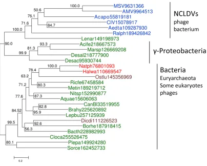
To reconstruct the evolutionary scenario for viral ligases, we used multiple alignments of the NAD-dependent and ATP-dependent ligases that included the respective pro-tein sequences from the NCLDV, other viruses, and repre-sentative archaea, bacteria, and eukaryotes (see Additional File 1 and Additional File 2, respectively), to build maximum likelihood (ML) phylogenetic trees. The tree for the NAD-dependent ligases contains an unequiv-ocally supported NCLDV clade (Figure 1). Statistical eval-uation of alternative tree topologies using the Approximately Unbiased (AU) test [22] indicated that trees with polyphyletic NCLDV effectively could be ruled out (all these alternative topologies had zero AU values). The NCLDV clade belonged to a branch that included mostly NAD-dependent ligases from gamma-proteobacte-ria along with some bacteriophage ligases one of which clustered with the NCLDV (Figure 1).
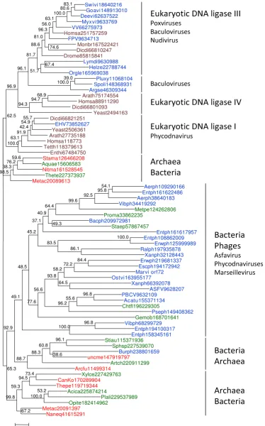
In a sharp contrast, the tree of ATP-dependent ligases showed a scattered distribution of the NCLDV branches (Figure 2). The chordopoxvirus ligases formed a strongly supported clade with eukaryotic ligase III as suggested by the previously noticed high sequence similarity between these proteins [17]. Although it has been claimed that ligase III was unique to vertebrates [14], our analysis
detected clear orthologs in insects, the choanoflaggelate
Monosiga brevicola and the social amoeba Dictyostelium dis-coideum, suggesting that ligase III evolved through a dupli-cation of the ligase IV gene at the onset of evolution of unikonts but was repeatedly lost in fungi and several ani-mal lineages (Figure 2 and Additional File 3). The NCLDV ligases clustered with the vertebrate homologs to the exclusion of the homologs from other unikonts (Figure 2 and Additional File 4). Interestingly, several non-NCLDV animal viruses with large DNA genomes from the families Baculoviridae and Nudiviridae also encode a ligase related to ligase III (Figure 2).
The ATP-dependent ligase of one of the phycodnaviruses,
Emiliana huxlei virus (representative of the genus Coccol-ithovirus), belonged to the eukaryotic ligase I branch (Fig-ure 2). The rest of the ATP-dependent ligases of the NCLDV, namely, those of African Swine Fever virus (the only available representative of the family Asfarviridae), phycodnaviruses and Marseillevirus, were scattered within a large cluster of ATP-dependent ligases of various bacteria and bacteriophages (Figure 2). The AU test indi-cated that the tree in which the latter 3 groups of viral ligases formed a clade could be effectively ruled out as an likely alternative to the tree in Figure 2 (the alternative topology with monophyletic NCLDV was associated with a zero AU value).
Evolutionary scenario for the DNA ligases of the NCLDV
NAD-depend-Table 1: DNA ligases of the NCLDV
Viral family Subfamily/genus Species ATP-dependent ligase
(GI/gene name)
NAD-dependent ligase (GI/gene name)
Poxviridae Chordopoxvirinae
Avipoxvirus Canarypox virus 40555999
Fowlpox virus 9634713/FPV043
Capripoxvirus Goatpox virus Pellor 148913010
Sheeppox virus 17077-99 21492584
Lumpy skin disease virus NI-2490 15150572
Cervidpoxvirus Deerpox virus W-848-83 62637522
Leporipoxvirus Myxoma virus 9633769
Rabbit fibroma virus 9633943
Molluscipoxvirus Molluscum contagiosum virus
Orthopoxvirus Vaccinia virus 66275973/A50R
Variola virus (smallpox virus) 9627683/J4R
Parapoxvirus Orf virus, complete genome
Bovine papular stomatitis virus
Suipoxvirus Swinepox virus 18640216
Yatapoxvirus Tanapox virus
Yaba monkey tumor virus
Yaba-like disease virus
unclassified Chordopoxvirinae Crocodilepox virus 115531716a
Entomopoxvirinae Amsacta moorei entomopoxvirus 9964513/AMV199
Melanoplus sanguinipes entomopoxvirus
9631366/MSV162
Ascoviridae Ascovirus Heliothis virescens ascovirus 3e
Trichoplusia ni ascovirus 2c
Spodoptera frugiperda ascovirus 1a
Asfarviridae Asfavirus African swine fever virus 9628207/NP419L
Iridoviridae Chloriridovirus Aedes taeniorhynchus iridescent
virus
(Invertebrate iridescent virus 3)
ent ligase gene was originally acquired by one of the NCLDV lineages, perhaps, the mimiviruses which repli-cate in amoebae where gene exchanges between viral and bacterial parasites and symbionts appear to be common [5,24,25]. This scenario implies that the ancestral NCLDV encoded no ligase, like many extant viruses. However, the monophyly of the NAD-dependent ligases from 3 distinct
NCLDV lineages (Figure 1), taken together with the polyphyly of the ATP-dependent ligases (Figure 2), favors the scenario of ancestral capture of the NAD-dependent ligase (Figure 3). Indeed, the alternative would require multiple gene transfers between viruses infecting phyloge-netically distant hosts, apparently, not a particularly plau-sible possibility.
Iridovirus
(small iridescent insect viruses)
Invertebrate iridescent virus 6 15078917/CIV205R
Lymphocystivirus Lymphocystis disease virus 1
Lymphocystis disease virus - isolate China
Megalocytivirus Infectious spleen and kidney necrosis virus
Ranavirus Singapore grouper iridovirus
Frog virus 3
Ambystoma tigrinum virus
Mimiviridae Mimivirus Acanthamoeba polyphaga mimivirus 55819181/MIMI_R303
Mamavirus Mamavirus unpublished
Phycodna-viridae
Chlorovirus Paramecium bursaria Chlorella virus
AR158
157953848
Paramecium bursaria Chlorella virus NY2A
157953038
Paramecium bursaria chlorella virus MT325
Acanthocystis turfacea Chlorella virus 1
155371134
Paramecium bursaria Chlorella virus FR483
Paramecium bursaria Chlorella virus 1
9632109/A544R
Coccolithovirus Emiliania huxleyi virus 86 73852627/EhV158
Phaeovirus Feldmannia species virus
Ectocarpus siliculosus virus 1
unclassified Phycodnaviridae
Ostreococcus virus OsV5 163955177
Marseille virus Marseille virus Unpublished
aA truncated protein missing OB-fold and HhH (helix-hairpin-helix) domains.
The specific sources of the NCLDV ligases are difficult to pinpoint with certainty but the trees in Figures 1 and 2 provide clues. The clustering of NAD-dependent viral ligases with homologs from (primarily) gamma-proteo-bacteria and bacteriophages (Figure 1) suggests that the viral ancestor of the NCLDV captured the ligase gene from a bacteriophage, perhaps, one that infected the mitochon-drial endosymbiont. This scenario is compatible with the hypothesis that the NCLDV evolved concomitantly with eukaryogenesis [4]. Indeed, the acquisition of the ligase
gene was most likely an early event considering the rapid degradation of the mitochondrial endosymbiont [26]. The tree of ATP-dependent ligases (Figure 1) suggests that they displaced the ancestral NAD-dependent ligase on several independent occasions and at different stages of viral evolution (Figure 3). Early replacements of the ances-tral NAD-dependent ligase by ATP-dependent ligases of bacterial or bacteriophage origin apparently occurred independently in phycodnaviruses, Marseillevirus, and asfarviruses. By contrast, later displacements of the
NAD-A maximum-likelihood phylogenetic tree of NNAD-AD-dependent DNNAD-A ligases
Figure 1
A maximum-likelihood phylogenetic tree of NAD-dependent DNA ligases. The numbers indicate the statistical sup-port (Expected-Likelihood Weights) of internal nodes. The supsup-port values are given as percentages (n = 1,000). Archaeal sequences are color-coded red, bacterial sequences green, eukaryotic sequences brown, and viral sequences blue. The NCLDV are shown in bold type. All proteins are denoted by their Genbank identification numbers (GIs). The truncated sequence of the NAD-dependent ligase of Crocodilepox virus was not used for phylogenetic analysis. Abbreviations: MSV, Melanoplus sanguinipes entomopoxvirus; AMV, Amsacta moorei entomopoxvirus; Acapo, Acanthamoeba polyphaga mimivirus; CIV, Chilo iridescent virus (Invertebrate iridescent virus 6); Aedta, Aedes taeniorhynchus iridescent virus (Invertebrate irides-cent virus 3); Ralph, Ralstonia solanacearum phage RSL1; Lenar, Lentisphaera araneosa HTCC2155; Acife, Acidithiobacillus ferrooxi-dans ATCC 23270; Marsp, Marinobacter sp. ELB17; Desal, Desulfatibacillum alkenivorans AK-01; Desac, Desulfuromonas
acetoxidans DSM 684; Natph, Natronomonas pharaonis DSM 2160; Halwa, Haloquadratum walsbyi DSM 16790; Ostlu, Ostreococ-cus lucimarinus CCE9901; Ricfe, Rickettsia felis URRWXCal2; Metin, Methylacidiphilum infernorum V4; Nitsp, Nitratiruptor sp. SB155-2; Aquae, Aquifex aeolicus VF5; CanBl, Candidatus Blochmannia floridanus; Brahy, Brachyspira hyodysenteriae WA1; Lepbu, Leptotrichia buccalis DSM 1135; Dicdi, Dictyostelium discoideum AX4; Borhe, Borrelia hermsii DAH; Bacth, Bacillus thuringiensis Bt407; Cloca, Clostridium carboxidivorans P7; Plepa, Plesiocystis pacifica SIR-1; Sorce, Sorangium cellulosum.
MSV9631366 AMV9964513 Acapo55819181
CIV15078917 Aedta109287930
Ralph189426842
Lenar149198973 Acife218667573
Marsp126669208 Desal218777900
Desac95930744 Natph76801093
Halwa110669547 Ostlu145356969 Ricfe67458584
Metin189219712 Nitsp152990877 Aquae15606063
CanBl33519955 Brahy225620892 Lepbu257125939
Dicdi111226523 Borhe187918415 Bacth228982993 Cloca255526475
Plepa149924280 Sorce162452733
100.0 78.4
80.3 63.2
87.3 71.2
82.8 95.9 77.6
92.6 56.3 84.52
99.5
93.3 81.3 99.9
84.7
100.0 50.6
76.1
71.6 100.0
90.0
80.1
0.2
ɶͲ
Figure 2 (see legend on next page) Swivi18640216 Goavi148913010 Deevi62637522 Myxvi9633769 VV66275973 Homsa251757259 FPV9634713 Monbr167522421 Dicdi66810247 Drome85815841 Lymdi9630988 Helze22788744 Orgle165969038 Pluxy11068104 Spoli148368931 Argse46309344 Arath75174554 Homsa88911290 Dicdi66801093 Yeast2494163 Dicdi66821251 EHV73852627 Yeast2506361 Arath27735188 Homsa118773 Tetth118379613 Enthi67484750 Stama126466208 Aquae15606583 Nitma161528545 Thete227373937 Metac20089613 Aerph109290166 Entph161622486 Aerph38640183 Vibph34419292 Metpe124262806 Proma33862235 Bacph209972981 Staep57867457 Entph161617957 Entph108862009 Erwph125999989 Ralph197935878 Xanph32128443 Erwph219681337 Escph194172942 Marvi orf72 Ostvi163955177 Xanph66392078 ASFV9628207 PBCV9632109 Acatu155371134 Chtfl196229305 Pseph149408362 Gemob168701641 Vibph68299729 Entph194100317 Entph158345161 Stiau115371936 Sphsp227539070 Burph238801659 uncme147919797 Artch220911299 Arcfu11499314 Xylce227429763 CanKo170289904 Thepe119719344 Acica225874214 Plali229537989 Opite182414962 Metac20091397 Naneq41615291 55.7 54.9 42.4 91.9 63.1 100.0 39.0 100.0 74.6 83.1 80.6 100.0 63.1 56.0 96.3 81.0 88.6 67.4 51.7 81.7 96.1 68.9 94.7 94.3 96.9 62.5 59.6 76.2 38.3 98.5 73.4 94.5 53.2 100.0 59.3 67.2 99.8 38.6 96.1 60.8 88.3 88.7 54.1 95.8 92.5 99.6 64.4 49.3 40.9 37.1 100.0 86.1 83.5 45.2 96.8 55.6 96.2 64.5 84.4 72.2 58.2 93.8 56.6 77.6 48.5 96.8 100.0 49.1 92.9 65.3 0.2
! " #dependent ligases with ATP-dependent ligases of eukaryo-tic origin seem to have occurred in Emiliana huxlei virus and in chordopoxviruses. In particular, ligase III appar-ently was acquired by chordopoxviruses not only after the radiation of the entompoxviruses and chordopoxviruses (probably concomitant with the radiation of the host ani-mals) but some time into the evolution of chordopoxvi-ruses themselves, given that the earliest branching group in this subfamily, crocodile pox virus, still has an NAD-dependent ligase (Table 1 and Additional File 1). Consid-ering the absence of genes for any ligases in many extant NCLDV and the lack of genomes encoding both an NAD-dependent and an ATP-NAD-dependent ligase, it seems likely that the displacements occurred via a ligase-less interme-diate as opposed to an intermeinterme-diate encoding both types of ligases.
Both the major form of bacterial and bacteriophage NAD-dependent ligase (LigA) and eukaryotic ligase III possess a C-terminal BRCT (BRCA1 C-terminal) domain that medi-ates interactions with components of various protein complexes involved in repair and cell cycle con-trol[27,28]. The BRCT domain is present in the NAD-dependent ligases of the mimiviruses and iridoviruses but is missing in the NAD-dependent ligases of entomopoxvi-ruses and the ATP-dependent ligases of chordopoxvientomopoxvi-ruses, the apparent origin of the latter from ligase III notwith-standing. Thus, acquisition of cellular ligase genes by dis-tinct NCLDV lineages was accompanied by independent but analogous truncations of the acquired gene.
Concluding remarks
The phylogenomic analysis described here led to unex-pected conclusions. Although ATP-dependent ligases are
more common among the NCLDV than NAD-dependent ligases, it is the latter that can be traced back to the last common ancestor of the NCLDV whereas the ATP-dependent ligases apparently were acquired by several viral lineages at different stages of their evolution. The most general message brought about by these findings is that phyletic patterns alone, at least, in some cases, are insufficient for an accurate evolutionary reconstruction and can lead to substantially oversimplified or even false scenarios.
More specifically, the study of the evolution of viral ligases has general implications for understanding the evolution of the NCLDV. The apparent acquisition of the NAD-dependent ligase gene at an early stage of evolution ante-dating the last common ancestor of the NCLDV is com-patible with the scenario of the origin of eukaryotic viruses by assembly of genes from diverse sources includ-ing bacteriophages and bacteria in the course of eukaryo-genesis [29]. The apparent independent displacement of the NAD-dependent ligase by ATP-dependent ligases of bacterial/bacteriophage origin in several NCLDV lineages implies that the primary radiation of the NCLDV occurred at the earliest stages of the evolution of eukaryotes, con-ceivably, prior to the radiation of eukaryotic supergroups and before the process of mitochondrial reduction was completed. A similar interpretation of the NCLDV evolu-tion was given previously on the basis of the examinaevolu-tion of the host ranges of the major lineages of these viruses [4]. Phylogenomic analysis of other conserved NCLDV genes has the potential to further clarify the evolutionary scenario and, possibly, the origin of this important class of viruses.
A maximum-likelihood phylogenetic tree of ATP-dependent DNA ligases
Figure 2 (see previous page)
Figure 3 (see legend on next page)
Ͳ
Ͳ
Ͳ
Ͳ
! " # $ % & %! # ' ( $ % & % ! # '
) * + # $ % & % ! # '
, & % !
Ͳ
# " !) * $ % & %! # ' - % . % $ % & % ! # '
Methods
The non-redundant protein sequence databases at the NCBI were searched using the BLASTP and PSI-BLAST pro-grams with the expectation (E) value for sequence inclu-sion in PSI-BLASt iterations set at 0.005 [30]. The sequences for phylogenetic analysis were aligned using the MUSCLE program with the default parameters [31]. To eliminate poorly aligned regions, each alignment col-umn was assigned a homogeneity value by scaling the sum-of-pairs score within the column between those of a homogeneous column (the same residue in all aligned sequences) and a random column (YIW, I. A. Seledtsov, K. S. Makarova, unpublished). Columns with homogeneity of less than 0.2 and/or with more than one-third of gap characters were removed from the alignment.
Maximum Likelihood (ML) phylogenetic trees were con-structed using the TreeFinder software [32], with the esti-mated site rates heterogeneity and the WAG (Whelan and Goldman) substitution model [33]. The Expected-Likeli-hood Weights [34] of 1,000 local rearrangements were used as confidence values of TreeFinder tree branches. The Approximately Unbiased (AU) test of tree topologies [22] was applied using TreeFinder [32].
Competing interests
The authors declare that they have no competing interests.
Authors' contributions
EVK initiated and planned the study, and wrote the man-uscript; NY analyzed data; the final manuscript was read and approved by both authors.
Reviewers' reports
Reviewer 1: Patrick Forterre, Institut Pasteur
The origin of viral genes is presently a hot topic of discus-sion. Some authors consider that all viral genes originated from cellular genes robbed by viruses whereas others emphasize that new genes can also appear in viral lineages and be transferred later on from viruses to cells. Phyloge-nies combining viral and cellular genes are indeed often difficult to interpret, especially the direction of transfer, and the interpretation can be strongly dependent of the prejudice of the author. This paper is an example of such case. Eugene Koonin has recognized in a previous paper the existence of specific viral genes (hallmark genes) that, according to him, even originated before cells, in a pri-mordial hydrothermal vent [29]. However, he is also a proponent of the idea that eukaryotes and their viruses originated after the emergence of Archaea and Bacteria from this primordial vent [29]. In this scenario, eukaryo-tes originated by the association of an ancient bacterium and an ancient archaeon, and eukaryotic viruses origi-nated by combining genes of bacterial and archaeal viruses together with eukaryotic genes from their hosts. In this scenario, genes present in eukaryotic viruses could not have originated directly from the ancestral viral world, but should have originated either from prokaryotic viruses or from the virus hosts. As a consequence, the authors systematically favour in this paper transfers from cells to viruses to explain the origin of NCLDV ligases.
I have a different prejudice. I think that most core genes of NCLDV are viral hallmark genes that originated directly in ancient viral lineages. Furthermore, I suspect that NCLDV proteins involved in replication might have played an important role in the origin of modern eukaryotic DNA genomes [35]. I thus favour the possibility of ancient LGT
Evolutionary scenario for the DNA ligases of the NCLDV
Figure 3 (see previous page)
from NCLDV to eukaryotes. The phylogenies presented in this paper do not really allow to decide between these dif-ferent interpretations but raises interesting questions.
An important point, in my opinion, is to determine, if possible, which ligase (s) was (were) present in the last common ancestor of each of the three domains. This is missing in the manuscript. To my understanding, the ancestral bacterium should have contained an NAD-dependent DNA ligase. It should thus be important to present a more exhaustive tree of bacterial NAD ligase in Figure 1, with their distribution among the various bacte-rial divisions, to clarify this point. For me, the tree in Fig-ure 1 suggests that several subfamilies of NAD ligases were already established before one of them was recruited in the bacterial domain. This tree suggests also the existence of several subfamilies presently encoded by bacteriovi-ruses and distantly related to the NCLDV DNA ligases. For me, it fits very well with the idea that the bacterial DNA ligase has a viral origin, and that Bacteria on one side, Archaea/Eukarya on the other, recruited independently their DNA replication machinery from different viruses.
In the case of ATP-dependent ligases, it should be impor-tant to determine which ligases were present in the LECA (the Last Eukaryotic Common Ancestor), in the ancestor of Archaea, and possibly in the common ancestor of Archaea and Eukarya. The trees suggest for me that ATP-dependent ligases were in fact recruited several times inde-pendently in the three domains. It should be also impor-tant to indicate on the tree where are the hosts of respective viruses. This can sometimes helps to polarize the direction of transfer. For instance, if the host is not located at its expected position in the cellular tree, one can suspect a transfer from the virus to the host.
In these phylogenies, when the authors detect a DNA ligase gene in Bacteria or Archaea, it would be important to determine if this is the bona fide ligase of the domain, or a ligase present in the genome of an integrated virus and/or plasmid [36]. In other words, it is important to dis-tinguish in cellular genomes those genes that are really cellular (they were already present in the ancestor of a par-ticular domain or recruited from another cell by LGT) and those that are in fact viral (present in integrated viruses or related elements). The confusion between these two kinds of genes in phylogenies can explain, in my opinion, the present confusion between networks and trees.
Finally, it should be interesting to have an idea of the structural differences between the various ligase sub-families studied here. In my opinion, cellular ligases (originating from the ancestor of a particular domain) should be very similar in terms of structure, as it is the case for instance for DNA gyrase in all Bacteria or Topo II in all
Eukarya, or else for Topo IB in both Archaea and Eukarya [37,38]. In contrast, families that diverged from ancient viral lineages before LUCA should exhibit more structural differences (as it is the case between DNA gyrase, T4 and eukaryotic Topo II or else between archaeal/eukayal Topo IB and bacterial or Poxvirus Topo IB) [37,38].
I thus encourage the authors to consider the two alterna-tive possibilities in the discussion of their results, either LGT from cells to viruses or from viruses to cells.
Authors' response
We greatly appreciate this review that puts the rather limited and specific study described in this article into the much more general context of evolution of viruses and cells, and while doing so, offers a perspective on these general issues that is orthogonal to our own view in some important aspects while congruent in others. There is no need to discuss these concepts as this was done in several previous publications [29,39,40].
However, a brief summary of the differences is due. The distinc-tion between our posidistinc-tion and that of Forterre that is of primary relevance for the conclusions of this study is the adoption of dif-ferent scenarios for the origin of eukaryotes. Our position is that the first eukaryotic cells emerged as a result of engulfment of an alpha-proteobacterium, the future mitochondrion, by an archaeon. To the best of our understanding, this scenario is best compatible both with comparative-genomic results and with more general considerations stemming from the parsimony principle [41-44]. This scenario, of course, has critical implica-tions for the origin of viruses infecting eukaryotes: these viruses are thought to have evolved in a « second melting pot of viral evolution », concomitant with eukaryogenesis, through amalga-mation of gene from viruses of prokaryotes, bacteria, archaea and the emerging eukaryote [29]. Under this scenario, the pos-sibility of the origin of any genes of the NCLDV directly from the primoridal pool of virus-like elements can be safely ruled out although some of the hallmarks could come from that pool through viruses of prokaryotes, having never been integral genomic components of cellular life forms. Acquisition of genes from the evolving NCLDV by the eukaryotic host remains a pos-sibility but there seems to be no compelling evidence that this was a major route of eukaryote evolution.
Forterre propounds a different scenario of evolution that includes a primordial virus world as well but considers eukary-otes to be one of the primary lines of descent in the evolution of cells [35,45-47]. It is further proposed that original bacterial, archaeal and eukaryotic cells might have possessed RNA genomes, whereas the DNA replication machineries were invented by viruses and independently grafted onto the 3 cell types [35]. Other authors also have developed evolutionary sce-narios under which eukaryotes represent a primordial cellular lineage, possibly, even the first type of cells to evolve [48,49].
cannot really agree that our adherence to the symbiogenetic sce-nario is a « prejudice ». Nevertheless, we do recognize that a definitive elucidation of the sequence of events that led to the emergence of eukaryotes is an extremely difficult task that requires much additional phylogenomic analysis and might not be attainable in the near future. Clearly, under the « primordial eukaryotic lineage » scenario, viral hallmark genes in the NCLDV could plausibly originate from the primordial virus world, and transfer of genes from these viruses to the eukaryotic hosts potentially could be a major route of evolution. Thus, interpretation of the phylogenomic analysis of viral genes, to a large extent, hinges on the choice between the two orthogonal scenarios for the origin of eukaryotes that cannot be definitively distinguished at this time.
Having acknowledged this uncertainty, we would like to point out that the history of the NCLDV ligases elucidated in this study is poorly compatible with the contribution of viral genes to the host genomes that is hypothesized by Forterre. Indeed, we show that viral NAD-dependent ligases are monophyletic and by implication were probably present in the common ancestor of the extant NCLDV. However, this class of ligases is only spo-radically represented in eukaryotic genomes (it cannot be ruled out that the respective genes were acquired from viruses although so far there are no direct evidence of such transfers). By contrast, ATP-dependent ligases are ubiquitous in eukaryo-tes but polyphyletic in the NCLDV, a finding that seems to effectively rule out the origin of eukaryotic ligases from viruses of this class. Moreover, there are two « smoking » guns of acqui-sition of ATP-dependent ligase genes from the hosts by distinct lineages of the NCLDV, namely, the ligase III homolog in pox-viruses and the ligase I homolog in Emiliana huxlei virus.
Finally, we should note that a more extensive (comprehensive) phylogenetic analysis suggested by Forterre is certainly of inter-est. However, in this paper, we focus specifically on the history of the NCLDV ligases; for this analysis, we used representative sets of ATP-dependent and NAD-dependent ligases from bacte-ria (and bactebacte-rial viruses), archaea, and eukaryotes, so we do not expect that inclusion of more exhaustive sequence sets (which complicates the construction of ML trees) would affect the message.
Reviewer 2: George V. Shpakovski,
Shemyakin-Ovchinnikov Institute of Bioorganic Chemistry, Russian Academy of Sciences, Moscow, Russia
The authors have performed a detailed phylogenomic analysis and reconstructed the history of the DNA ligases in Nuclear-Cytoplasmic Large DNA Viruses of eukaryotes (NCLDV). The study reveals a quite complex evolutionary trajectory of the enzyme involved in NCLDV' genome rep-lication: although ATP-dependent ligases are approx. 3 times more often present in different NCLDVs and previ-ous studies (Ref. 1 & Ref. 4 in the paper) regarded this type of enzyme as the one which was present in the ancestral
NCLDV genome (alone or together with the NAD-dependent DNA ligase form), the last common ancestor of the NCLDV probably contained an NAD-dependent enzyme only (Fig. 3). The novel conclusion reported in the manuscript is based mostly on the monophyly of both the NCLDV as a group and all viral NAD-dependent ligases. The story is nicely presented and interpreted but, in my opinion, the scenario suggested is not the only one which could be inferred. Because of clearly demonstrated polyphyletic origin of the viral ATP-dependent ligases (Fig. 2), the presence of enzyme of this type in the ances-tral NCLDV genome can be effectively ruled out. But the fourth remaining possibility (that the ancient, primordial NCLDV genome did not contained its own DNA ligase, but employed host analogs instead, like 19 out of 45 cur-rently known viruses do) cannot be excluded. Following this assumption it could be possible to effectively decrease the total number of evolutionary events (exemplified by 'lost' or 'gain' of the DNA ligases - see Fig. 3 of the manu-script) from 17 events (as it is now in the authors' inter-pretation) to only 10, which will be more in concord with the Occam's razor principle. Additional data and further research will probably clarify the matter.
Authors' response
The hypothesis that the ancestral NCLDV encoded no ligase was implicit in the original manuscript but in the revision we made it explicit. However, we also indicate that this scenario is not particularly plausible, given the monophyly of the NAD-dependent ligases of the NCLDV, because it would require mul-tiple gene transfers between viruses infecting taxonomically dis-tant hosts. Put another way, although the number of the events under this scenario indeed could be smaller than under the ancestral NAD-dependent ligase scenario that we favor, the nature of the inferred events also should be taken into account. As some classes of events (such as the well established gene loss) are more likely than other (such as the dubious gene transfer between viruses from distant hosts).
Reviewer 3: Igor Zhulin, Joint Institute for Computational Sciences, University of Tennessee - Oak Ridge National Laboratory
ATP ligases from NCLDV genomes and examined their distribution patterns as well as their relationships to homologous enzymes in selected Bacteria, Archaea, and Eukaryotes. Phylogenetic analysis of the proteins shows that all viral NAD-dependent ligases are monophyletic, whereas viral ATP-dependent enzymes show sporadic dis-tribution throughout the trees. This information along with distribution patterns mapped onto a viral species tree built from conserved NCLDV genes support that NAD-dependent ligases were present in the NCLDV common ancestor, but were displaced by ATP-dependent ligases on multiple independent occasions in viral evolution.
This is a very clearly organized manuscript. It addresses a specific question, and the results support the conclusions. I'm not as excited about the results as are the authors, but that is because I do not know much about viruses.
I have one minor question that is more of a personal curi-osity than a criticism. The species tree (Fig. 3) is a consen-sus of the individual trees of conserved NCLDV genes, and I wonder how this would compared to a tree built from a concatenated alignment of these genes/proteins?
Authors' response
This issue is addressed elsewhere [5,6]. The differences in the tree topologies are minor.
My only issue with the paper is the methods section on sequence identification and alignment. It is too short and contains no detail whatsoever. There is nothing I can say that is not illustrated by simply copying it below:
"Protein sequence databases at the NCBI were searched using the BLASTP and PSIBLAST programs [30]. The sequences for phylogenetic analysis were aligned using the MUSCLE program [31]. Poorly conserved positions and positions including gaps in more than one-third of the sequences were removed prior to tree computation".
What databases? What cutoffs and parameters? How were the poorly conserved positions identified?
Providing the necessary methodology details is essential, especially for a computational paper.
Authors' response: The details are included in the revised manuscript.
Additional material
Acknowledgements
The authors thank Bernard Moss for providing results prior to publication and critical reading of the manuscript, and Tatiana Senkevich for helpful dis-cussions. The authors' research is supported by the DHHS (National Library of Medicine, National Institutes of Health) intramural research pro-gram.
References
1. Iyer LM, Aravind L, Koonin EV: Common origin of four diverse families of large eukaryotic DNA viruses. J Virol 2001,
75(23):11720-11734.
2. Katsafanas GC, Moss B: Colocalization of transcription and translation within cytoplasmic poxvirus factories coordi-nates viral expression and subjugates host functions. Cell Host Microbe 2007, 2(4):221-228.
3. Condit RC: Vaccinia, Inc.--probing the functional substructure of poxviral replication factories. Cell Host Microbe 2007,
2(4):205-207.
4. Iyer LM, Balaji S, Koonin EV, Aravind L: Evolutionary genomics of nucleo-cytoplasmic large DNA viruses. Virus Res 2006,
117(1):156-184.
5. Boyer M, Yutin N, Pagnier I, Barrassi L, Fournous G, Espinosa M, Rob-ert C, Azza A, Sun S, Rossmann MG, Suzan-Monti M, La Scola B, Koonin EV, Raoult D: Giant Marseillevirus highlights the role of amoebae as a melting pot in emergence of chimaeric micro-organisms. Proc Natl Acad Sci USA 2009 in press.
6. Yutin N, Wolf YI, Raoult D, Koonin EV: Eukaryotic large nucleo-cytoplasmic DNA viruses: Clusters of orthologous genes and reconstruction of viral genome evolution. Virol J 2009,
6(1):223.
Additional file 1
Multiple sequence alignment of NAD-dependent ligases used for phy-logenetic analysis.
Click here for file
[http://www.biomedcentral.com/content/supplementary/1745-6150-4-51-S1.TXT]
Additional file 2
Multiple sequence alignment of ATP-dependent ligases used for phyl-ogenetic analysis.
Click here for file
[http://www.biomedcentral.com/content/supplementary/1745-6150-4-51-S2.TXT]
Additional file 3
A maximum-likelihood phylogenetic tree of fungal ATP-dependent DNA ligases.
Click here for file
[http://www.biomedcentral.com/content/supplementary/1745-6150-4-51-S3.PPT]
Additional file 4
A maximum-likelihood phylogenetic tree of ATP-dependent DNA ligases including multiple vertebrate species.
Click here for file
Publish with BioMed Central and every scientist can read your work free of charge "BioMed Central will be the most significant development for disseminating the results of biomedical researc h in our lifetime."
Sir Paul Nurse, Cancer Research UK
Your research papers will be:
available free of charge to the entire biomedical community peer reviewed and published immediately upon acceptance cited in PubMed and archived on PubMed Central yours — you keep the copyright
Submit your manuscript here:
http://www.biomedcentral.com/info/publishing_adv.asp
BioMedcentral
7. Koonin EV: Comparative genomics, minimal gene-sets and the last universal common ancestor. Nat Rev Microbiol 2003,
1(2):127-136.
8. Charlebois RL, Doolittle WF: Computing prokaryotic gene ubiq-uity: rescuing the core from extinction. Genome Res 2004,
14(12):2469-2477.
9. Koonin EV, Mushegian AR, Bork P: Non-orthologous gene dis-placement. Trends Genet 1996, 12(9):334-336.
10. Snel B, Bork P, Huynen MA: Genomes in flux: the evolution of archaeal and proteobacterial gene content. Genome Res 2002,
12(1):17-25.
11. Mirkin BG, Fenner TI, Galperin MY, Koonin EV: Algorithms for computing parsimonious evolutionary scenarios for genome evolution, the last universal common ancestor and domi-nance of horizontal gene transfer in the evolution of prokaryotes. BMC Evol Biol 2003, 3(1):2.
12. Paran N, De Silva FS, Senkevich TG, Moss B: Cellular DNA Ligase I is Recruited to Cytoplasmic Vaccinia Virus Factories and Masks the Role of the Vaccinia Ligase in Viral DNA Replica-tion. Cell Host Microbe 2009, 6(6):563-569.
13. Kerr SM, Smith GL: Vaccinia virus DNA ligase is nonessential for virus replication: recovery of plasmids from virus-infected cells. Virology 1991, 180(2):625-632.
14. Ellenberger T, Tomkinson AE: Eukaryotic DNA ligases: struc-tural and functional insights. Annu Rev Biochem 2008, 77:313-338. 15. Martin IV, MacNeill SA: ATP-dependent DNA ligases. Genome
Biol 2002, 3(4):REVIEWS3005.
16. Pascal JM: DNA and RNA ligases: structural variations and shared mechanisms. Curr Opin Struct Biol 2008, 18(1):96-105. 17. Wang YC, Burkhart WA, Mackey ZB, Moyer MB, Ramos W, Husain
I, Chen J, Besterman JM, Tomkinson AE: Mammalian DNA ligase II is highly homologous with vaccinia DNA ligase. Identifica-tion of the DNA ligase II active site for enzyme-adenylate formation. J Biol Chem 1994, 269(50):31923-31928.
18. Shuman S, Schwer B: RNA capping enzyme and DNA ligase: a superfamily of covalent nucleotidyl transferases. Mol Microbiol 1995, 17(3):405-410.
19. Aravind L, Koonin EV: Gleaning non-trivial structural, func-tional and evolutionary information about proteins by itera-tive database searches [In Process Citation]. J Mol Biol 1999,
287(5):1023-1040.
20. Wilkinson A, Day J, Bowater R: Bacterial DNA ligases. Mol Micro-biol 2001, 40(6):1241-1248.
21. Aravind L, Koonin EV: Prokaryotic homologs of the eukaryotic DNA-end-binding protein Ku, novel domains in the Ku pro-tein and prediction of a prokaryotic double-strand break repair system. Genome Res 2001, 11(8):1365-1374.
22. Shimodaira H: An approximately unbiased test of phylogenetic tree selection. Syst Biol 2002, 51(3):492-508.
23. Benarroch D, Shuman S: Characterization of mimivirus NAD+-dependent DNA ligase. Virology 2006, 353(1):133-143. 24. Benarroch D, Claverie JM, Raoult D, Shuman S: Characterization
of mimivirus DNA topoisomerase IB suggests horizontal gene transfer between eukaryal viruses and bacteria. J Virol 2006, 80(1):314-321.
25. Filee J, Siguier P, Chandler M: I am what I eat and I eat what I am: acquisition of bacterial genes by giant viruses. Trends Genet 2007, 23(1):10-15.
26. Gross J, Bhattacharya D: Mitochondrial and plastid evolution in eukaryotes: an outsiders' perspective. Nat Rev Genet 2009,
10(7):495-505.
27. Bork P, Hofmann K, Bucher P, Neuwald AF, Altschul SF, Koonin EV:
A superfamily of conserved domains in DNA damage-responsive cell cycle checkpoint proteins. FASEB J 1997,
11(1):68-76.
28. Rodriguez MC, Songyang Z: BRCT domains: phosphopeptide binding and signaling modules. Front Biosci 2008, 13:5905-5915. 29. Koonin EV, Senkevich TG, Dolja VV: The ancient Virus World
and evolution of cells. Biol Direct 2006, 1:29.
30. Altschul SF, Madden TL, Schaffer AA, Zhang J, Zhang Z, Miller W, Lip-man DJ: Gapped BLAST and PSI-BLAST: a new generation of protein database search programs. Nucleic Acids Res 1997,
25(17):3389-3402.
31. Edgar RC: MUSCLE: multiple sequence alignment with high accuracy and high throughput. Nucleic Acids Res 2004,
32(5):1792-1797.
32. Jobb G, von Haeseler A, Strimmer K: TREEFINDER: a powerful graphical analysis environment for molecular phylogenetics.
BMC Evol Biol 2004, 4:18.
33. Whelan S, Goldman N: A general empirical model of protein evolution derived from multiple protein families using a maximum-likelihood approach. Mol Biol Evol 2001,
18(5):691-699.
34. Strimmer K, Rambaut A: Inferring confidence sets of possibly misspecified gene trees. Proc Biol Sci 2002, 269(1487):137-142. 35. Forterre P: Three RNA cells for ribosomal lineages and three
DNA viruses to replicate their genomes: a hypothesis for the origin of cellular domain. Proc Natl Acad Sci USA 2006,
103(10):3669-3674.
36. Cortez D, Forterre P, Gribaldo S: A hidden reservoir of integra-tive elements is the major source of recently acquired for-eign genes and ORFans in archaeal and bacterial genomes.
Genome Biol 2009, 10(6):R65.
37. Brochier-Armanet C, Gribaldo S, Forterre P: A DNA topoisomer-ase IB in Thaumarchaeota testifies for the presence of this enzyme in the last common ancestor of Archaea and Eucarya. Biol Direct 2008, 3:54.
38. Forterre P, Gribaldo S, Gadelle D, Serre MC: Origin and evolution of DNA topoisomerases. Biochimie 2007, 89(4):427-446. 39. Koonin EV: The origin of introns and their role in
eukaryogen-esis: a compromise solution to the introns-early versus introns-late debate? Biol Direct 2006, 1:22.
40. Koonin EV: On the origin of cells and viruses: primordial virus world scenario. Ann N Y Acad Sci 2009, 1178:47-64.
41. Martin W, Muller M: The hydrogen hypothesis for the first eukaryote. Nature 1998, 392(6671):37-41.
42. Mans BJ, Anantharaman V, Aravind L, Koonin EV: Comparative genomics, evolution and origins of the nuclear envelope and nuclear pore complex. Cell Cycle 2004, 3(12):1612-1637. 43. Martin W, Koonin EV: Introns and the origin of nucleus-cytosol
compartmentation. Nature 2006, 440:41-45.
44. Yutin N, Wolf MY, Wolf YI, Koonin EV: The origins of phagocyto-sis and eukaryogenephagocyto-sis. Biol Direct 2009, 4:9.
45. Forterre P, Philippe H: Where is the root of the universal tree of life? Bioessays 1999, 21(10):871-879.
46. Forterre P: The two ages of the RNA world, and the transition to the DNA world: a story of viruses and cells. Biochimie 2005,
87(9-10):793-803.
47. Forterre P: The origin of viruses and their possible roles in major evolutionary transitions. Virus Res 2006, 117(1):5-16. 48. Poole A, Jeffares D, Penny D: Early evolution: prokaryotes, the
new kids on the block. Bioessays 1999, 21(10):880-889. 49. Kurland CG, Collins LJ, Penny D: Genomics and the irreducible
nature of eukaryote cells. Science 2006, 312(5776):1011-1014. 50. Martin W, Dagan T, Koonin EV, Dipippo JL, Gogarten JP, Lake JA: