LONDON
'^ O N 5f.;,MICROGLIAL-NEURONAL
INTERACTIONS
A thesis submitted for the Degree Doctor of Philosophy
at the Faculty of Medicine, University of London.
Paui Kingham
Ceil Signalling Laboratory
Department of Neurochemistry
ProQuest Number: U538031
All rights reserved
INFORMATION TO ALL USERS
The quality of this reproduction is dependent upon the quality of the copy submitted.
In the unlikely event that the author did not send a complete manuscript and there are missing pages, these will be noted. Also, if material had to be removed,
a note will indicate the deletion.
uest.
ProQuest U538031
Published by ProQuest LLC(2016). Copyright of the Dissertation is held by the Author.
All rights reserved.
This work is protected against unauthorized copying under Title 17, United States Code. Microform Edition © ProQuest LLC.
ProQuest LLC
789 East Eisenhower Parkway P.O. Box 1346
ABSTRACT
Recent evidence suggests that microglia, the resident macrophages of the CNS, play a
role in neurodegeneration. In the Alzheimer’s brain there are increased levels of both
activated and apoptotic microglia, Chromogranin A (CGA), is a peptide upregulated in
Alzheimer’s disease and may be a physiological activator of microglia. The signalling
pathways involved in the response to CGA were investigated in cell cultures of
microglia as well as the subsequent effect of secreted factors on neuronal cultures.
Following exposure to CGA, cultured microglia displayed apoptotic changes - pyknotic
nuclei and DNA fragmentation. Activated microglia produced nitric oxide prior to a
collapse in mitochondrial membrane potential (A\|/m) which could be blocked with
cyclosporin A suggesting the involvement of the mitochondrial permeability transition.
Nitric oxide synthase inhibitors prevented both the fall in Av|/m and downstream
apoptosis whereas the caspase inhibitor, YVAD-CHO, only prevented the latter. The
translocation of cytochrome c from the inner mitochondrial membrane to cytosol was
not necessary for caspase activation. CGA activated microglia also released glutamate,
attenuation o f which prevented the microglial death as did the metabotropic glutamate
receptor antagonist, MSPG. Cultured neurones treated with conditioned medium from
CGA activated microglia underwent apoptosis which was blocked by z-DEVD-fmk, a
different class of caspase inhibitor. Neuronal death was in part mediated by activation
of ionotropic glutamate receptors and also by proteins released from activated
microglia. Exposure of neuronal cultures to cathepsin B, which was secreted by CGA
activated microglia, resulted in neuronal apoptosis which could also be blocked with
z-DEVD-fink. Single cell fluorescence imaging of intracellular calcium ([Ca^^i]) in
neurones revealed a small elevation in [Ca^^]i following the addition of conditioned
medium which may constitute an early step in the apoptotic pathway. The discovery of
these distinct signalling pathways could allow for new therapeutic strategies in the
CONTENTS
Title...1
Abstract... 2
Table o f contents...3
List o f figures...6
List o f tables...10
Acknowledgements... 10
Abbreviations... ... 11
1. INTRODUCTION...13
1.1 General Introduction to Microglia... 14
1.1.1 The CNS as an immunologically privileged site... 14
1.1.2 Phenotypic characteristics o f microglia and their origin...15
1.1.3 Microglial cell markers... 17
1.1.4 The Facial Nerve Lesion to study microglial-neuronal interactions...19
1.1.5 Isolation and cell culture o f microglia...21
1.1.6 Microglial activators and the channels and receptors involved in microglial function... 23
1.1.6.1 Cytokines... 24
1.1.6.2 Ion channels...25
1.1.6.3 Microglial activators signalling through Ca^^i... 25
1.1.7 Chromogranin A, activation o f microglia and role in disease states... 27
1.1.8 Microglia secrete mediators o f cell function and survival...29
1.1.8.1 Nitric oxide... 29
1.1.8.2 Glutamate and NMDA receptor agonists... 31
1.1.8.3 Cytokines and other neurotoxic proteins... 32
1.1.8.4 Growth factors/neurotrophins...33
1.1.9 Role o f microglia in disease states: in vivo observations...34
1.1.9.1 Alzheimer’s disease... 35
1.1.10 Summary...36
1.2 Cell death in the CNS - Apoptosis or N ecrosis?... 37
1.2.1 Characterisation o f apoptosis: morphological features...39
1.2.2 Cell death in Caenorhabditis elegans: a model for mammalian apoptosis 40 1.2.3 Caspases: structure and function... 41
1.2.3.1 Caspase-1... 43
1.2.3.2 Caspase-3 (CPP32/apopain)...44
1.2.4 Death signals and caspase activation... 46
1.2.5 Caspases and the execution o f apoptosis...48
1.2.6 Are mitochondria involved in apoptosis?...49
1.2.7 Mitochondrial membrane potential, permeability transition and apoptosis.... 50
1.2.8 Mitochondria release death proteins... 54
1.2.8.1 Cytochrome c ...55
1.2.8.2 AIF...: ...56
1.2.9 Apoptosis and mitochondria in neurodegenerative disease...59
1.3 Models to study neuronal cell death...60
1.3.1 The cerebellum and its circuitry... 62
1.3.2 The cerebellum and pathology...63
1.3.3 Cerebellar granule cells in vitro...64
1.3.3.1 Isolation and morphological development...64
1.3.3.2 Developmental changes... 65
1.3.3.3 Apoptotic cell death in culture... 67
1.3.3.4 Glutamate: The neurotransmitter o f CGCs...68
1.3.4. Methods in Ca^^ imaging... 69
1.3.4.1 Fluorescent dyes...70
1.3.4.2 Imaging systems...71
1.3.5 Definition o f excitotoxicity...71
1.3.5.1 Glutamate and Ca^^ influx... 73
1.3.5.2 Ca^^ dependent effectors of cell death... 74
1.3.6 Apoptotic cell death in excitotoxic conditions... 75
1.3.7 Oxidative neuronal death... 76
1.3.8 The hippocampus...77
1.3.8.1 Hippocampal organisation and circuitry... 77
1.3.8.2 The hippocampus and memory disorders...78
1.3.8.3 HT22 cells: a mouse hippocampal cell line... 79
2. MATERIALS AND METHODS... 81
2.1 Materials... 82
2.2 Microglial cell culture... 83
2.2.1 Method 1...83
2.2.2 Method 2 ...84
2.2.3 N9 microglial cell line...85
2.3 Neuronal cell culture...85
2.3.1 Rat cerebellar granule cell culture... 85
2.3.2 HT22 hippocampal cell line... 87
2.4 Immunocytochemistry... 87
2.5 Assessment o f cell viability and apoptosis... 88
2.6 DNA fragmentation analysis...90
2.7 Nitrite and nitrate measurement... 91
2.8 Determination o f glutamate content in microglial supernatants... 92
2.9 Measurement o f lactate dehydrogenase activity in culture supernatants... 93
2.10 Measurement o f cellular ATP/ADP levels...94
2.11 Measurement o f mitochondrial membrane potential... 96
2.12 Cell lysis and protein preparation... 97
2.13 Bradford protein assay... 98
2.14 SDS-polyacrylamide gel electrophoresis... 98
2.15 Protein transfer and immunoblotting...99
2.16 Single cell Ca^^ fluorescence imaging... 101
3. CGA MEDIATED MICROGLIAL ACTIVATION: THE ROLE OF NITRIC OXIDE AND GLUTAMATE IN CELL DEATH... 103
3.1. Introduction...104
3.2 A morphological and immunocytochemical characterisation of unstimulated
and CGA exposed primary rat brain microglia cultures... 107
3.3 CGA activated microglia release nitric oxide and subsequently die... 110
3.4 CGA activated microglia release glutamate, attenuation o f which prevents cell death...119
3.5 CGA activated microglia secrete cathepsin B to the culture media... 123
3.6 Discussion... 124
3.6.1 Microglia and NO... 124
3.6.2 Microglia and glutamate...127
3.6.3 Conclusions... 131
4. MICROGLIAL APOPTOSIS: THE ROLE OF MITOCHONDRIA AND CASPASES... 132
4.1 Introduction... 133
Summary o f results...135
4.2 CGA induced microglia apoptosis involves caspase-1 activation... 136
4.3 CGA induces mitochondrial depolarisation in microglia... 142
4.4 Mitochondrial permeability transition, NO and microglial apoptosis... 147
4.5 Cytochrome c release is dependent on the nature of apoptotic stimulus...151
4.6 Discussion...157
4.6.1 Microglial apoptosis involves NO and caspase activation...157
4.6.2 Mitochondrial depolarisation and the permeability transition...160
4.6.3 NO and the permeability transition... 162
4.6.4 Microglial apoptosis and cytochrome c ... 163
4.6.5 Conclusions... 165
5. SOLUBLE FACTORS RELEASED FROM CGA ACTIVATED MICROGLIA INDUCE NEURONAL APOPTOSIS... 167
5.1 Introduction... 168
Summary o f results... 170
5.2 CGA activated microglia induce neuronal apoptosis... 171
5.3 Mechanism o f microglial induced neuronal apoptosis...180
5.4 Intracellular Ca^^ responses to neurotoxic factors in granule cells...184
5.5 Cathepsin B induces neuronal apoptosis... 187
5.6 Discussion...193
5.6.1 Microglial conditioned medium is neurotoxic... 194
5.6.2 Microglial conditioned medium induces apoptosis and mitochondrial depolarisation in cerebellar granule neurones... 196
5.6.3 Neurotoxicity is mediated by glutamate receptor agonists and proteins...197
5.6.4 Microglial conditioned medium raises [Ca^^]i in CGCs... 201
5.6.5 Microglial conditioned medium and cathepsin B are neurotoxic to HT22 neurones... 203
6. GENERAL DISCUSSION...206
6.1 Tissue culture - a suitable system to investigate microglial-neuronal
interactions... 207 6.2 Microglial cell death in culture: a model for in vivo control o f activated cells?. 209 6.3 Activated microglia in culture are toxic to neurones: a link betvyeen CGA
and neurodegenerative disease?... 214 6.4 General conclusions...218
7. REFERENCES ...220
Publications relevant to thesis
LIST OF FIGURES C hapter 1
Figure 1.1.10.1 A summary o f microglial reactions... 37
Figure 1.2.3.1 Schematic o f processed caspase-1...44
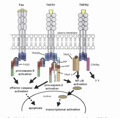
Figure 1.2.4.1 Pathways activated by the death receptor ligands. Fas and TNF... 47
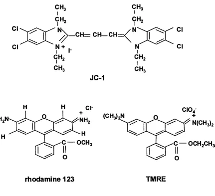
Figure 1.2.7.1 The chemical structures o f potentiometric dyes used to study
mitochondria... 52
Figure 1.2.7.2 Schematic o f the components of the mitochondrial PT pore and factors that influence its opening...54
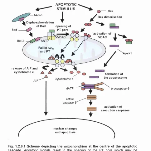
Figure 1.2.8.1 Scheme depicting the mitochondrion at the centre o f the
apoptotic cascade... 58
Figure 1.3.1.1 Neurones in the cerebellum showing excitatory and inhibitory synapses... 63
Figure 1.3.4.1 Schematic o f an imaging system and inset the structure of
Fura 2 ... 72
Figure 1.3.8.1 The neuronal circuits in the hippocampal formation... 79
C h ap ter 2
Figure 2.7.1 Typical linear standard curves for nitrite and nitrate following conversion to nitrite...92
Figure 2.9.1 A typical linear standard curve for serial dilutions o f lactate
dehydrogenase... 94
Figure 2.10.1 Typical standard curve for the chemiluminescence produced
with serial dilutions of ATP...95
Figure 2.13.1 A typical linear protein standard curve for serial dilutions o f BSA 98
C hapter 3
Figure 3.2.1 Primary cultures o f neonate rat brain microglia are sensitive
to L-leucine methyl ester... 108
Figure 3.2.2 Immunochemical staining of cultures with the macrophage/
microglia specific markers, 0X 42 and ED-1... 109
Figure 3.3.1 CGA induces cell death in primary cultures o f rat brain microglia. I l l
Figure 3.3.2 Chromogranin A toxicity of microglial cells is dose dependent... 112
Figure 3.3.3 Dose dependent and time course profile o f nitric oxide generation by microglia activated with chromogranin A ... 114
Figure 3.3.4 Stimulation of the N9 microglial cell line with CGA or LPS results in the increased expression o f iNOS protein... 115
Figure 3.3.5 Genistein inhibits CGA induced tyrosine phosphorylation,
nitric oxide production and cell death in the N9 cell line...116
Figure 3.3.6 CGA induced microglial cell death is prevented by NOS
inhibitors...117
Figure 3.3.7 Effect o f the nitric oxide donor, SNAP, on nitrite accumulation
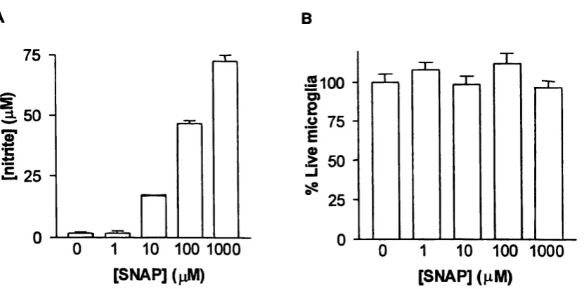
and cell death in primary cultures o f rat brain microglia...118
Figure 3.4.1 Chromogranin A activated microglia release glutamate...121
Figure 3.4.2 Blockade of glutamate release attenuates microglial cell death... 122
Figure 3.5.1 Stimulation of microglia with CGA results in the extracellular
release o f cathepsin B ...123
C hapter 4
Figure 4.2.1 Chromogranin A induces apoptosis in microglia... 136
Figure 4.2.2 Time course of CGA induced microglial apoptosis... 138
Figure 4.2.4 ATP levels remain high until late time points during CGA
induced microglial apoptosis... 141
Figure 4.3.1 CGA induces mitochondrial depolarisation in microglia... 142
Figure 4.3.2 Complete mitochondrial depolarisation following CGA activation. 144
Figure 4.3.3 CGA causes reduced mitochondrial membrane potential as
measured with TMRE... 145
Figure 4.3.4 CGA activation o f microglia causes reduced mitochondrial
membrane polarisation as measured with rhodamine 123...146
Figure 4.4.1 Changes in mitochondrial morphology following CGA activation. 148
Figure 4.4.2 Microglia activated with CGA undergo the mitochondrial
permeability transition...148
Figure 4.4.3 Nitric oxide dependent mitochondrial depolarisation...149
Figure 4.4.4 The nitric oxide donor, SNAP, sensitizes microglia to the
toxic effects of glutamate... 150
Figure 4.5.1 CGA activation o f microglia causes increased mitochondrial cytochrome c expression rather than translocation to the
cytosol... 152
Figure 4.5.2 Whole cell lysates of CGA treated microglia reveal increased
levels o f cytochrome c ...153
Figure 4.5.3 Staurosporine causes the release o f cytochrome c to the
cytosol... 154
Figure 4.5.4 Staurosporine induced microglial apoptosis involves mitochondrial depolarisation... 155
Figure 4.5.5 ATP levels fall following the induction o f microglial apoptosis
by staurosporine ... 156
Figure 4.6.1 Distinct apoptotic pathways evoked by CGA and staurosporine....158
C hapter 5
Figure 5.2.1 Conditioned medium from activated microglia induces cell death in primary cultures of rat cerebellar granule neurones... 172
Figure 5.2.2 The toxic effect o f conditioned medium is dependent upon the
Figure 5.2.3 The effect of microglial activation status on the toxicity o f
conditioned medium... 174
Figure 5.2.4 Neurotoxicity is dependent on the amount of conditioned medium added and the concentration o f CGA used to activate microglia. 174
Figure 5.2.5 Neurotoxicity is dependent on the microglial activation time and length of exposure to conditioned medium... 176
Figure 5.2.6 Neurones exposed to medium from microglia activated with CGA display apoptotic nuclei... 177
Figure 5.2.7 Neurotoxicity evoked by conditioned medium can be blocked
by a caspase-3 inhibitor...178
Figure 5.2.8 Conditioned medium induces DNA fragmentation and
mitochondrial depolarisation in cerebellar granule neurones... 179
Figure 5.3.1 Cerebellar granule neurones treated with microglial conditioned medium do not generate significant levels of nitric oxide...180
Figure 5.3.2 Attenuation of neuronal death induced by conditioned media
from CGA exposed microglia...181
Figure 5.3.3 Glutamate and NMDA mediated neurotoxicity...183
Figure 5.3.4 The effect of cytokines on neuronal cell viability... 183
Figure 5.4.1 Effect o f conditioned medium on [Ca^^]i in cerebellar granule
neurones... 185
Figure 5.4.2 The role of glutamate receptors in the [Ca^^], elevation...186
Figure 5.4.3 Effect of conditioned medium on KCl evoked calcium signals
in neurones... 187
Figure 5.5.1 Microglial conditioned medium is neurotoxic to the HT22
cell line... 189
Figure 5.5.2 Cathepsin B induces apoptosis in HT22 cells and CGCs... 190
Figure 5.5.3 Time course of HT22 neuronal death and caspase activation... 191
Figure 5.5.4 Neither mitochondrial depolarisation or nitric oxide production are involved in conditioned medium or cathepsin B induced
HT22 cell death...192
Figure 5.5.5 The effect of kinase inhibitors on HT22 cell death evoked by
C hapter 6
Figure 6.4.1 Schematic o f the role CGA may play in neurodegenerative
disease... 219
LIST OF TABLES C hapter 1
Table 1.1.3.1 Cellular markers for microglia... 18
Table 1.2.3.1 Characteristics o f the caspase family o f proteins... 42
C hapter 3
Table 3.2.1 Cell yields and purity of different microglial culture technique.... 108
ACKNOWLEDGEMENTS
I would like to thank my Principal supervisor, Dr Jennifer Pocock, for her guidance and
continual enthusiasm throughout the course of these studies. I am also grateful to Prof.
Louise Cuzner. Thanks also to Gareth and Amanda for fun times both in the lab and at
the Flyer and Goose, Anne for those all too frequent trips to “the dogs” and Sarah for
ABBREVIATIONS
mitochondrial membrane potential [Ca^^]i: intracellular free calcium
AD: Alzheimer’s disease
AIF: apoptosis inducing factor
AMT-HCl : 2-amino-5,6-dihydro-6-methyl-4H-1,3 -thiazine hydrochloride ANT: adenine nucleotide transporter
Apaf-1 : apoptosis protease activating factor APC: antigen presenting cells
AraC: cytosine arabinoside ATP: adenosine 5’-triphosphate
BBB: blood brain barrier
BDNF: brain derived neurotrophic factor bFGF: basic fibroblast growth factor
BM: physiological bathing medium
BSA: bovine serum albumin
CGA: chromogranin A
CGC: cerebellar granule cell
CNQX: 6-cyano-7-nitroquinoxaline-2,3-dione CNS: central nervous system
CsA: cyclosporin A
CSF: cerebrospinal fluid
DAB: diaminobenzidine tetrahydrochloride
DCG4: (2S, 2 ’R, 3’R)-2-(2’,3’-dicarboxycyclopropyl) glycine
DD: death domain
DIV: days in vitro
DMEM: Dulbecco’s modified eagle medium
EAA: excitatory amino acid
EAE: experimental allergic encephalomyelitis
ECL: enhanced chemiluminescence
ECM: extracellular matrix EGF : epidermal growth factor
EGTA: ethylene glycol-bis(P-aminoethyl ether) N, N, N ’, N ’-tetra acetic acid FACS: fluorescence activated cell sorting
F ADD: fas-associated death domain
Fak: focal adhesion kinase
FCCP: carbonyl cyanide p-(tri-fluoromethoxyl)phenylhydrazone
FCS: foetal calf serum
Fura-2: fura-2 acetoxymethyl ester GalC: galactocerebroside
GFAP: glial fibrillary acidic protein HRP: horse radish peroxidase
ICE: interleukin Ip converting enzyme
IF : interferon
IL: interleukin
iNOS: inducible nitric oxide synthase INT: iodonitrotetrazolium
KGDHC: a-ketoglutarate dehydrogenase complex
LDH: lactate dehydrogenase
L-NNA: N°-Nitro-L-arginine
LPS: lipopolysaccharide
MNCM: microglial non activated conditioned medium MACM: microglial activated conditioned medium MCSF: macrophage colony stimulating factor
MEM: modified eagle medium
mGluR: metabotropic glutamate receptor
MHC: major histocompatability complex
MK-801: (+)-5-methyl-10,11 ,-dihydro-5H-dinezo [a,d]cyclohepten-5,10-imine hydrogen maleate
MSPG: (RS)-a-methyl-4-sulphonophenylglycine
NGF: nerve growth factor
NMDA: N-methyl-D-aspartate
NO: nitric oxide
NOS: nitric oxide synthase
NSE: non specific esterase
PAF: platelet activating factor
PAGE: polyacrylamide gel electrophoresis
PARP: poly-(ADP-ribose)-polymerase
PBS: phosphate buffered saline
PD: Parkinson’s disease
PDGF: platelet derived growth factor
PT: permeability transition
PVDF: polyvinylidene difluoride
ROS: reactive oxygen species
SBTI: soybean trypsin inhibitor
SNAP: S-nitrosopenicillamine
TGF: transforming growth factor
TMRE: tetramethylrhodamine ester
TNF: tumour necrosis factor
TTBS: tween tris buffered saline
TUNEL: terminal transferase in situ end labelling VDAC: voltage dependent anion channel
YVAD-CHO: Tyr-Val-Ala-Asp-aldehyde
1. General introduction
The central nervous system (CNS) consists o f a network o f approximately 10^^
neurones, the sole purpose of which is to transmit the impulses responsible for allowing
an organism to communicate with the external environment (Alberts et a l, 1989).
Since the neuronal cell plays such an active role, the other 90% o f cells present in the
CNS known as glia have been considered as the passive elements o f the nervous system.
More recently, evidence has been accumulating to suggest that the neurone-glia unit
may determine neuronal function and signalling. Three principal types o f glia exist; the
star shaped astrocytes, the myelinating oligodendrocytes and microglia (Nissl, 1891 as
cited in Barron, 1995). Microglia share many common characteristics with
macrophages and have come to be considered the resident macrophage o f the CNS (Del
Rio-Hortega, 1932 as cited in Barron, 1995). In the normal brain, microglia assume a
resting/ramified process bearing state but they have the ability to become activated and
adopt a form that resembles the mature macrophage. During this transition, factors may
be released which can affect neuronal signalling and survival. Such processes may
underlie many neurodegenerative diseases. The aim o f this study has been to
investigate these microglial-neuronal interactions using a tissue culture system. Some
of the important considerations are now discussed.
1.1. The CNS a s an immunologically privileged site
The existence o f the blood brain barrier (BBB) allows the exclusion o f leukocytes from
the brain and suggests that immune reactions are unlikely to occur in the CNS (Fabry et
a l, 1994). However, recent evidence suggests that regions o f the CNS can connect
directly to lymph nodes thus allowing the possibility o f CNS antigen presentation to
cells of CNS have the capacity to act as antigen-presenting cells (APC) since they
express the Major Histocompatability Complex (MHC) antigens necessary to activate
lymphocytes and produce an immune response. In vitro data suggested that astrocytes
were responsible for this but the observations could not be confirmed in vivo (Fontana
et al., 1984). Instead it was found that microglia and perivascular cells expressed MHC
in vivo (Matsumoto and Fujiwara, 1986; Hickey and Kimura, 1988; Streit et al., 1989).
In particular, a subset o f microglia known as peri-vascular microglia are the more
effective antigen presenters (Ford et al., 1995) and these have been shown to be
associated with blood vessels (Graeber et al., 1989a) thereby providing a link between
the CNS and external environment. In addition, the other parenchymal microglia can
exhibit MHC II antigens but this expression is probably less constitutive and more
inducible. This suppression of immune activity in the brain probably functions as a
protective mechanism since such tissue is more sensitive to immune mediated cellular
damage. For instance under pathological conditions MHC class II expression becomes
apparent on microglia thus allowing them to become effective APCs (Fabry et al., 1994)
and parenchymal microglia are likely to mediate autoimmune diseases such as multiple
sclerosis (Hayes et al., 1987). Thus, since microglia have a similar immunological
profile to other macrophages they have formed the basis o f neuroimmunological
studies.
1.1.2 P henotypic c h aracteristics of microgiia an d their origin
Microglia were first decribed in the 1930’s using a silver carbonate staining technique
(Del Rio Hortega, 1932). With subsequent technical advances in microscopy the
structure of this cell has been examined in detail. The resting or ramified microglia
surround. At the surface there are many cell processes which become highly branched
giving a distinct morphology to that of resting macrophages in other tissues. It is these
resting microglia which constitute approximately 10% of the glial population in the
mature brain (Vaughan and Peters, 1974). Though the numbers vary greatly in different
regions of the brain they are more predominant in gray matter than white (Lawson et a i,
1990). Ameboid microglia are observed during development and may also be actively
phagocytic. They have a more abundant transparent, cytoplasm containing numerous
vacuoles and large dense bodies (lysosomes). The surface membrane emits many fine
filopodia. These cells may also show increases in the expression of immuno-markers
such as MHC antigens (Streit et al., 1989). Also under conditions o f brain injury the
resting microglia may convert into a reactive macrophage in which the “activated”
microglia shows upregulated levels of these macrophage proteins but do not undergo a
change in morphology (Graeber et al., 1988a). The opposite can also occur (see section
1.1.4). Thus the context in which the term “activated microglia” is used should always
be defined.
The origin o f ramified microglia has been highly debated (extensively reviewed in
Cuadros and Navascues, 1998). It has been proposed that either microglia are derived
from the neuroepithelium as with neurones and other glia cells, or they are of
haematopoietic origin i.e. derived from monocytes which is more consistent with the
mononuclear phagocyte system. There is evidence for both theories but it is the latter
one that is more generally accepted. During development there is a decrease in
blood-derived ameboid cells which correlates strongly with increases in ramified microglia
(Ling and Wong, 1993). However, there has been mixed success in attempts to show
(Hickey et al., 1992) with as few as 10% o f the cells derived from bone marrow
transplants (De Groot et al., 1992) whereas other reports showed a continuing influx of
genetically tagged haematopoietic cells into the brain (Eglitis and Mezey, 1997). In
contrast, in vitro studies show that microglia and astroglia may have a common
progenitor cell and thus microglia may be o f neuroectodermal origin (Fedoroff et al.,
1997). Despite this, microglia contain most known macrophage specific antigens
(Flaris et al., 1993). Furthermore, there is a subset o f microglia known as peri-vascular
microglia that are associated with blood vessels (Graeber et al., 1989a) and these are
regularly replaced with cells from the bone marrow (Hickey and Kimura, 1988; Hickey
et ah, 1992).
1.1.3 Microglial cell m arkers
Initial studies by Rio-Hortega used a silver staining technique to identify microglia
which is now known to be non specific. Thus techniques have been developed in an
attempt to selectively identify microglia but it is impossible to distinguish between
macrophages and microglia. Instead only quantitative differences in the expression of
antigenic markers between these different cell populations can be made. These can be
examined using fluorescence activated cell sorting (FACS). For instance, microglia
have been shown to have immunophenotypes which are CD45^°^CD1 Ib/c^ and other
macrophages are CD45^'^"'CD1 Ib/c^ (Sedgwick et al., 1991; Ford et al., 1995).
Microglia can be visualised in culture or sections using either immunohistochemistry,
enzymatic labelling or lectin binding techniques. Monoclonal antibody staining is the
most commonly used but there is still no antibody that can distinguish between
monocytes/macrophages and microglia (Williams et al., 1992). Furthermore, there are
(Tsuchihasi et a i, 1981 ; Hume et al., 1983). This may be due to the activation states of
the cells. Since many antibodies are raised to macrophage specific antigens they are
likely to preferentially label, active, ameboid microglia with only faint staining of the
ramified forms e.g. EDI-3 (Dijkstra et al., 1985; Graeber et al., 1989a). However,
microglia have been shown to express CR3 complement receptors throughout
development and this allows the identification o f resting cells in rat (OX-42), mouse
(MAC-1) and human (EBMl 1) (Graeber et al., 1989a; Matsumoto et aL, 1985; Hayes et
al., 1987). The F4/80 antibody will also stain both ameboid and ramified microglia
(Perry et al., 1985). Additionally, antibodies directed against Fc receptor may also be
useful in identifying microglia (Perry et al., 1985).
Marker Ligand/Monoclonal Antibody Microglial form labelled
Silver carbonate Not specific Ramified and Ameboid
Enzyme histochemistry Non-specific esterase (NSE) Ameboid
Sugar/glycan receptors Lectins I-B4 and RCA-1 Ramified and Ameboid
Monoclonal antibody against
97 Kd cytoplasmic antigens EDI (rat) Ameboid
Unknown membrane antigen ED2, ED3 (rat) Ameboid
Complement receptor (CRl) Ebmll (human) Ramified and Ameboid
Complement receptor (CR3) OX-42 (rat), MAC-1 (mouse) Ramified and Ameboid
Unknown antigen F4/80 (mouse) Ramified and Ameboid
Fc receptor 2.4G2 (mouse), MAh 10.1 (human) Ramified and Ameboid
Table 1.1.3.1 Cellular m arkers for m icroglia
Non-specific esterase (NSE) activity is absent in resting cells but associated with
ameboid microglia (Hayes et al., 1987). However, this marker should be used with
caution when identifying cultured microglia as it is a characteristic of blood monocytes
which do not bind to neurones or other glial cells can also be used to label all microglial
cell morphologies (Streit and Kreutzberg, 1987). Nevertheless since they are directed
against D-galactose they will also stain macrophages. In conclusion, the presence of
many macrophage specific antigens in microglia suggests a common origin but it may
also be the result of the similar phagocytic role o f these cells functioning in specific
areas (Thomas, 1990).
1.1.4 The Facial Nerve Lesion to study microgiiai-neuronai interactions
Activation of microglia following CNS injury occurs as the result of signalling
molecules released by dying neurones. Evidence from ischaemic lesions suggests that
neuronal damage does not have to be lethal to elicit a microglia response since the
penumbric region contains surviving neurones in association with reactive glial cells
(Morioka et aL, 1993). Once activated, microglia may secrete either toxic or
neurotrophic factors depending on the nature of the injury. During irreversible injury
microglia could produce neurotoxic molecules to kill the neurones in a rapid manner
whereas they may produce neurotrophins in situations where recovery is possible. Such
reactions have been studied in vivo.
An in vivo microglial reaction can be elicited following disruption to the blood brain
barrier. However, in this situation it is impossible to distinguish the events mediated by
resident microglia and those occurring as the result of macrophages derived from the
bloodstream. Thus, a model known as facial nerve transection is used, in which
neuronal damage is evoked at a site distant to the region of injury (Streit et al., 1988)
thus overcoming the problem of damage caused by surgery. It is characterised by
up-regulate the macrophage antigens of CR3 receptors and MHC and also the intermediate
filament protein vimentin (Graeber et al., 1988a and b; Streit et aL, 1989). They
increase the secretion o f a number of cytokines (Frei et aL, 1988; 1991). Activated
microglia then surround the neurone and remove nerve endings from the cell body thus
freeing the cell from any other neural input. The majority o f the neurones survive and
regrow their axons suggesting that this is a beneficial stage (Blinzinger and Kreutzberg,
1968). It may be that neuronal-microglial contact is needed for transfer o f neurotrophic
factors. Despite activation o f the microglia they do not become phagocytic in this
model. However if the motor neurone is induced to degenerate by addition o f a toxic
lectin from Ricinus communis, the microglia become more rounded and have phagocytic
activity clearing neuronal debris (Streit and Kreutzberg, 1988). This step can be blocked
by the addition o f anti-metabolites such as adriamycin and cytosine arabinoside (AraC)
(Graeber et aL, 1989c; Svensson and Aldskogius, 1993) suggesting that it is the resident
microglia that become phagocytic rather than any invading macrophages. It is probable
that this phagocytic phase is a late stage in the microglial activation cascade and is
unnecessary for axon stripping.
There are differences in how microglia respond to neuronal damage in the developing
and mature CNS. The intensity of microglial activation is lower after neonatal injury
with correspondingly reduced MHC expression (Milligan et aL, 1991; Morioka and
Streit, 1991). This may be due to differences in the immunological markers found at
this stage in microglial development (Ling et aL, 1990) but also to the fact that the
microglia are likely to be responding to apoptotic rather than necrotic neuronal cell
death (Ashwell et aL, 1989). Apoptosis occurs after nerve transection in the new-born
animal as neurones are deprived o f vital neurotrophins. Microglia respond to changes
diffusible factors released lytically by dying adult neurones (Savill et al., 1993). In this
instance microglia show minimal proliferation and the inflammatory response is limited.
Thus activation o f the microglia is not a simple one step process but occurs in a graded
fashion involving multiple signals (Kreutzberg, 1996). It was originally proposed that
microglia would progress from a ramified resting morphology through reactive
phenotype to the phagocytic stage but this may also involve an intermediate step during
which microglia become hyper-ramified, a morphology often observed in the aged brain
(Streit et a l, 1988). Each stage occurs in a defined period o f time with associated
biochemical changes i.e. cell enlargement (hypertrophy) and cytokine production is an
early event within 24 h whereas proliferation and upregulation o f MHC II antigens
occurs over a time scale of weeks suggesting different factors are required to signal each
stage. Determining the nature o f these molecules and their regulation o f microglial
activation may hold the key to controlling the CNS immune response. Many candidate
factors have been hypothesised from studies o f cultured microglia.
1.1.5 isolation and cell culture of microglia
The signal transduction pathways and molecules involved in microglial activation can
be studied by using in vitro models of microglia. Microglia may be isolated from other
cells o f the CNS in a number of ways following mechanical and/or enzymatic disruption
o f brain tissue. After plating, the purity of the culture can be monitored using
immunocytochemical markers such as the aforementioned macrophage specific
monoclonal antibodies along with those to detect cells expressing glial fibrillary acidic
protein (GFAP) and galactocerebroside (GalC) respective indicators o f astrocytes and
1983; Benjamins et a l, 1987). Any method o f isolation can be subject to contamination
from circulating monocytes which cannot be distinguished from microglia with
antibodies. One way of reducing such contamination is to perfuse the animal prior to
removal of the brain. The initial step in any of the techniques is dissection of the tissue
and subsequent removal of the meninges and any visible blood vessels. It may be first
necessary to mechanically disrupt the tissue. Next a mixture of enzymes, trypsin and
collagenase is used digest the tissue and free the cells, and DNase to prevent the
solution becoming gelatinous. Alternatively gentle homogenisation can be used to free
the cells, overcoming the need for enzymatic disruption to surface membrane
molecules. Next the microglia need to be separated from the mixed population of cell
types and three methods have been reported for this.
The original method described, exploits differences in the adherence of glial cells to
tissue culture flasks (McCarthy and de Vellis, 1980). Simply the suspension is plated
onto flasks and the glia allowed to proliferate for 7 days. The loosely attached
microglia along with oligodendrocytes are then shaken off leaving behind astrocytes.
This mixture of cells is then transferred to new flasks and using different rotation speeds
the oligodendrocytes removed leaving only microglia attached to the flasks. These can
then be removed by trypsinisation and re-plated. The cultures are typically maintained
at 37°C in an atmosphere of 5%C02/95% air. Each shaking stage needs be monitored
immunocytochemically to ensure the correct population of cells detaches. This is
especially important when using cells from different stages o f development since foetal
microglia are less adherent and adult microglia more adherent than astrocytes. The
disadvantage with this technique is the length of time it takes to obtain the microglia
cells meaning the phenotype may not be a true reflection of cells freshly isolated.
Furthermore with numerous stages involved the yields obtained may be as low as 10%
of the total microglial population.
Other methods use discontinuous Percoll gradients to separate the microglia from the
mixed glia suspension (Sedgwick et aL, 1991; Havenith et aL, 1998). Cells may need to
be isolated from myelin on an initial gradient and then microglia can be separated from
other cells by using different density layers o f Percoll. The cell yield from this
technique is approximately 5 x 10^/rat brain. Finally the last method takes advantage of
the fact that microglia express the Fc receptor. Mixed glia can be incubated with
erythrocytes to generate rosetted microglia. These are then separated from myelin and
other cells by centrifugation through a single layer o f Percoll and the microglia freed
from erythrocytes using a protocol of hypotonic lysis (Hayes et aL, 1988). Cell yields
are similar to those for the discontinuous gradient method. All three techniques have
the inherent problem that they result in some activation of these cells. However, once in
culture, microglia adopt the ramified and ameboid morphological forms seen in vivo.
Thus in vitro studies can be used to investigate the functions of microglia.
1.1.6 Microglial activ ato rs and th e channeis and recep to rs involved in microgliai function
Cultured microglial are seen to undergo the morphological change from resting state to
ameboid form when exposed to a number o f factors. The most favoured activator for
studying microglia in vitro is lipopolysaccharide (LPS). However, whether this
functions as an endogenous activator is debatable since this molecule would not be
present in situtions o f injury without infection. Thus other potential microglial
1.1.6.1 Cytokines
Like other cells of the immune system, microglia can respond to cytokines. These
soluble polypeptides include interleukin (IL), interferon (IF), tumour necrosis factor
(TNF), macrophage colony stimulating factor (M-CSF) and transforming growth factor
(TGF). Each o f these classes may contain two or more molecular forms. Such
cytokines are generally believed to control the processes o f microglial activation and
proliferation (reviewed in Gehrmann et aL, 1995). Furthermore, they affect both the
immunological profile (e.g. increased MHC class II molecule expression) and the
functional characteristics of these cells. Interferon-y and IL-4 are two T-cell derived
cytokines that can up-regulate Fc receptors and MHC class II expression and also cause
proliferation o f microglia (Colton et aL, 1992). Colony stimulating factors (CSF) such
as granulocyte macrophage-CSF (GMCSF) and macrophage-CSF (MCSF) may be
released by astrocytes and promote microglial proliferation as well as maturation and
differentiation during development (Giulian and Ingeman, 1988). These ligands bind to
tyrosine kinase receptors which are upregulated in activated pre-mitotic microglia
(Raivich and Kreutzberg, 1994). There is also evidence o f an increase in receptor
number in the facial nerve model where the presence o f ligand influences the expression
o f surface adhesion molecules necessary for the microglial reaction. Other cytokines
including pi and IL-IO are known to down-regulate microglial activation.
TGF-pi may also be produced by microglia themselves and thus serve to control the
1.1.6.2 Ion channels
Patch clamp studies on cultured microglia and tissue slices suggest that microglia
express a large number of ion channels (reviewed in Eder, 1998). The best studied of
these are the channels and it is now known that six different types exist including an
inward rectifier, delayed rectifier, HERG-like, G-protein activated and voltage
dependent and independent Ca^^-activated channels. In comparision with
macrophages, the inward and delayed rectifiers are likely to play a role in regulating
membrane potential (Gallin and Sheehy, 1985). This is important for the functional
state of the microglia since hyperpolarisation may be a requirement for initiating
microglial functions such as proliferation and secretion o f molecules. Microglia also
contain channels which may be important in maintaining membrane potential and pH
o f the cell following phagocytosis (DeCoursey and Cherny, 1994; Henderson and
Chappell, 1996). Cl channels are thought to play a role in proliferation induced by
M-CSF treatment (Schlichter et a i, 1996) and the ramification o f microglia (Eder et al.,
1998). Microglia also express Na^ channels and voltage gated Ca^^ channels, the
functions o f which are unknown (Korotzer and Cotman, 1992; Colton et al., 1994).
Microglial ion channels may be modulated by stimulation with EPS and thus the pattern
o f expression may be dependent on the functional state o f the cell.
1.1.6.3 Microglial activators signalling through [Ca^*]i
Cultured microglia undergo a morphological change when treated with adenosine
5’-triphosphate (ATP) suggesting it could be a physiological modulator o f activation
(Ferrari et al., 1997). ATP has long been known to act as neurotransmitter in the CNS
phospholipase C (Pi receptors) or ATP specific (P2 receptors) which may be either
ionotropic (Pix and Pzz) or metabotropic (Piy, Pzu, Pzt, and Pid) (Zimmermann, 1994;
Bumstock and Wood, 1996). Evidence exists that immune cells such as microglia also
contain the ionotropic receptors (Walz et a l, 1993; Haas et al., 1996) and they are
responsible for the direct influx o f Ca^^ into the cell following ATP treatment (Ferrari et
al., 1996). The activation of these receptors leads to microglial apoptosis (Ferrari et al.,
1997). In contrast the activation of metabotropic receptors probably instigates InsP]
induced Ca^^ release from internal pools and it has been suggested might result in
capacitative Ca^^ entry (Moller et a i, 1997a).
Another activator of microglia is platelet activating factor (PAF). Originally identified
for its action on platelets (Benveniste et al., 1979) it has a diverse role in the CNS
(Feuerstein et al., 1990; Bazan, 1994). Specific receptors for this molecule have been
found in both primary cultures of microglia (Mori et al., 1996) and immortalised
microglial cells (Rhigi et al., 1995). PAF treated microglia release arachidonic acid,
suggesting this might be an important physiological activator o f microglia (Mori et al.,
1996). Furthermore, PAF can induce [Ca^^Jj elevation in these cells through both
internal Ca^^ release and Ca^^ influx. Other factors such as endothelin and complement
have been shown to cause changes in intracellular Ca^^ in microglia but whether they
are involved in the transition from a resting phenotype to active state remains to be
determined. Endothelin receptors were first reported in astroglia (Hosli and Hosli,
1991) but have more recently been studied in microglia (Moller et al., 1997b). Both
endothelin 1 and endothelin 3 cause an increase in intracellular Ca^^ and probably work
through the ETg receptor since BQ788 can block their effects. These endothelin
receptors may play a role in pathology especially during glial proliferation and have
fragments C5a and C3a trigger a transient rise in [Cd?^]\ (Moller et al.y 1997a) which
probably arises from an initial stimulation of the metabotropic receptor followed by
release of Ca^^ from internal stores and consequent capacitative influx. Also, recently
glutamate has been shown to have an effect on microglia acting through the
metabotropic receptors (Biber et al.y 1999). The peptides p-amyloid and chromogranin
A (CGA) can cause changes in Ca^^ and thus proposed as activators o f microglia (Silei
et a i, 1999; Taupenot et a i, 1996). Since CGA was used throughout the course of
these studies it will now be reviewed in greater detail.
1.1.7 Chrom ogranin A, activation of microgiia and role in d ise a se s ta te s
Chromogranin A (CGA) was first discovered in the catecholamine containing
chromaffin granules of the adrenal medulla but has subsequently been found to exist in
the secretory, large dense core vesicles of neuronal and neuroendocrine cells (Eiden et
aiy 1987). There is a complex mechanism controlling the transcription o f the CGA
encoding gene (located on chromosome 14) thus restricting its expression to these cells
(Wu et aiy 1991; Nolan et a i, 1995). The primary structure o f the protein has been
resolved and indicates a very hydrophilic protein with an abundance o f charged amino
acids (Benedum et a i, 1986; Helman et a i, 1988). The corresponding molecular
weight is 48-52 kDa but this may be proteolytically cleaved to smaller peptides, some of
which have biological activity. Cleavage is likely to occur at highly conserved regions
where there are two or more adjacent, basic, amino acids (Benedum et a i, 1986;
Helman et a i, 1988). A number of functions for CGA in the neuroendocrine system
have been proposed. It may act as the precursor for pancreastatin, a peptide which
inhibits insulin and glucagon secretion from the endocrine pancreas (Tatemoto et a i,
by sites for proteolytic cleavage (lacangelo et al., 1988). Other peptide derivatives of
CGA have been found, including vasostatin (Aardal et al., 1993) and chromostatin
(Galindo et al., 1991) suggesting processing of CGA to be important. In addition, CGA
may exert a regulatory function within the neuroendocrine cell, directing peptides to
defined secretory pathways (Seidah et al., 1987).
Using immunochemical technqiues it has been shown that CGA is widely expressed
throughout the CNS and found in a variety of both neurones and glia (O’Connor et al.,
1984; Munoz et al., 1990). Detection of CGA mRNA in various brain regions, with a
particularly high expression in pyramidal neurones of the human cortex has further
confirmed these findings (Mahata et al., 1991). The glycosylated form o f CGA is more
vridespread in the CNS than the periperhal neuroendrocine system suggesting that CGA
may have distinct functions in the different systems (Schober et al., 1989). For
instance, it is known that CGA can directly influence synaptic activity in the dopinergic
system (Brudzynski and Munoz, 1994). However, secreted CGA may also play a role
in neuropathology. CGA can be detected in the cerebrospinal fluid originating from the
CNS (O’Connor et al., 1993). In this study, it was observed that CGA levels were
stongly decreased in the CSF of Parkinson’s disease (PD) patients but were unaffected
in Alzheimer’s disease (AD). In contrast, other studies show there are changes in the
levels and processing o f CGA in the CSF from AD (Sekiya et al., 1994; Blennow et al.,
1995) and increases in CGA from PD patients (Eder et a l, 1998). Thus CGA may be
important in these diseases. In the case of PD this is further suggested by the fact that
CGA is found concentrated in the Lewy bodies o f the substantia nigra (Nishimura et a l,
1994). CGA has also become a focus of AD reseach since it can accumulate in the
senile plaques (Munoz, 1991). Furthermore, since there is synaptic pathology in AD
be a direct effect or mediated through effects on other cell types. In particular,
increased numbers of activated microglia are seen in AD (see section 1.1.9.1) and
recently it has been shown that CGA can activate microglia in culture, resulting in the
release o f NO (Taupenot et al., 1996). Furthermore during the course of these PhD
studies, the same group showed that conditioned medium from CGA activated
microglia could cause cell death in neuronal cultures (Ciesielski-Treska et al., 1998).
However, the nature of the molecules responsible was not resolved. Thus CGA
provides a novel, physiological activator of microglia that can be used to study
microglial-neuronal interactions.
1.1.8 Microglia sec re te m ediators of cell function and survival
Treatment of cultured microglia with inflammatory stimuli has allowed insights into the
nature o f some o f the molecules released by activated microglia and their role in
mediating neuronal survival and function. A number o f candidate factors are discussed
below and the relevance of the in vitro observations is correlated with those seen in
vivo.
1.1.8.1 Nitric oxide
Nitric oxide (NO) is an inorganic free radical gas generated from L-arginine through an
enzymatic reaction catalysed by nitric oxide synthase (NOS). Three isoforms of the
enzyme exist, two of which are constitutive (cNOS or type I NOS) and found in
neurones (nNOS) or endothelial cells (eNOS) and one which is inducible (iNOS or type
II NOS) and expressed in numerous cell types including macrophages and microglia
(Nathan, 1992; Vincent, 1994). As the name suggests iNOS is only expressed only after
o f in contrast to cNOS (Nathan, 1992; Vincent, 1994). The NO produced by
cNOS is involved in the regulation o f blood pressure and neurotransmission
(Lowenstein and Snyder, 1992) whereas that released by iNOS may play a role in
cytotoxicity since levels remain elevated for long periods. NO can react with free
radical species such as superoxide (O2") to generate toxic molecules such as
peroxynitrite. Microglia are probably the main contributors o f NO in the CNS and this
may be responsible for mediating neuronal degeneration occurring in ischaemic and
neurodegenerative disorders (Dawson et al., 1991; Boje and Arora, 1992; Chao et al.,
1992; Meda et al., 1995). Expression of the iNOS protein in macrophage/microglia is
upregulated in conditions such as EAE (Van Dam et al., 1995) and increased NOS
mRNA is associated with demyelinating regions in MS (DeGroot et al., 1997). It may
be that microglial released NO damages oligodendrocytes in these diseases (Merrill et
al., 1993). The relevance of NO in human disease remains controversial. Despite the
fact that human macrophages express the iNOS gene (Reiling et al., 1994) there are
many reports showing that cultures of human microglial cells fail to release significant
quantities of NO upon stimulation with cytokines (Lee et al., 1993; Peterson et al.,
1994; Janabi et al., 1996). This may be due to differences between the species in the
signal transduction pathways mediating iNOS gene transcription. In addition to EPS
and other cytokines, chromogranin A and p-amyloid protein can induce NO release
from microglial cultures (Taupenot et a l, 1996; Meda et a l , 1995; Goodwin et a l,
1995). Finally it may be that the NO causes the release of other toxic molecules such as
glutamate from microglia that may be responsible for neuronal death
1.1.8.2 G lutam ate and NMDA receptor ag o n ists
Glutamate is the principal excitatory neurotransmitter in the CNS and is toxic at high
concentrations. This excitotoxicity may be involved in the neuronal damage observed
in diseases such as Alzheimer’s. Macrophages are able to produce large quantities of
glutamate (Newsholme et al., 1987) and therefore microglia may play a role in this
process. Glutamate can be released through a low affinity Na^ independent transporter,
Xc’, which is able to exchange extracellular cystine for intracellular glutamate
(Watanabe and Bannai, 1987). Furthermore, in culture, microglial cells have been
shown to release large quantities of glutamate by this mechanism and this can cause
NMDA receptor mediated toxicity to neurones in vitro (Piani et aL, 1991; Lees et a/.,
1993; Piani and Fontana, 1994; Klegeris et al., 1997; Klegeris and McGeer, 1997). In
addition, microglia may secrete other NMDA receptor agonists. Microglia release
prostaglandins and arachidonic acid following treatment with LPS (Gebicke-Haerter et
a l, 1989; Minghetti and Levi, 1995). Arachidonic acid has been shown to potentiate
NMDA receptor currents and may thus contribute to neurotoxicity (Dumuis et al., 1988)
and prostaglandin levels are elevated in the CSF o f patients with neurological disorders
(Griffin et al., 1994). Quinolinic acid levels are also increased in the CSF. This has
been shown to be released from microglia (Espey et a l, 1997) and acts as an NMDA
receptor agonist to induce cell death (Brew et al., 1995). Furthermore, microglia treated
with zymosan can secrete an unidentified heat stable, proteinase resistant molecule of
low molecular weight (Giulian et a l, 1994). Whilst the neurotoxic action of this
molecule can be blocked by the administration o f NMDA receptor antagonists such as
APV it appears to be fimctionally and structurally distinct from glutamate or other
agonists such as quinolinic acid. Similar observations have been made in HIV-infected
1.1.8.3 Cytokines and other neurotoxic proteins
Microglia can secrete a number of cytokines which are able to induce apoptosis by
binding to death ligands and inducing caspase activation (see section 1.2.4). TNF-a
was initially identified for its toxic effect on tumour cells (Imamura et al., 1987) but can
cause damage to other cells. In culture microglia activated with LPS release a cytotoxic
factor that is inactivated by anti-TNF-a antibody (Frei et al., 1987) and it has
subsequently been shown that TNF-a may kill oligodendrocytes in vitro (Merrill, 1992).
This suggests that microglial derived TNF-a may play a role in demyelinating lesions.
TNF-a also works in conjunction with IL-1 enhancing its production from macrophages
thereby creating a cytokine cascade. IL-1 is produced by macrophages and microglia
and is associated with many diseases of the CNS including Alzheimer’s and multiple
sclerosis (Morganti-Kossmann et al., 1992). In the case of the latter, IL-1 acts to
inhibit oligodendrocyte proliferation whilst TNF-a has a direct cytotoxic effect on the
oligodendrocytes (Merrill, 1992). IL-1 released by microglia may also act as a
astrocytic mitogen and stimulate reactive gliosis in brain trauma (Giulian et ah, 1986).
Additionally, IL-6 and the anti-inflammatory cytokines, TGF-p and IL-10, are released
by immunostimulated microglia (Kiefer et al., 1993). Other molecules found in
conditioned medium from activated microglia include elastase like proteases and
urokinase plasminogen activator which are responsible for degradation of myelin basic
protein in culture (Nakajima et al., 1992). Microglia can also secrete metalloproteases
(Qiu et al., 1997) and the cysteine proteinase cathepsin B (Ryan et al., 1995) which may
have implications for neurodegeneration and other unidentified proteins which can
1.1.8.4 Growth factors/neurotrophins
Besides the aforementioned cytotoxic molecules, in vitro studies suggest that microglia
can secrete growth factors including platelet derived growth factor (PDGF), epidermal
growth factor (EGF), insulin like growth factors and basic fibroblast growth factor
(bFGF) which may enhance survival and neurite outgrowth o f CNS neurones (Rappolee
et al., 1988; Shimojo et a l, 1991; Araujo and Cotman, 1992). Microglial conditioned
medium has also been shovm to stimulate myelination in culture (Nagata et al., 1993).
Activated microglia can also release nerve growth factor (NGF, Mallat et al., 1989)
which may be beneficial, though one study suggests it causes cell death via p75 in the
developing retina (Frade and Barde, 1998). Different populations o f microglia may
exist expressing different members of the NGF family (Elkabes et al., 1996). NT-3 can
induce phagocytic activity in microglia and it has been proposed that the release of the
neurotrophins, BDNF and NT-3, is responsible for a continuing cycle o f microglial
activation. Thus many of these growth factors may aid regeneration. In normal
circumstances the CNS cannot regenerate following injury which is in part due to
oligodendrocytes which produce molecules that inhibit nerve elongation. However,
through the secretion of growth factors and the release of neurotoxic molecules such as
NO that kill oligodendrocytes, regeneration may be possible (Thanos et al., 1989).
In conclusion, data from in vitro experiments tend to indicate that when microglia are
activated they are more likely to release cytotoxic than beneficial factors. This is likely
to mirror the process of activation in vivo since the morphological changes that occur
are similar. In addition to eliciting neuronal toxicity some o f these factors may be
responsible for causing microglial cell death. This may function to limit the degree of