Nanotechnology, Science and Applications
The effect of nanoparticle uptake on cellular
behavior: disrupting or enabling functions?
Alice Panariti
Giuseppe Miserocchi Ilaria Rivolta
Department of Experimental Medicine, University of Milano Bicocca, Monza, Italy
Correspondence: Ilaria Rivolta Department of Experimental Medicine, University of Milano Bicocca,
Via Cadore, 48, 20900 Monza, Italy Tel +39 2 6448 8319
Fax +39 2 6448 8086 Email [email protected]
Abstract: Nanoparticles (NPs) are materials with overall dimensions in the nanoscale range. They have unique physicochemical properties, and have emerged as important players in current research in modern medicine. In the last few decades, several types of NPs and microparticles have been synthesized and proposed for use as contrast agents for diagnostics and imaging and for drug delivery; for example, in cancer therapy. Yet specific targeting that will improve their delivery still represents an unsolved challenge. The mechanism by which NPs enter the cell has important implications not only for their fate but also for their impact on biological systems. Several papers in the literature discuss the potential risks related to NP exposure, and more recently the concept that even sublethal doses of NPs may elicit a cell response has been proposed. In this review, we intend to present an overall view of cell mechanisms that may be perturbed by cell–NP interaction. Published data, in fact, emphasize that NPs should no longer be viewed only as simple carriers for biomedical applications, but that they can also play an active role in mediating biological effects.
Keywords: nanoparticles, uptake, intracellular trafficking, bio compatibility
Introduction
The document of the Scientific Committee on Emerging and Newly Identified Health
Risks1 (September 28–29, 2005) expressed an opinion concerning “The appropriateness
of existing methodologies to assess the potential risks associated with engineered and adventitious products of nanotechnologies.” The main concern of this document was that “Very little is known about the physiological responses to nanoparticles.” In fact, knowledge of the perturbation induced by nanoparticles (NPs) on basic cellular func-tions appears crucial to establish a potential hazard to health. This comment applies also to NPs that proved not to be toxic according to standard regulations. Considering the consequences of the exposure of cells to NPs, one should distinguish between several degrees of effects. One thing is certain: NPs, depending on their type and concentration, may cause either cell death or side effects; both these situations can be lumped together in the term “cytotoxicity.” Alternatively, NPs can be well tolerated by the cells. Anyway, it should be borne in mind that being “well tolerated” does not imply that NPs do not affect cellular own pathways. Given this consideration, we wish to include all the well-tolerated cellular responses induced by the interaction between cells and NPs within the term “latent toxicity” since they may occur either before the onset of classical signs of cytotoxicity or simply represent an intracellular reaction in response to an extracellular event. The broad spectrum of different NP–cell interac-tions definitely includes the impact of NPs on the plasma membrane, NP intracellular
Dove
press
R E V I E w
open access to scientific and medical research
Open Access Full Text Article
Nanotechnology, Science and Applications downloaded from https://www.dovepress.com/ by 118.70.13.36 on 24-Aug-2020
For personal use only.
Number of times this article has been viewed
This article was published in the following Dove Press journal: Nanotechnology, Science and Applications
trafficking, and NP-dependent perturbation of basic cellular functions.
This review is intended to look over the major cell physiological mechanisms involved in cellular uptake and intracellular trafficking in an attempt to unravel the impact of NPs that share the same pathways on physiological cel-lular response.
Intracellular transport
Intracellular uptake depends upon an orchestrated series of events that requires a coordinated function of lipids and pro-teins of the plasma membrane to happen. Beside membrane dynamics, the role of cytoskeleton also needs to be considered as it is pivotal in intracellular trafficking.
Pathways of cellular uptake
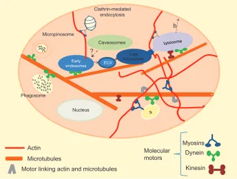
The uptake of extracellular material across the plasma membrane of eukaryotic cells (Figure 1) is well described and characterized by a variety of mechanisms going from
passive diffusion to active transport.2 The former (Figure 1a),
described by Fick’s law, is defined as a concentration
gradient–driven mass transport of a compound.2 The latter
is in charge of taking up larger molecules and molecular complexes, and is known, in general terms, as endocytosis. In this process, localized regions of the plasma membrane
wrap around the material to be internalized, fold in and detach to form endocytotic vesicles. Endocytosis occurs both consti-tutively and as a response triggered by extracellular signals and in general occurs by multiple mechanisms that fall into two broad categories – “pinocytosis” and “phagocytosis.” Pinocytosis is a constitutive process that occurs in most of the cells, continuously and independently from the needs of the cell. With pinocytosis, we can distinguish at least four basic mechanisms: macropinocytosis (Figure 1b), clathrin-mediated endocytosis (Figure 1c), caveolae-clathrin-mediated endo-cytosis (Figure 1d), and clathrin- and caveolae-independent endocytosis (Figure 1e). Phagocytosis (Figure 1f) is instead typically restricted to specialized mammalian cells like macrophages and it is a process requiring activation of recep-tors that transmit the signal inside the cell and trigger the
response.3 The differences between phago- and pinocytosis
internalization pathways rely on the size of the endocytotic vesicles: the former involves the ingestion of large particles
by large vesicles called phagosomes (diameter . 250 nm),
while the latter involves the ingestion of fluids and solutes through small pinocytotic vesicles (diameter from a few up to hundreds of nm), except for macropinocytosis that
involves formation of vesicles bigger than 1 µm up to 5 µm.
In general, when fluid is internalized, the process is also
called fluid-phase endocytosis.4
(f) Phagocytosis (a) Passive
diffusion
Pinocytosis
(b) Macropinocytosis
(c) Clathrin-mediated endocytosis
(d) Caveolae-mediated endocytosis
(e) Clathrin-and caveolae-independent
endocytosis
X = Opsonized target = Actin
Early endosomes
= Dynamin
Caveosomes
Lysosomes (pH 4,5)
Late endosomes
(pH 5)
= Caveolin = Clathrin
Phagosome
Nucleus
Figure 1 Summary of pathways of cellular uptake. For clarity, most of the intracellular organelles have been omitted. In this image, clathrin-mediated and receptor-mediated endocytosis share the same pathway.
Notes: Vesicles originated by a given pathway are not scaled; in fact, the dimension of macropinosome can reach 5 µm, pits coming from the clathrin-mediated pathway about 150 nm, and clathrin- and caveolae-independent vesicles about 50 nm.
Dovepress
Panariti et al
Nanotechnology, Science and Applications downloaded from https://www.dovepress.com/ by 118.70.13.36 on 24-Aug-2020
The phagocytic process starts after opsonization of the target by opsonins in bloodstream, in order to make the foreign substance visible to macrophages. Opsonized particles attach to the macrophage surface through specific
receptor–ligand interactions.4 Receptor ligation is the
begin-ning of a signaling cascade that triggers actin assembly, forming cell surface extensions (pseudopodia) that zipper up around the particle and engulf it. The resulting phago-some will ferry its content throughout the cytoplasm. As actin is depolymerized from the phagosome, the newly denuded vacuole membrane becomes accessible to early endosomes. Through a series of fusion and fission events, the vacuolar membrane and its contents will mature, fusing with late endosomes and ultimately lysosomes to form a
phagolysosome.5
Clathrin-mediated endocytosis occurs in specialized regions enriched in clathrin proteins that cover 2% of the total area of the plasma membrane. The polymerization of this protein in basket-like structures induces the formation of a coated pit of 120–150 nm. Dynamin is required for the formation of vesicles that deliver the inner material to early endosomes and fuse with prelysosomal vesicles to give rise to late endosomes and finally to a lysosome. The formation of clathrin vesicles is also common to receptor-mediated endocytosis. In this case, a specific molecule recognizes its transmembrane receptor located on the plasma
mem-brane, and the complex forms clathrin-coated vesicles.4 The
receptor-mediated endocytosis is a selective mechanism of concentration that increases by more than 1000 times the efficiency of internalization of ligands avoiding, in particular, the simultaneous internalization of large volumes of
extracel-lular fluid.3 During the process, the uncoating of the vesicles
allows recycling of clathrin units, receptors, and ligands. In the case of polarized cells, the recycled molecules can either return to the membrane from which they were internalized, or they can cross the cell and deliver their content to the opposite
membrane in a process called transcytosis.3
In addition to clathrin-coated vesicles, there are other mechanisms by which cells can form pinocytotic vesicles. One of these pathways starts at the level of caveolae, whose main structural proteins are the caveolins. Such proteins, being integral membrane ones, unlike those of clathrin-coated vesicles, do not dissociate from the vesicles after endocytosis. Caveolae detach from the plasma membrane using dynamin and bring their content to compartments similar to
endo-somes, called caveosomes.3,6 The caveosomes’ content avoids
lysosomes, and is therefore protected from exposure to low pH and hydrolytic enzymes.
Macropinocytosis is one of the most important types of clathrin and caveolae-independent endocytosis pathways, occurring in many cells, including macrophages. Actin, as in phagosomes, drives the formation of membrane protru-sions that collapse onto and fuse with the plasma membrane, forming large endocytotic vesicles called macropinosomes. Such vesicles can either fuse with acidic cell compartments
like lysosomes or recycle their content to the surface.4 Other
clathrin- and caveolae-independent endocytosis pathways involve cholesterol-rich microdomains called “rafts,” which have a 40–50 nm diameter. Vesicles budding from lipid rafts may be internalized without caveolins and carry their cargo delivered to caveosomes.
Degradation of intracellular and extracellular material is crucial to maintain cellular and organism homeostasis. Lysosomes are the intersection where traffic flows converge intracellularly for degradation. As mentioned above, the most important degradation pathway that occurs in the pres-ence of endocytotic vesicles involves early endosomes that
mature into late endosomes (pH = 5) and finally fuse with
acid compartments of lysosomes (pH = 4.5). A second route
of degradation in lysosomes is a process called autophagy. Autophagosomes are referred as double-membrane vesicles that can enwrap intracellular substrates in a nonspecific fashion during bulk turnover of cytoplasm or specifically target damaged organelles, protein aggregates, or specific proteins for lysosomal degradation or secretion. Following phagocytosis or macroendocytic engulfment mechanisms, signals are transmitted across the vacuole to recruit and acti-vate molecules involved in the formation of autophagosomes. The formed autophagosome undergoes maturation and fusion
with lysosomes.7
Intracellular trafficking
Microtubules, actin filaments, and intermediate filaments are components of the cytoskeleton of the majority of eukaryotic cells. Cytoskeleton is crucial for many cellular functions, such as mitosis, cytokinesis, cell motility, muscle contrac-tion, maintenance of cell shape, endocytosis, intracellular trafficking, and protein secretion.
During the last two decades, much attention has been focused on the regulation of membrane traffic by the actin and microtubule cytoskeletal network, including their molecular motors – myosins – which move on actin filaments, and dyneins and kinesins, which use microtubules as tracks.
All three motor classes use the energy derived from adenosine triphosphate (ATP) hydrolysis to translate it into movement, a step along the track. Moreover, motors may link
Dovepress Nanoparticle uptake and cellular behavior
Nanotechnology, Science and Applications downloaded from https://www.dovepress.com/ by 118.70.13.36 on 24-Aug-2020
the microtubule and actin systems as well. Motor-dependent movement is proposed to increase the probability of vesicle
collisions and therefore promote fusion events.8
Research on organelles transport demonstrates that some organelles can switch tracks and move on either microtu-bules or actin filaments. Some myosins drive short-range movements through actin-rich regions before (as in endocy-tosis) or after (as in exocytosis or recycling) transport along
microtubules.3 Recent evidence suggests that microtubules
themselves, in the absence of motors, can move around
cellular structures inside the cells.9
As far as cellular uptake and intracellular trafficking are concerned, actin polymerization is a determinant for the formation of phagosomes and macropinosomes, and several myosin isoforms are involved in these processes. Microtubules facilitate the fusion of these large vesicles with endosomes and the transport towards the nucleus is mostly dynein-dependent. In clathrin-mediated endocytosis, actin polymerization and class I myosins generate membrane invagination, and coated-pit formation, conscription, and vesicle scission. Myosin VI and some adaptor proteins are essential for the transport of the nascent endocytotic vesicles from clathrin membrane–rich regions to early endosomes. In mammalian cells, actin but not myosin participates in the formation and the internalization of caveolae, but it remains unclear how caveosomes reach early endosomes. Cargoes travel from early to late endosomes, and those that are des-tined for degradation reach lysosomes, whereas others are recycled either to the plasma membrane (through recycling
endosomes) or to Golgi apparatus (trans–Golgi network).5,10
Independently, on the different theories that state that either late endosomes derived from early ones by a maturation process (unlikely), or that they are preexisting cellular com-partments connected by transport vesicles, or, again, that they are part of a common tubular network, the progress of endocytosed material is directed following a centripetal route from the membrane to the nuclear region and requires
an intact microtubule network.11 The fusion between early
and late endosomes seems not to be a direct process, but is mediated by shuttles termed endosomal carrier vesicles, also
microtubule-dependent.11 There is no experimental evidence
that the delivery between late endosomes and lysosomes may be driven by vesicular transport; instead, there is clear
evidence for direct fusion of these organelles.12 Lysosomes
move on microtubules, and their movement has been shown to require cytoplasmic dynein and kinesin. More recently, it has been suggested that the F-actin network and associated molecular motors such as myosins might be involved in
this movement controlling the trajectories of organelles on
microtubules.13,14 The contribution of the actin and
microtu-bules in vesicular trafficking is not restricted to the direction from plasma membrane towards lysosomes: there is some evidence that these systems cooperate in the exocytotic route, starting from sorting endosomes. In this scenario, kinesins play an important role, and once vesicular cargo has reached the cell periphery by microtubule transport, myosin V has a prominent role in the transport of the cargo component on the cell surface (see Figure 2b). Interestingly, dynein shows the capability of moving bidirectionally: this suggests that molecular motors can regulate the balance between recycling
and degradation.6,8,10
Intermediate filaments have been always associated with mechanical properties able to provide structural resilience, but it has become clearer now that intermediate filaments play an important role in membrane traffic and movements of organelles and motor cargoes. Although intermediate filaments have a nonpolar structure and no specific asso-ciated motor proteins, they are well integrated with actin and microtubule cytoskeletons, by a direct interaction with kinesin, dynein, and myosin.
Why should nanoparticles
enter cells?
For at least three decades NPs have been thoroughly studied because of the properties that make them suitable for diag-nosis and delivery. They may be intended as a toolbox to be used to exploit one or more functions at the same time in what is now called the nanomedicine field. They offer the unique possibility to overcome cellular barriers in order to improve the delivery of various drugs and drug candidates, including promising therapeutic biomacromolecules such as nucleic acids, antisense oligonucleotides, small interfering RNA (siRNA), and plasmid DNA, that can only exert their function once inside the cells, and that otherwise may not be delivered. In fact, as polar molecules, they cannot permeate the lipid bilayer of plasma membrane or other biological membranes (blood–brain, air–blood, gastrointestinal barriers). In terms of drug delivery, one central advantage is the concept of the possibility to load NPs with a high concentration of the desired drug. In carrying a large payload, nanocarriers can favorably modulate biodistribution and pharmacokinetic profiles of the drug formulations. They may be also used as carriers for contrast agents in vivo magnetic resonance
imaging or, again, as an all-in-one system.15 In fact, through
their multimodal loading capability, the surface or core of the NPs may be loaded with multiple agents, so that treatment
Dovepress
Panariti et al
Nanotechnology, Science and Applications downloaded from https://www.dovepress.com/ by 118.70.13.36 on 24-Aug-2020
Kinesin Myosins Dynein
Motor linking actin and microtubules
... ...
Micropinosome
Nucleus
Early endosomes
Caveosomes
?
Phagosome
Clathrin-mediated endocytosis
Late endosomes
ECV
Actin Microtubules
Molecular motors
b
Figure 2 (a and b) Intracellular trafficking. The cell environment is considerably more complex than is represented in this simplified image. Many of the intracellular organelles are not represented for clarity. Also, we do not represent the centrosome, form which arise most of the microtubules. (a) Vesicle or organelle switching track; (b) Transport towards the cell surface; kinesins and myosins play an important role in directing vesicles backward to the plasma membrane.
and imaging of treatment can occur simultaneously.16,17 More
recently, they also have been applied in real-time live-cell monitoring of a number of cell parameters such as
intracel-lular oxygen concentration.18
NPs can be made of different inorganic and organic materials: they can be lipid-based (such as liposomes,
micelles, solid lipid, or lipoprotein-based NPs),18–21 polymeric
(such as polylactic acid, poly[lactic-co-glycolic acid], and
poly[alkylcyanoacrylate]),22,23 chitosan-based,24 quantum
dots,25 silicon-based26 gold NPs,27 and magnetic NPs.17
Since the intrinsic characteristics and thus the relevant applications of NPs are closely related to their size, shape, and surface properties, great efforts have been devoted to control the synthesis of NPs. Moreover, NPs have been engineered in different ways to solve problems related to their stability in physiological medium. However, the aim of this review is not intended for discussing such very complex problems, nor will a comprehensive discussion of the toxicity issue be presented here. Thus, for these issues, we invite readers to
refer to more appropriate reviews.28,29
Methods to analyze intracellular
behavior of nanoparticles
The tracking of NPs in living cells provides new insights into understanding the NP uptake processes, intracellular transport,
and their complex behaviors. To this end, the fluorescence microscope is widely used. This implies that NPs are labeled with a fluorescent probe, a molecule that absorbs light at a certain wavelength and emits it to another longer one. If such a compound is illuminated at the wavelength of absorption and then observed through a filter that allows passage of the single wavelength emission, it is observed to shine against a dark background. The two most commonly used fluorescent dyes are fluorescein, which emits an intense green
fluores-cence (λ emission maxima ∼520 nm) when excited with
blue (λ absorption maxima ∼490 nm), and rhodamine, which
emits a deep red fluorescence (λ emission maxima ∼580 nm)
when excited with light yellow-green (λ absorption
maxima ∼550 nm). Originally, fluorescence microscopy
required the cells to be fixed and eventually immunostained, eg, with antibodies for intracellular organelles, for colocal-ization studies. This approach can cause fixation artifacts and does not easily provide information on the kinetics of colocalization, as opposed to live-cell imaging. Due to the advent of fluorescent proteins and technical developments in fluorescence microscopy, time-lapse imaging of live cells is becoming the preferred method. However, even this procedure is not free of disadvantages: the prolonged exposure to laser emission may in fact cause the photobleaching of the samples, and thus a decrease in the observed signal.
Dovepress Nanoparticle uptake and cellular behavior
Nanotechnology, Science and Applications downloaded from https://www.dovepress.com/ by 118.70.13.36 on 24-Aug-2020
An alternative approach to be used for purely experi-mental purposes is to mark the NPs with radioactive iso-topes to study the biodistribution in cells or tissues. The resolution of these experiments is often increased by using protocols named “pulse-and-chase,” in which the radioac-tive material (pulse) is added only for a very short period of time and then removed and replaced by nonradioactive molecules (chase). Such experiments are combined with autoradiography on sections of cells or tissues to detect the radioactive signal.
One last possibility, very often used, is transmission electron microscopy (TEM) that can detect structures much smaller than those seen with visible light. In theory, the TEM resolution would be 0.002 nm, ie, 100,000 times that of the optical microscope. The practical resolving power, however, is in fact 0.1 nm. The sample in this case must be colored with an electron-dense material and the image is observed
on a screen or on a photographic plate.30
Physicochemical properties
of nanoparticles affect their
cellular uptake
There are various mechanisms of nanocarrier cell internaliza-tion that are highly influenced by NPs’ physicochemical
prop-erties, such as size, shape, and chemistry.4 The NPs, in fact,
have to be soluble in physiological solutions, then, depending on the route of administration chosen (oral, intravenous), at a certain point they interact with the cell’s plasma membrane and, eventually, gain access to the cells and to the appropriate
organelle where the biological target is located.4 As the
number of available NP typologies increases, it becomes crucial to understand how NPs’ physicochemical properties determine the interaction with biological systems and enter the intracellular space in order to optimize the potential use of nanodelivery. Yet the definition of the absolute profile to match a specific endocytotic pathway is extremely difficult.
In fact, if phagocytosis is confined to a particular type of cell and occurs only as a consequence of a well-described process, thus making it possible to outline the best features of NPs most suitable for this process of internalization, the great majority of cell types can simultaneously utilize more than one internalization pathway (see Figure 2). This is because cells can express different levels of target receptors or, for polarized cells, present differences related to the environment of the apical and basolateral membranes.
Particle size can affect the biodistribution, the efficiency (ie, how many NPs are found inside the cell at a given time point), and the cellular uptake pathway for liposomes,
quantum dots, polymeric, gold, and silica NPs by influencing
their adhesion and interaction with cells.4,31,32 In some NP
applications, the first aim is to avoid clearance by the reticu-loendothelial system, thus prolonging the circulation time in the blood and increasing the bioavailability at the target site. The clearance rate increases with increasing size of NPs: NPs
in a range of 250 nm to 3 µm have been shown to have an
optimal in vitro phagocytosis, while NPs with a size limit of around 200 nm preferentially involve other uptake routes, like clathrin- or caveolin-mediated endocytosis.
Together with size, shape is one of the primary parameters that requires special attention. The vast majority of NPs developed for drug delivery have a spherical shape, but other forms such as cube-shaped (so-called cubosomes), cylindri-cal, ellipsoids, and disks have recently been proposed as new
drug nanocarriers.4 It seems, though, that macrophages fail
to internalize NPs when they present too large a surface area, as they are spread around because of the complexity of actin
structure required to initiate the process of phagocytosis.33
When we consider nonphagocytic pathways, there is not yet a general tendency to prefer one shape over others: some works, considered by Hillaireau and Couvreur, have shown that spherical NPs had a higher and faster rate of endocytosis compared to rods, disks, or gold NPs, while other studies have suggested preferential uptake of rod-shaped or cylindrical
particles.4 The shape of NPs also influences the trafficking of
nanomaterial inside the cells: hexagonal shapes are retained in the cytoplasm, while the rod-like ones are moved towards
the nucleus by microtubules.34
NP rigidity seems to be a further significant factor
influ-encing the entry pathway. The study of Beningo and Wang35
shows that macrophages tend to present a strong preference for rigid particles because soft ones are unable to stimulate the formation and closure of phagosomes. Banquy et al demonstrated that soft hydrogel NPs were internalized via macropinocytosis, stiff NPs by a clathrin-dependent mecha-nism, and NPs with intermediate elasticity exhibited multiple
uptake mechanisms.36
When we characterize NPs in terms of surface charge, it is well established that due to the negatively charged character of the cell plasma membrane, cationic NPs are internalized more efficiently than neutral and anionic NPs.
It is quite clear now that surface chemistry properties criti-cally affect the way NPs interact with each other, with their surrounding environment, and with cells. In this respect, NP aggregation in cell culture media represents the most impor-tant limitation for medical device performance. Cantow and Battaglia reported that studies have shown that in presence of
Dovepress
Panariti et al
Nanotechnology, Science and Applications downloaded from https://www.dovepress.com/ by 118.70.13.36 on 24-Aug-2020
serum, the size of the nanocarriers had a strong influence on opsonin adsorption, and therefore on phagocytosis as well as
for other internalization routes.32 The interaction between NPs
and serum protein induces the formation of a protein corona that can quickly cover the entire NP surface. The principal driving forces for protein adsorption are hydrophobic and elec-trostatic interactions, combined with an increase in entropy caused by the protein unfolding. In fact, adsorption can alter protein structure, exposing segments normally buried in the native protein conformation, thus modifying signaling, and in
some cases causing fibril formation.32 In order to reduce this
phenomenon, in recent years different strategies of coating the surface with a polymer brush able to generate steric forces have been developed. Mainly, these include poly(ethylene
glycol), polysaccharides (such as dextran), 266 poly(N-vinyl
pyrrolidone), poly(vinyl alcohol), poly(2-methyl-2-oxazoline), poly(2-ethyl-2-oxazoline), poly(2-methacryloyloxyethyl
phos-phorylcholine), and poly(sulfobetaine methacrylate).32
Despite the growing body of knowledge in this field, the understanding of how all these factors can be combined to define the best characteristics for specific NP–cell interaction remains a great challenge.
Nanoparticle uptake
According to the physicochemical characteristics of the nano-carrier and the nature of the target cells, two main internal-ization pathways may occur: either a non–energy- dependent
interaction with plasma membrane21,37 or the energy- dependent,
endocytotic pathways4 (see Figures 1 and 2).
As far as the non–energy-dependent pathway is con-cerned, we have demonstrated that coumarin solid-lipid NPs (SLNs) interact with the plasma membrane after a few minutes of exposure. During this kind of interaction, due to SLN strong chemical and structural similarity with the plasma membrane, they undergo a structural modification that may be explained by a direct mixing or exchange of phospholipids between the target cell plasma membrane and particle. This chemico-physical passive interaction process where SLN structural lipids may merge with cell membranes
could facilitate drug delivery into the interior of the cell.21
This pathway may be shared among all the lipid-based NPs. For all the other types of NPs, the uptake occurs mainly via
one of the different forms of endocytosis.29
At the moment, there is no evidence that NPs, once close to the cell membrane, are taken up individually, and one may doubt the technical ability to capture this event. Especially for very small NPs, it is more likely that only when a critical density is reached locally, they can be taken up and the entire
cluster proceed to completion.38,39 As we have already said,
it is also stated that different types of uptake by endocytosis strongly depend on NP size.
Though it is not possible to generalize the pathway uptake for NPs with given characteristics, we briefly report that NPs with a diameter up to several hundreds of nanometers preferentially enter the cells via pino- or macropinocytosis, the clathrin-dependent uptake is the preferred route for NPs on whose surface are adsorbed serum proteins, and lastly, negatively charged nano-objects enter mostly likely by caveolin- and/or clathrin-mediated endocytosis, but not
by macropinocytosis.29 Regardless of the fact that the
first-generation, thus naked, NPs, may enter cells by their own way, an improvement of their efficacy and specificity was required. For this reason, a second generation of nanovectors have been developed, offering a higher degree of sophisti-cation compared to their predecessors, since they present environmentally sensitive components or targeting moieties
in order to improve NP uptake.40 In these terms, one could
have only NP surface charge playing a role: positive NPs show quicker and higher internalization, while negative ones have a higher uptake rate constant, thus fewer NPs enter and
at a slow rate.32,41
Additionally, NPs can be targeted to a definite cell population by the attachment of molecules that are mainly recognized by selected cells and thus ameliorate selective drug delivery to specific cell types. When the specific pep-tide is bound on NP surface, it will be recognized by the receptors located on the plasma membrane that are unique to certain cells or related to certain diseases. This typical ligand-receptor recognition will exploit the mechanism of
receptor-mediated endocytosis (see Figure 1c).17 One of the
receptors that has frequently been used as a tool to increase the concentration of drugs within the cell is the folate recep-tor or the scavenger receprecep-tor class B type I, since these are
overexpressed in many types of cancer cells.16,42 Transferrin
is another example of how receptors can be exploited for
passing the blood–brain barrier43 and entry to the
endo-somal pathway. Another approach is the use of fusogenic or cell-penetrating peptides. Among the most frequently used peptides is Tat, which has been shown to facilitate the uptake of various cargoes and deliver it directly to the endosome compartments.
Nanoparticle intracellular trafficking
Once inside the cells, NPs, as aggregates, initiate their traf-ficking, which can be distinct in its random, diffusive motion guided by thermal agitation and active movement. The latter
Dovepress Nanoparticle uptake and cellular behavior
Nanotechnology, Science and Applications downloaded from https://www.dovepress.com/ by 118.70.13.36 on 24-Aug-2020
depends on cytoskeleton components, namely microtubules, actin, and intermediate filaments. These are not static struc-tures, but highly dynamic since they undergo time-dependent restructuring. It is worthwhile considering that beside these two extremes, motor activity may have consequences on diffusion motion. In other words, the net motion reflects a contribution from the specific cytoskeleton-associated pro-cess to which the cargo is coupled, in addition to nonspecific neighboring processes that can give rise to fluctuations of the cytoskeleton itself.
The dynamics of the cytoskeleton components are so relevant that in the absence of rearrangement of both micro-tubules and actin, the intracellular movements resemble
Brownian motion.44 While the active forces that drive
non-thermal fluctuations in cells act primarily through the cytoskeleton, these fluctuations can have important con-sequences for the transport and stirring of small particles not associated with the cytoskeleton, such as organelles and perhaps even individual proteins or protein complexes, due to mechanical resistance of the surrounding medium. This is because of the fundamental hydrodynamic coupling of cytoskeletal filaments to the surrounding cytoplasm: the motion of the cytoskeleton does not occur in a stationary cytoplasmic fluid, but drags this fluid along. Thus, fluid dynamics dictates that the large, active displacements seen for microtubules must be accompanied by a very vigorous stirring and fast transport within the cytoplasm. This “active diffusion,” in some cases, could be the dominant motion of
individual proteins.45
In general, after cellular uptake, exogenous material of various origins tends to accumulate in the perinuclear
region.21,46–48 This accumulation is against a diffusion
gradi-ent and favored probably by a local energy generated by the
cytoskeleton dynamics,37 suggesting that the integrity of the
actin filaments may play a role in the intracellular distribution of NPs, maybe generating a force that helps the concentration of the substance in the perinuclear region.
Specific intracellular nanoparticle
targeting
Another important issue to consider is the necessity, in some cases (ie, drug delivery), to direct NPs, or their cargo, selec-tively to one type of cell organelle as a specific target. In this case the functionalization of the NPs plays a central role in the redirection of the particle to specific cell subcompartments, thus interfering with physiological intracellular trafficking.
The use of stimuli-responsive nanocarriers offers an interesting opportunity for drug and gene delivery in the
optimization of therapies. An example of a biological stimu-lus that can be exploited to target drugs and genetic material
is pH.49 Cellular components such as the cytoplasm,
endo-somes, lysoendo-somes, endoplasmic reticulum, Golgi apparatus, mitochondria, and nuclei are known to maintain their own characteristic pH values, which range from 4.5 in the lyso-some to about 8.0 in the mitochondria. Moreover, pH value is greatly affected by diseases: the hypoxic environment in cancer leads to an increase in production of lactic acid and hydrolysis of ATP, both contributing to acidification. In fact, most solid tumors have lower extracellular pH (pH 6.5) than
the surrounding tissues (pH 7.5).50 By selecting the right
material composition, it is possible to engineer nanocarriers that can exploit these pH differences and allow the release of the delivered drugs or genes to the selected target site.
pH-sensitive poly(β-amino ester), a biodegradable cationic
polymer, in acidic microenvironment undergoes rapid
dis-solution and releases its content all at once,51 thus it may
represent a good scaffold to deliver anticancer drugs. Other strategies involve the presence of acid-sensitive spacers
like poly(vinylpyrrolidone-co-dimethyl maleic anhydride)
between the drug and the polymer that enable, after endo-cytosis, drug release in endosomes or lysosomes of tumor
cells.52 In this scenario, NPs designed to be pH-responsive
that undergo physicochemical changes to release enclosed drugs at acidic pH conditions are promising vehicles for
antitumor drug delivery.53
If the target is not the lysosome or in general the acidic compartments of the cells, the low pH environment and various lysosomal enzymes result in the degradation of endocytosed components, thus the loss of the therapeutic effect. This happens unless there are specific mechanisms for the payload to escape out of the lysosomes and maxi-mize the efficiency of various treatments. One example to pursue the “lysosomal escape” strategy is represented by liposomes containing pH-responsive polyanionic poly-mers and lipids such as dioleoylphosphatidylethanolamine (DOPE). At low pH, the polyanionic polymers and DOPE change conformation, disrupting the endosomal membrane and causing translocation of its contents into the cytosol. Similarly, some bacterial toxins that unfold in the low pH environment insert in the membrane and translocate into the cytosol; various drug systems have been based on the ability of diphtheria toxin to transfer from endosomes into
the cytosol in a pH-dependent manner.54,55 The N-terminus
of hemagglutinin subunit HA-2 of the influenza virus has been exploited for the endo/lysosomal escape of several drug-delivery systems. These peptides may also be used in synergy,
Dovepress
Panariti et al
Nanotechnology, Science and Applications downloaded from https://www.dovepress.com/ by 118.70.13.36 on 24-Aug-2020
as with Tat: this latter speeds up the cell internalization and HA-2 that escapes lysosome targeting if, for example, the cargo is pH-sensitive or has to be delivered to organelles
other than lysosomes.56
Another interesting approach is photochemical internal-ization, which uses light to facilitate the release of endocy-tosed macromolecules into the cytoplasm. The endosomal/ lysosomal membranes are highly damaged by using amphiphilic photosensitizers, such as disulfonated aluminium phthalocyanine and meso-tetraphenylporphine disulfonate, and the result is the subsequent release of entrapped drugs into the cytosol. Photochemical internalization has demon-strated a broad range of biological applications, but the most promising is the delivery of siRNA to silence oncogenes and
related tumor-inducing genes in cancerous cells.57 Lysosomal
escape may also be achieved by modulating, again, the surface charge: chitosan-based NPs or polyamines such as poly(ethylene imine) positively charged, escape
lyso-some probably due to the “proton-sponge effect.”58,59 These
molecules behave as lysosomotropic agents, weak bases that penetrate in the lysosome in protonated form and increase the intracellular pH and affect cleavage events. Once inside the acid compartment, the amine groups of these molecules can absorb protons generated by the ATPase with a consequent
lysis of endosomes caused by increasing osmolarity.60
Other interesting targets for specific therapies are the mitochondria and the nuclei. Mitochondrial dysfunction is associated with a variety of human disorders, such as neurodegenerative and neuromuscular diseases, obesity and diabetes, ischemia-reperfusion injury, cancer, and inherited
mitochondrial diseases.61,62 The design of nanosystems for
this purpose requires great effort since reaching intracellular subcompartments, such as mitochoudria, need to undertake more complicated routes than is required for a cytosol delivery. In fact, two more key obstacles to overcome for mitochondrial drug delivery are the outer and the inner mitochondrial barriers. First of all, as previously reported, for those particles that enter cells via the endosomal path-way, they must escape the endosome before fusion with the lysosome to prevent content degradation. Based on the mito-chondrial membrane features, physicochemical properties
of NPs have been well characterized.63 Outer mitochondrial
membrane can permeate NPs up to 3 nm in size and mol-ecules with a molecular weight less than 5000 daltons through voltage-dependent anion channels. If the therapeutic target is within the mitochondrial matrix, the drug will also have to cross the inner membrane, which has a strong negative potential, thus drug lipophilicity, charge, and polar surface
area positively influence the delivery that may be achieved also by integrating mitochondrial translocation ligands and/ or positively charged ligands into the NP design.
Lastly, nuclei represent the main target for gene delivery therapy, and numerous polymers, peptides, liposomes have recently been developed for this purpose. Keeping in mind that as a general rule, positive NPs more easily approach
the nucleus,64 small molecules enter the nucleus via the
nuclear pore complex across the nuclear membrane, while large molecules require appropriate targeting signals. The most innovative approach involves peptides containing spe-cific nuclear localization sequences. These sequences have stretches of highly basic amino acids: either one cluster or two clusters of basic residues separated by 10–12 neutral residues. Nuclear localization sequences can be covalently coupled to the DNA to prevent the loss of the sequences’ signature and the exposure of the peptide on the surface of
the nanostructure.65,66
Effects of nanoparticles
on cellular physiology
Although numerous novel nanomedicine-related applications are under development or nearing commercialization, the process of converting basic research in nanomedicine into
commercially viable products is still long and difficult.67
In spite of the abundant literature (thousands of publications may be found on Medline, Web of Science, or INSPEC), the practical use of many NPs is limited by the lack of knowledge concerning their potential toxicity. Since ethical, experimen-tal, and economic issues unquestionably call for the use of the smallest possible numbers of animals in scientific research, a large series of in vitro cellular models are employed to predict
the effects of NPs in biological systems.68 Here we review
different cell behaviors that are altered in the presence of NPs without causing cytotoxicity, as classically intended. One phenomenon easy to find in many cell lines is the reduction in the ability to produce formazan deposits even when NPs
are incubated at a sublethal dose.69–71 This result is intended as
an index of mitochondrial stress, since the reaction involved in the formazan formation is catalyzed by a mitochondrial reductase. It has been demonstrated that a perturbation of the normal mitochondrial activity is correlated with an increase
in reactive oxygen species (ROS) production.72
Oxidant stress at high levels is known to lead to cell injury
and death.73 In endothelial and epithelial cells, incubation with
NPs may elicit an oxidative stress, measured by an increase in ROS or in nitric oxide production. These two messengers modulate inflammatory reactions, which can also be indicated
Dovepress Nanoparticle uptake and cellular behavior
Nanotechnology, Science and Applications downloaded from https://www.dovepress.com/ by 118.70.13.36 on 24-Aug-2020
by an increase in the transcription and transduction of tumor
necrosis factor-alpha74 or of transcription factor-2, which
is a member of the basic region–leucine zipper transcrip-tion factor family that regulates the expression of genes in
response to various stress signals.75
ROS formation is also interconnected with cytoskeleton disorganization or damage, such as it has been hypoth-esized that ROS release due to mitochondrial disruption and cytoskeleton dysfunction may act as an interconnected systems. Decrease in actin dynamics leads to reduced mito-chondrial membrane potential via open voltage-dependent anion channels, which increases mitochondrial ROS release
and cell apoptosis sensitivity.76
The resulting reduction of mitochondrial activity induces a decrease in ATP production that is necessary for many cellular functions, including cell motility and intracellular trafficking. This point is of particular interest, since the different components of cytoskeleton interact to control multiple functions, including migration, cell morphology
and structural integrity,77 division, deformation, intracellular
transport,44 and tissue organization.73
Any modification in cytoskeleton architecture can also lead to alteration in the expression of cytoskeleton-associated
proteins.77 In some cases, these are related to
neurode-gerative disease such as amyotrophic lateral sclerosis or Huntington’s disease. Magnetoliposomes affect actin struc-ture and formation of focal adhesion complexes and impair
cell proliferation.77 Iron oxide NPs are reported to interfere
with actin and tubulin structures,73 inducing cell retraction,
rounding, and deposition of massive dense filament matters
adjacent to the nucleus and vacuoles in the cytoplasm.78
Disruption of cytoskeleton structures caused by magnetic NPs contributes to cell detachment and the failure of dif-ferentiation, since all these processes are coordinated by
cytoskeleton.78
In general, ROS production, together with actin and microtubule damage, affects mechanical properties of many cell types, increasing cell permeability on vascular and epi-thelial districts. Endoepi-thelial cell permeability is critical, since it controls the passage of macromolecules, cells, and fluid
from the blood into the vascular wall and tissues.73 The actin
cytoskeleton is connected to cell tight junction and plays an important role in the organization and maintenance of these
structures79 regulating epithelial permeability. More recently,
some evidence suggests that microtubules are also important in tight-junction assembly and stability playing a part in the
regulation of paracellular permeability.80 Several papers, in
fact, report that NP exposure impairs the barrier function of
epithelial or endothelial monolayers, monitored as a decrease in the transepithelial/endothelial electrical resistance, without
altering the viability of the cells.81,82 One important mediator
in the alteration of endothelial permeability in the opening of paracellular permeability pathways interfering with adherens
junction is intracellular free calcium.83
Incubation of cells with titanium oxide NPs caused an increase in the intracellular calcium concentration. Calcium is an important second messenger involved in a multitude of intracellular signaling pathways. Enhanced intracellular
free calcium concentration (Ca2+
i) levels are known to lead to
the activation of protein kinase C, which is involved in many
intracellular signaling pathways.84 Despite little influence on
cell viability, incubation of cells with zinc oxide and cerium oxide NPs also changes the resting intracellular calcium with-out compromising the homeostatic mechanisms that maintain
cytosolic calcium concentrations at low levels.85 These results
suggest that cerium oxide NPs have the potential to induce intracellular oxidative stress and increase the intracellular
Ca2+
i level, but these influences are small.86
An increase of intracellular free calcium in response to a cellular stress or a cytoskeleton alteration promotes autophagy
or mitophagy (mitochondrial specific autophagy).87,88
Autophagy is a fundamental process in the quality and quantity regulation of intracellular biological function. The activation of autophagic pathways in stressed cells serves to prevent the accumulation of potentially harmful damaged proteins and organelles, resulting in cytoprotection against
cellular stresses. Incubation of cells with iron oxide NPs89 or
carbon nanotubes88 has been demonstrated to promote this
physiological defense mechanism.
Another important chapter related to the influence of NPs on physiological cell functions deals with the ionic cur-rents across the plasma membrane. Evidence emphasizes a direct connection of cytoskeleton with mechanosensitive ion channels. In fact, tangential forces on the plasma membrane are transmitted either directly to stretch activated channels, or to the cytoskeleton that, in turn, may lead to increased ten-sion or curvature of the cell membrane, and thus even small alterations in the cytoskeleton have the potential to regulate
ion-channel activity.90 One important example in this context
is the cystic fibrosis transmembrane conductance regulator
(CFTR) that is activated by membrane stretch.91 McCarthy
et al92 recently demonstrated that polystyrene NPs direct
activation of CFTR channels in a monolayer of Calu-3, a human airway submucosal cell line, and completed these data also in baby hamster kidneys engineered to express the
wild-type CFTR gene, thus confirming that NPs have the
Dovepress
Panariti et al
Nanotechnology, Science and Applications downloaded from https://www.dovepress.com/ by 118.70.13.36 on 24-Aug-2020
ability to act as modulators of ion-channel function in human airway epithelial cells.
With regard to “latent toxicity,” different types of engi-neered NPs were found to increase the heart frequency
in guinea pig Langendorff perfused heart.93 The authors
explained this observed increase in heart rate with two mecha-nisms not mutually exclusive. One possibility hypothesizes an NP-induced release of catecholamines from the neural endings, the other that NPs evoke a release of endothelin from endothelial cells in the heart and endothelin acts directly at chromaffin cells existing in the sympathetic nerve, leading to catecholamine release.
Other examples of the effects of NPs on excitable cells are highlighted by studies conducted on neurons. In fact, even if the brain is protected by a highly selective barrier with very little permeability – the blood–brain barrier – and therefore it is not easy for nonfunctionalized NPs to cross the blood–brain barrier and reach the central nervous system, such studies are of interest because they still show how NPs can modulate the activity of ion channels. Voltage-gated sodium channels mediating the very rapid rising phase and its initial component of the falling phase of action potentials in excitable cells and silver NPs, despite having no effect on the firing rate of hippocampal neurons, showed a signifi-cant reduction in channel peak amplitude, as well as in the overshoot and voltage threshold of the evoked single action
potential.94 On the other hand, zinc NPs’ increase of neuronal
excitability resulted from the enhancement of both sodium
and potassium current amplitudes.95 The same type of NPs
enhance olfactory neuron responses, probably facilitating the coupling of odorant receptors and an olfactory neuron–
specific G protein involved in odorant signal transduction.96
In hippocampal neurons, it has been found that exposure to cadmium–selenium quantum dots in concentration that do not cause cell death is followed by an increase in the intra-cellular calcium due both to the store release and calcium entry from membrane channels. Moreover, an impairment
was found in the voltage-gated sodium current, which implies a reduction in the fraction of the available channels
in the window of physiological potentials.97 Copper oxide
NPs on CA1 pyramidal neurons reduce the availability of potassium channels at physiological potential, thus it can be speculated that they may impact on the duration of the action potentials, since voltage-gated potassium currents play crucial roles in modifying neuronal cellular and network
excitability.98
Conclusion
Within this review, we took the opportunity to summarize some of the cellular biology pathways related to uptake of extracellular matters and their intracellular trafficking. We discussed only the cellular pathways that are present in the literature as mechanisms of NP uptake and traffick-ing, and thus that are of interest in the nanomedicine field. What is clear is that below the cytotoxicity threshold, the broad spectrum of different NP–cell interactions impacts on many different cellular physiology function levels (mitochondria, ROS production, cytoskeletal, intracellular calcium, and membrane currents) and elicits a spectrum of tissue responses. These findings provide strong evidence that nanostructures per se not only passively interact with cells but also actively engage and mediate the molecular processes that, usually, are essential for regulating cell func-tions. These perturbed activities may or may not, late in time, reveal as stressful for the cells; for this reason, we like to call them, as an ensemble, “latent toxicity.” If some of these effects may be tolerated because in certain situations (cancer diagnostic imaging or chemotherapy deliver) a risk/benefit ratio has to be taken into account, this cannot be accepted for continuous use.
One strategy to trick the cells and to obtain the NP- mediated desired effect could be to try to camouflage NPs in a way to exploit a sort of biomimesis and let the physiological pathway chaperone the pharmacological cargo. Despite this possibility, though, we should keep in mind, at least from the energetic point of view, that the uptake and the escort of a cargo that mainly occurs as active processes should imply an increase in fuel consumption. The other possibility is that NP uptake occurs by passive processes and the intracellular trafficking utilizes some energy gradients preexisting inside
the cells, due to cell structure itself (cytoskeleton).37 In other
words, NP management may either amplify the cell request for energy or saturate the preexisting routes. These aspects must be taken into consideration regardless of the widespread idea of cytotoxicity. Going back to the title of the review,
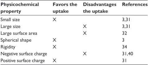
Table 1 Summary of the influence of the physicochemical
properties considered in this review on nanoparticle uptake
Physicochemical property
Favors the uptake
Disadvantages the uptake
References
Small size X 3,31
Large size X 3,31
Large surface area X 32
Spherical shape X 3
Rigidity X 34
Negative surface charge X 31,40
Positive surface charge X 31
Dovepress Nanoparticle uptake and cellular behavior
Nanotechnology, Science and Applications downloaded from https://www.dovepress.com/ by 118.70.13.36 on 24-Aug-2020
one could hypothesize that NPs stimulate some cellular functions instead of disrupting them, in the sense that NP uptake implies an increase in working level of the cellular machinery. We saw in fact an increase in ROS production, an increase in intracellular calcium, and in general an enhance-ment of cellular activity.
However, the general term of “cellular activity” should be regarded either as activation or inhibition of specific pathways mediated by NPs. Clearly, the actual balance between detrimental and beneficial effects is a critical point in therapy. This is the case for NPs loaded with siRNA or anti-microRNAs that specifically interfere or silence a cel-lular pathway involved in tumor initiation, progression, and
in chemotherapy resistance.99–101 On one side, a potential
detrimental effect of NPs per se should be balanced with the potential beneficial effect of the loaded cargo. On the other side, one can recall, independently of the presence of cargo, that a detrimental effect of some polymeric NPs on cancer cells favors their autophagy as stress cell response,
thus remaining metabolically active.102
Finally, one last consideration should be reserved for the importance of cooperation among several competencies to analyze the various aspects of NP–cell/tissue interaction, including material science, surface chemistry, physics, mathematical modeling, biochemistry, molecular biology, pharmacology, cellular physiology, and integrated function physiology.
Acknowledgments
Alice Panariti is supported by the PhD Program in Trans-lational and Molecular Medicine (DIMET) of the Univer-sity of Milano–Bicocca. Giuseppe Miserocchi and Ilaria Rivolta were partially funded by the European Community’s Seventh Framework Programme (FP7/2007–2013) under grant agreement no 212043 (NAD) and partially from the Italian Ministry of Education, Universities and Research.
Disclosure
The authors report no conflicts of interest in this work.
References
1. Modified opinion (after public consultation) on the appropriateness of existing methodologies to assess the potential risks associated with engineered and adventitious products of nanotechnologies by Scientific Committee on Emerging and newly identify Health Risks (SCENIHR), September 28–29, 2008.
2. Sugano K, Kansy M, Artursson P, et al. Coexistence of passive and carrier-mediated processes in drug transport. Nat Rev Drug Discov. 2010;9:597–614.
3. Soldati T, Schliwa M. Powering membrane traffic in endocytosis and recycling. Nat Rev Mol Cell Biol. 2006;7:897–908.
4. Hillaireau H, Couvreur P. Nanocarriers’ entry into the cell: relevance to drug delivery. Cell Mol Life Sci. 2009;66:2873–2896.
5. Caviston JP, Holzbaur EL. Microtubule motors at the intersection of trafficking and transport. Trends Cell Biol. 2006;16:530–537. 6. Mellman I, Nelson WJ. Coordinated protein sorting, targeting and
dis-tribution in polarized cells. Nat Rev Mol Cell Biol. 2008;9:833–845. 7. Florey O, Overholtzer M. Autophagy proteins in macroendocytic
engulfment. Trends Cell Biol. 2012;22(7):374–380.
8. Schliwa M, Woehlke G. Molecular motors. Nature. 2003;422:759–765. 9. Howard J Hyman AA. Dynamics and mechanics of the microtubule
plus end. Nature. 2003;422;753–758.
10. Weisz OA, Rodriguez-Boulan E. Apical trafficking in epithelial cells: signals, clusters and motors. J Cell Sci. 2009;122:4253–4266. 11. Aniento F, Emans N, Grittiths G, Gruenberg J. Cytoplasmic
dynein-dependent vesicular transport from early to late endosomes. J Cell Biol. 1993;123:1373–1387.
12. Luzio JP, Rous BA, Bright NA, Pryor PR, Mullock BM, Pier RC. Lysosome-endosome fusion and lysosome biogenesis. J Cell Sci. 2000;13:1515–1524.
13. Cordonnier NM, Dauzonne D, Louvard D, Coudrier E. Actin filaments and myosin i alpha cooperate with microtubules for the movement of lysosomes. Mol Biol Cell. 2001;12:4013–4029.
14. Taunton J, Rowning BA, Coughlin ML, et al. Actin-dependent propul-sion of endosomes and lysosomes by recruitment of N-WASP. J Cell Biol. 2000;148:519–530.
15. Chen T, Shukoor MI, Wang R, et al. Smart multifunctional nanostructure for targeted cancer chemotherapy and magnetic resonance imaging. ACS Nano. 2011;5:7866–7873.
16. Ng KK, Lovell JF, Zheng G. Lipoprotein-inspired nanoparticles for cancer theranostics. Acc Chem Res. 2011;44:1105–1113.
17. Breunig M, Bauer S, Goepferich A. Polymers and nanoparticles: intelligent tools for intracellular targeting? Eur J Pharm Biopharm. 2008;68:112–128.
18. Fercher A, Borisov MS, Zhdanov AV, Klimant I, Papkovsky DB. Intra-cellular O2 sensing probe based on cell-penetrating phosphorescent nanoparticles. ACS Nano. 2011;5:5499–5508.
19. Attama AA. SLN, NLC, LDC: state of the art in drug and active delivery.
Recent Pat Drug Deliv Formul. 2011;5:178–187.
20. Gobbi M, Re F, Canovi M, et al. Lipid-based nanoparticles with high binding affinity for amyloid-beta1-42 peptide. Biomaterials. 2010;31: 6519–6529.
21. Rivolta I, Panariti A, Lettiero B, et al. Cellular uptake of coumarin-6 as a model drug loaded in solid lipid nanoparticles. J Physiol Pharmacol. 2011;62:45–53.
22. Mahapatro A, Singh DK. Biodegradable nanoparticles are excel-lent vehicle for site directed in-vivo delivery of drugs and vaccines.
J Nanobiotechnology. 2011;9:55.
23. Danhier F, Ansorena E, Silva JM, Coco R, Le Breton A, Préat V. PLGA-based nanoparticles: an overview of biomedical applications.
J Control Release. 2012;161(2):505–522.
24. Yue ZG, Wei W, Lv P, et al. Surface charge affects cellular uptake and intracellular traff icking of chitosan-based nanoparticles.
Biomacromolecules. 2011;12:2440–2446.
25. Probst J, Dembski S, Milde M, Rupp S. Luminescent nanoparticles and their use for in vitro and in vivo diagnostics. Expert Rev Mol Diagn. 2012;12:49–64.
26. Mann AP, Tanaka T, Somasunderam A, Liu X, Gorenstein DG, Ferrari M. E-selectin-targeted porous silicon particle for nanoparticle delivery to the bone marrow. Adv Mater. 2011;23:H278–H282. 27. Rippel RA, Seifalian AM. Gold revolution – gold nanoparticles for
modern medicine and surgery. J Nanosci Nanotechnol. 2011;11: 3740–3748.
28. Wu L, Zhang J, Watanabe W. Physical and chemical stability of drug nanoparticles. Adv Drug Deliv Rev. 2011;63:456–469.
29. Zhao F, Zhao Y, Liu Y, Chang X, Chen C, Zhao Y. Cellular uptake, intracellular trafficking, and cytotoxicity of nanomaterials. Small. 2011;7:1322–1337.
Dovepress
Panariti et al
Nanotechnology, Science and Applications downloaded from https://www.dovepress.com/ by 118.70.13.36 on 24-Aug-2020