R E S E A R C H A R T I C L E
Open Access
Clinical map document based on XML (cMDX):
document architecture with mapping feature for
reporting and analysing prostate cancer in radical
prostatectomy specimens
Okyaz Eminaga
1*, Reemt Hinkelammert
1, Axel Semjonow
1, Joerg Neumann
2, Mahmoud Abbas
2, Thomas Koepke
1,
Olaf Bettendorf
3, Elke Eltze
4, Martin Dugas
5Abstract
Background:The pathology report of radical prostatectomy specimens plays an important role in clinical decisions and the prognostic evaluation in Prostate Cancer (PCa). The anatomical schema is a helpful tool to document PCa extension for clinical and research purposes. To achieve electronic documentation and analysis, an appropriate documentation model for anatomical schemas is needed. For this purpose we developed cMDX.
Methods:The document architecture of cMDX was designed according to Open Packaging Conventions by separating the whole data into template data and patient data. Analogue custom XML elements were considered to harmonize the graphical representation (e.g. tumour extension) with the textual data (e.g. histological patterns). The graphical documentation was based on the four-layer visualization model that forms the interaction between different custom XML elements. Sensible personal data were encrypted with a 256-bit cryptographic algorithm to avoid misuse. In order to assess the clinical value, we retrospectively analysed the tumour extension in 255 patients after radical prostatectomy.
Results:The pathology report with cMDX can represent pathological findings of the prostate in schematic styles. Such reports can be integrated into the hospital information system.“cMDX”documents can be converted into different data formats like text, graphics and PDF. Supplementary tools like cMDX Editor and an analyser tool were implemented. The graphical analysis of 255 prostatectomy specimens showed that PCa were mostly localized in the peripheral zone (Mean: 73% ± 25). 54% of PCa showed a multifocal growth pattern.
Conclusions:cMDX can be used for routine histopathological reporting of radical prostatectomy specimens and provide data for scientific analysis.
Background
Prostate Cancer (PCa) is the most commonly diagnosed cancer in men and one of the leading causes of cancer deaths in Germany [1]. As therapeutic approach, many patients choose total removal of the prostatic gland (radical prostatectomy). Pathology reports of radical prostatectomy specimens include clinically relevant information as well as clinically essential information
derived from the macroscopic examination and micro-scopic evaluation, which play a supporting role in clini-cal decision making and prognostic evaluation of PCa [2,3]. Consequently, diverse standardized sectioning and documentation protocols of radical prostatectomy speci-mens are described [4-7]. In our center, we use the stan-dardized pathologic report according to Bettendorf (Figure 1) [6]. This report includes a diagrammatic representation of histopathological findings in the pros-tate gland. It is a practicable method which documents tumour extension, extracapsular tumour growth, and * Correspondence: [email protected]
1
Prostate Center, Dept. of Urology, University Hospital Muenster, Albert-Schweitzer-Str. 33, D-48149 Muenster, Germany
Full list of author information is available at the end of the article
the status of surgical margins in radical prostatectomy specimens.
To our knowledge, there is no established electronic standard for graphical documentation of PCa that meets clinical and research requirements. These requirements include a flexible documentation of PCa extension with an anatomical schema that can be used for clinical and
research purposes. To provide an analyzable data acqui-sition model for anatomical schemas in electronic form,
we propose a document architecture called “clinical
Map Document based on XML”(cMDX). The
develop-ment of this docudevelop-ment architecture depends on clear definitions of domain terminologies, functional and data hierarchies, as well as assessment rules. It is intended to
improve information consistency and data integrity of routine data in hospital information systems and clinical studies.
This article describes a data model based on schematic diagrams for documentation and analysis of prostatect-omy specimen reports.
Methods
To develop a data acquisition model for the histopatho-logical examination report, the requirements for the documentation system were collected by unstructured interviews with three urologists and three pathologists. Additionally, twenty reports in paper form were ana-lysed. Thereafter, a multiple-layer model based on XML (Extensible Markup Language) specifications and vector graphics was designed in order to generate similar reports in electronic form. Figures 2, 3, 4, 5 and 6 illus-trate the data structure that is explained in detail in the following sections. Table 1 describes the presentation attributes; Tables 2 and 3 show attributes and textual information included in this model. In this paper, terms written inboldor italicrepresent either XMLelement
or attribute names. Element and attribute names are concatenated with the first letter of each word capita-lised (UpperCamelCase) or with a dash (String-Upper-CamelCase). Supplementary tools for cMDX were written in C# with Microsoft©Visual Studio 2008. The electronic Hospital Information System (HIS) Orbi-s™used in the University Hospital Muenster is provided by AFGA Health Care©. All patient records are currently stepwise transformed from paper documentation to entirely electronic documentation.
Analysis of the pathology report
The report of radical prostatectomy specimens (Figure 1) contains text and graphic data. Personal and pathologic information are textual. The anatomical scheme of the prostate gland is a template for the graphical documen-tation of histopathological findings. As previously published by Bettendorf et al. [6], the anatomical schema consists of both seminal vesicles and the pros-tate sectioned into eight defined slices. For each slice, a
“slice factor” is attributed to estimate the tumour
volume in relation to the total prostate volume. Symbols and icons are added to facilitate identification of the pathological findings. The report template was primarily designed for the documentation of PCa and High Grade Prostatic Intraepithelial Neoplasia (HGPIN), a presum-able precursor for PCa [8,9]. PCa was graded according to Gleason [10] and Helpap [11] and staged using the TNM staging system (2002) [12].
The TNM-classification system [12] is a well-known pathological documentation system that codes tumour spread (T), lymph node invasion (N) and metastases (M). Parameters like residual (R), venous invasion (V) and lymph vessel invasion (L) provided additional clini-cal information about PCa. The Gleason score system is a standard applied to assess the histological pattern and spread of PCa [10]; Helpap is a method for the histolo-gical evaluation of cell differentiation and the existence of atypical prostatic cells [11].
Document architecture of“cMDX”
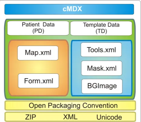
cMDX document architecture was designed to meet the Open Packaging Convention (OPC) [13] (Figure 2 and additional file 1). Data were divided into template data and patient data (Figure 2 and 3). Each XML element in cMDX was declared by a corresponding class library. The element declaration was performed with the XML namespace“xmlns”.
Template data (TD)
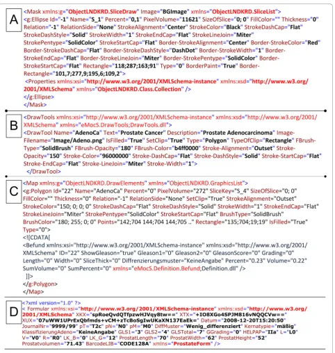
The template data is stored in addition to the Back-ground Image component (BGImage) in two different XML files: Mask.xml and Tools.xml. Tools.xml stores information about drawing tools applied to sketch pathological changes in scheme styles (e.g. freehand drawing) and provides parameters for the drawing tools.
The xml container Tools.xml contains DrawTool
ele-ments. Each xml element DrawToolhas two types of
attributes:
(1) Explanatory attributes for pathological changes like
Description, Name of a disease written in short form,
Image-Filename for the symbolic representation of the disease, andTextfor the full name of the disease.
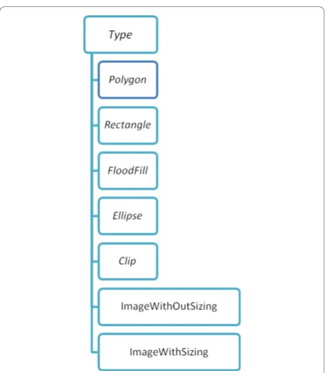
(2) Attributes for graphical representation (Table 1), which are used for building a new graphic object to be displayed onscreen (Figure 4).
In addition, the optionalProperties Element enables the interaction of the drawing tools with the application. Mask.xml stores configuration data to define the appearance of the graphical template including a
back-ground Image, which is stored in BGImage, and vector
shapes with clip function. These shapes represent the drawing surfaces onscreen. Three shapes were defined: (1)Polygon (2)Rectangle (3) Ellipse. Every shape ele-ment, whose tag name is the applied shape name, was stored inside the root elementMask. For instance, the elementEllipse represents a slice of the prostate. It has identification (e.g. Id, Name) and representation attri-butes (Table 1), which, for example, provide information
about thePercentage volume of the slice in relation to the whole prostate volume (slice factor), the PixelVo-lumeof the slice, and theSizeOfSlicefor storing the real dimension of the prostate slice.
The component“BGImage”includes an Image
repre-senting the prostate and the seminal vesicles in sche-matic style and can be changed by the user. Various image formats (TIFF, BMP, WMF, PNG, JPEG, GIF and SVG) are hereby supported.
Patient Data (PD)
Data acquisition from the prostatectomy specimen con-sists of two types of information: (1) morphometrical information about histological changes (e.g. PCa, HGPIN, positive surgical margin) with additional textual descriptions (Gleason Score, length of positive surgical
margin), and (2) clinical and personal data in text form. Accordingly, two xml containers were designed: Form. xml and Map.xml. Map.xml captures morphometrical information about histological findings to reconstruct them in schematic styles. Diverse graphic objects were
defined and stored in the root xml element Map
(Fig-ures 3 and 5). The representation of the spread pattern of PCa and HGPIN was based on vector graphics. The real dimension of a cancer area (LengthandWidth) as well as the slice thickness (SliceThick) can be optionally given for special scientific queries. In addition to the representation attributes, attributes like Relationand
RelationSlideprovide the topographical location of PCa foci in relation to each other. An XML Element called
Befund was integrated into the CDATA-Section of a
graphic object showing a PCa focus.Befund captures
information about histological patterns of the corre-sponding PCa focus (Table 2).
Form.xml contains the element Formularwith
attri-butes containing personal data; pathological findings were categorised as textual information not directly related to the morphometrical data like prostate length, width and volume (ProstatLength, ProstatWidth, Pros-tatvolumen) (Table 3). Sensible personal data was encrypted with Rijndael 256-bit Cryptography [14] to avoid misuse by unauthorized users (Figure 5).
Visualization Model
A Four-Layer model for visualization of graphical infor-mation was designed (Figure 6): (1) The Input Layer registers the cursor movement onscreen and the draw-ing tools selected by the user for data acquisition. (2) The Mask Layer defines the drawing surface. (3) The Draw Layer represents the drawing elements resulting from the Layers 1 and 2. (4) The Background Layer con-tains schematic diagrams with graphical descriptions as images.
Clinical evaluation of cMDX for estimating tumour volume and multifocal PCa
Two-hundred and fifty-five patient records were chosen randomly for a retrospective study, in which their reports of radical prostatectomy specimens were digita-lized and converted into cMDX reports. We analysed PCa foci within the prostate boundary. Therefore, a C#-based tool was implemented to analyse cMDX documents. Results including mapping, tumour volume, multifocality and incidence of PCa in each slice were exported as CSV files and analysed with PASW© Statis-tics version 18 (SPSS Inc., Chicago, United States). At the same time, an experienced pathologist estimated tumour volume and multifocality of PCa by eyeball judgment alone. These results were then compared with the results of the analysis tool.
Cancer volume estimate
In the pathology institute the prostate volume ( Prostat-volumen) was calculated after formalin fixation by weighing the prostate specimen without the seminal vesicles. For the purpose of our study, the prostate weight in grams is considered roughly equivalent to its volume in cubic centimeters (cm3); the tumour/entire gland ratio is then used to calculate the volume of the tumour in cm3. In addition, the diameters of prostate specimens were recorded (ProstatLength,ProstatWidth). A correction factor for tissue shrinkage after formalin fixation was not applied. The computational tumour volume estimate was performed on the basis of the Bettendorf’s scheme [6]: The tumour area in each slice in the diagram is estimated by counting the pixels which contain PCa. The cancer area is divided by the slice area (pixel) and then multiplied with the slice fac-tor to calculate the relative cancer volume. Finally, the total relative cancer volume is multiplied with the pros-tate volume to estimate the cancer volume in cm3.
Results
cMDX Editor
The cMDX Editor assists users in generating cMDX documents. Figure 7 illustrates the user interface of this
application. The graphical presentation of the schematic diagram was processed according to the Four-Layer model described above (Figure 6). Each pathological finding was assigned to a drawing tool element (Table 4 and Figure 8). For example, the user may create a poly-genic shape to present the boundary of a PCa focus. The magic wand tool based on the 8-flood fill algorithm was provided to indicate PCa foci marked on the back-ground image. The user can change the backback-ground
image and the properties of the defined drawing sur-faces. An overview of the pathological parameters and estimation of the tumour volume is included in the elec-tronic report (Additional file 2).
Clinical Evaluation of cMDX
The cMDX documentation system was successfully con-nected to the local electronic Hospital Information Sys-tem (HIS). The cMDX Editor can generate reports in
Portable Document File (PDF) format to be imported into the corresponding electronic patient record. The complete electronic documentation took on average about eleven minutes (mean: 11 ± 2 min STD). The electronic documents were preferred by pathologists and
urologists because of the clear and unambiguous presen-tation of the pathological findings.
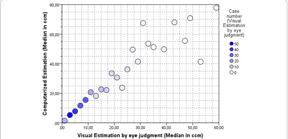
Two-hundred and fifty-five reports of radical prosta-tectomy specimens were digitalized using the cMDX Editor. The computerized estimation of tumour volume showed that the mean tumour volume was 17 ± 16.4 cm3. In comparison, the mean tumour volume estimated by eyeball judgment was 10 ± 10.3 cm3. Thus there is a significant dissimilarity in tumour volumes between the two different estimation methods (mean difference = 6.6 ± 8.7 cm3, T = 12.02, DF = 254, p < 0.0001) (Figure 9). In our patient population, 75% of PCa were staged as pT2c or pT3a. Statistically, there is a highly significant
Figure 6Visualization Model: Four-Layer Model. The workflow of the Four-Layer model: (1) The Input Layer registers the cursor movement and the drawing tool selected by the user, thereby calling functions for user guidance or data acquisition. (2) The Mask Layer defines the drawing surface. (3) The Drawing Layer represents the drawing elements resulting from layers 1 and 2. (4) The Background Layer contains schematic diagrams with graphical descriptions. The red lines in the Mask Layer are defined by attributes beginning with the word“Border”(see table 1). These red lines show the boundary of a drawing surface on which shapes and symbols can be drawn.
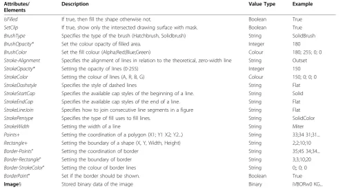
Table 1 List of presentation attributes Attributes/
Elements
Description Value Type Example
IsFilled If true, then fill the shape otherwise not. Boolean True
SetClip If true, show only the intersected drawing surface with mask. Boolean True
BrushType Specifies the type of the brush (Hatchbrush, Solidbrush) String SolidBrush
BrushOpacity* Set the colour opacity of filled area. Integer 180
BrushColor Set the fill colour (Alpha;Red;Blue;Green) Colour 180; 255; 0; 0
Stroke-Alignment Specifies the alignment of lines in relation to the theoretical, zero-width line String Outset
StrokeOpacity* Setting the opacity of lines (0-255) Integer 150
StrokeColor Setting the colour of lines (A, R, B, G) Colour 150; 0; 0; 0
StrokeDashstyle Specifies the style of dashed lines String Flat
StrokeStartCap Specifies the available cap styles of the beginning of a line. String Solid
StrokeEndCap Specifies the available cap styles of the end of a line. String Flat
StrokeLineJoin Specifies how to join consecutive line segments in a figure String Flat
StrokePentype Specifies the type of fill uses to fill lines. String SolidColor
StrokeWidth Setting the width of a line String Miter
Points+ Setting the coordination of a polygon (X1; Y1 X2; Y2...) String 33;34 31;31...
Rectangle+ Setting the boundary of a shape (X, Y, Width, Height) String 2;2;10;10
Border-Points° Setting the coordination of border String 35;45 34;34...
Border-Rectangle° Setting the boundary of border String 3;3;10;20
Border-StrokeColor° Setting the colour of border lines String 0;; 0; 0
BorderPaint° Set if the border should be shown. Boolean True
Image§ Stored binary data of the image Binary iVBORw0 KG...
Available (*) only in ToolML, (+) in VGraphML and MaskML, (°) in MaskML and (§) in VGraphML.
Table 2 List of attributes for textual information: the
“Befund”element describes histological patterns of
the PCa
The root element: Befund
Attributes Description
ID Identification number of PCa area
ShowGleason Show the Gleason score in the pathology report
Gleason1 Primary Gleason pattern
Gleason2 Secondary Gleason pattern
GleasonScore The Gleason Score
Grading Gleason Grading
correlation between T-staging and tumour volume (spearman rho = 0.513, p < 0.01). The cumulative dia-gram of 1615 PCa foci revealed that prostate cancers are mostly localized in the peripheral zone (median: 83.5%, mean: 73 ± 25%) and toward the base PCa foci seem to diverge laterally (Figure 10 and Table 5). 52.5% of PCa were multifocal. The analysis tool could detect 90.3% of Table 3 List of attributes for textual information: the
“Form”element
The root element: Form
Attributes Description
pT, pN, pM TNM-Classification
Datum Date of the report
Kernatypie Atypical changes in cell nucleus
DiffMuster Grade of cell differentiation
KlassifizierungAdeno Classification of adenocarcinoma
GLS1 Primary Gleason pattern
GLS2 Secondary Gleason pattern
GLSTotal Total Gleason score
GGrading Gleason grading
HELPAP HELPAP specification
L Lymph vessel invasion
V Venous vessel invasion
R Status of resection margin
LK-B Number of lymph nodes with metastases
LK-G Total number of extirpated lymph nodes
ProstatLength, ProstatWidth Prostatvolumen
Prostate diameters (Optional) Prostate volume
BarcodeLIB Defining the barcode type (e.g. CODE128A)
Figure 7The Graphical User Interface of cMDX Editor. A: tool bar of the cMDX Editor (new template, open, save, save as...); B: drawing tools for documenting histological changes; C: input field for personal and clinical data; D: window for viewing the histological properties of the selected PCa area. E: the drawing surface.
Table 4 List of drawing tools
Symbol Description DrawTool (Type)
High grade Prostatic Intraepithelial Neoplasia
ImageWithoutSizing
Adenocarcinoma Polygon
Adenocarcinoma FloodFill
Capsular invasion Clip (TypeOfClip:
Polygon)
Extracapsular extension Clip (TypeOfClip: Polygon)
PCa identified as multifocal by eyeball judgement (Table 6). This analysis of 255 cMDX reports had taken
20 ± 2 seconds on a notebook with Intel® T5600 Core™
Duo 1.83, RAM: 4 GB and Nvidia® Geforce® Go 7300 128 MB.
Discussion
cMDX is a data acquisition model for graphical and tex-tual information. Sensible data were encrypted with 256-bit cryptographic algorithms to enable data analysis
compliant with data privacy regulations. The
Figure 8How to draw A: a PCa area, B: capsular invasion, and C: modify the extent of capsular invasion. D: Magic Wand Tool. A: The pathologist sketches the boundary of the PCa area. After clicking“accept”, the boundary is completed automatically. B: The user sketches lines to describe a capsule invasion. C: The user can modify the extent of the capsule invasion by selecting and moving the blue squares shown above. D: The“magic wand tool”allows selecting areas marked on the background image by selecting“only outside contour,”the tool marks only the outside contour of the focus; otherwise, the inside contours are included.
visualization model based on the Four-Layer Model combined a graphical overview of tumour growth in a schematic style with the related textual information. In this manner, the electronic documentation of histo-pathological information usually given by reports in paper form is realised.
XML enables to extract clinical data and graphical schemes, and to transmit them into different formats like binary graphics (PNG, JPEG) vector graphics (SVG), Portable Document Files (PDF), text files (CVS), and Client Report Definition (rdlc) [15]. Therefore, cMDX can be converted into a data format supported by the HIS. The file architecture is compatible with open
package conventions and therefore standard documents like HL7 CDA can be integrated into cMDX [16].
The software prototype of the cMDX Editor can gen-erate and modify pathology reports according to the clinical requirements of pathologists and urologists. The integration of reports of radical prostatectomy speci-mens into the HIS improves clinical utilities and accep-tance by the clinicians.
The application provided an estimate of the tumour volume, which may be an important prognostic indica-tor for predicting prostate cancer recurrence following surgery [17]. In the literature several methods to deter-mine tumour volume of PCa can be found. These include visual estimation, computer planimetry with
Figure 10The cumulative diagram of 1615 PCa foci in 255 prostatectomy specimens. The cumulative diagram of 1615 PCa foci shows that prostate cancers are mostly localized in the peripheral zone, and that toward the base PCa foci seem to diverge laterally. PCa seems to be frequently located in the dorsal half of the peripheral zone of the prostate. The frequency of tumour foci in the various locations are coded by colours, blue = low, red = high frequency.
Table 5 The spread pattern of PCa in PZ
Section Level of Prostate PC Localized in PZ
Base 36%
Middle 1 80%
Middle 2 87%
Apex 89%
Median 83.5%
Mean 73% +/- 25
Table 6 Multifocality of PCa & detection rate of multifocal PCa
Control
PCa Unifocal Multifocal Total (%) Analysis Tool Unifocal 121 13 134 (52.5)
Multifocal 0 121 121 (47.5)
Total (%) 121 (47.5) 134 (52.5) 255 (100)
computer-assisted image analysis. Computer planimetry of the full set of slides of a serially sectioned prostate is believed to be the best technique for an accurate mea-surement of tumour volumes; however, this technique is expensive and time consuming and is therefore not applied in routine clinical practice [18]. The estimation of tumour volume by eyeball judgment is simple and less time consuming, and cheap but shows less precision of measurement [18], it is performed directly under the microscope or after the transformation of tumour areas into the reconstruction map of the prostate in a sche-matic style [6]. Our results show that the estimation by eyeball judgment alone resulted in lower tumour volumes as compared with cMDX estimation. The com-puterised estimation of tumour volume with the cMDX Editor seems to be more accurate than the estimation by eyeball judgment. We anticipate an increase in accu-racy of eyeball judgement with a grid method getting close to the accuracy of the computational volume esti-mation. Nevertheless further investigations are needed here. However, these estimation methods are not accu-rate in comparison to the computer planimetry because they exclude the real dimensions of the prostate and the PCa foci.
The anatomical representation of the prostate in sche-matic style facilitates collecting and analysing the spread pattern of PCa in the prostate. For instance, such infor-mation can be helpful to assess the biopsy strategy in order to increase the detection rate of PCa [19-21]. Our approach enables to analyse cMDX reports and export the results into programs like SPSS© or Excel©. Our results confirm the consensus that the major tumour mass of PCa is located in the peripheral zone (PZ) [22]. According to Chen et al., 74% of PCa foci were localized in the PZ and toward the base, diverged laterally [23]. We found a significant correlation between tumour volume and pathological stage of the specimens, thus confirming the findings of Nelson et al. [24]. The cur-rent analysis tool can detect multifocal PCa with a sensi-tivity of 90.3%. The multifocality rate of PCa was 52.5%, which is consistent with prior series that reported multi-focality rates of 50 to 87% [25]. The accuracy to differ-entiate multifocal from unifocal PCa depends on the applied algorithm. The transformation of the tumour area into a rectangular shape increases the size of the tumour area and increases the probability of one focus to overlap with another adjacent focus in the vicinity, this way reducing the detection rate of multifocal PCa.
A limitation of our documentation model is the docu-mentation time necessary, cMDX failed to shorten the documentation time. cMDX is still developing and we will focus on this drawback in future versions.
An electronic documentation standard for pathologic findings of prostate specimens has not yet been
introduced. We conducted a Pubmed search using the key words“electronic documentation prostate”,“ electro-nic report prostate”and“electronic documentation
pros-tatectomy.” Only one software program similar to the
cMDX Editor could be found: PixelProstate (freeware), developed by Nickels [26], mainly focuses on the mea-surement of tumour volume. PixelProstate has an inter-nal database to store clinical data and provides a simplified 3 D illustration of the prostate and PCa foci. To document the patterns of the tumour spread, the PCa foci are drawn using multiple circles. The documentation of histological patterns of each PCa focus is not sup-ported. In contrast, the cMDX Editor enables to convert a pathologic report into a portable file format and to draw PCa foci with the freehand drawing tool. In addi-tion, capsular invasion, extracapsular extension, positive surgical margins, and histological patterns of a PCa focus can be documented, but a 3 D illustration model similar to that of PixelProstate is not available. cMDX is a free open source document architecture. The applications based on cMDX (cMDX Editor and Analysis tool) are still prototypes (Beta-Version). We plan to develop two versions of these applications (commercial and non-com-mercial) and make them available as soon as possible.
cMDX offers an extensible document architecture by adding a new drawing tool and by providing additional class libraries. If needed, additional pathological para-meters or characteristics can be added. For instance, recording the Gleason score of a PCa focus is feasible by adding the class library“eMocS.Definition.Befund”from the corresponding XML part of cMDX. Furthermore, this feature could extend the spectrum of the clinical applications of cMDX to various medical applications, e. g. for protocols for cardiac catheterisation or extension estimates of burn injuries in emergency surgery.
Conclusions
cMDX can be applied to store clinical data in schematic styles for reporting and analysing pathological para-meters in radical prostatectomy specimens. It facilitates to evaluate routine data for scientific purposes.
Additional material
Additional file 1: An example cMDX file“Example.cMDX”. Please open the file using a zip programme to view the document architecture.
Additional file 2: An example of a report in electronic form “Example_ElectronicForm.pdf”. The paper version of this report is depicted in Figure 1.
Acknowledgements
Author details
1Prostate Center, Dept. of Urology, University Hospital Muenster,
Albert-Schweitzer-Str. 33, D-48149 Muenster, Germany.2Prostate Center, Gerhard-Domagk Institute for Pathology, University Hospital Muenster, Gerhard-Domagkstr. 17, D-48149 Muenster, Germany.3Institute of Pathology and Cytology,
Technikerstrasse 14, D-48465 Schüttorf, Germany.4Institute for Pathology,
Saarbrücken-Rastpfuhl, Rheinstrasse 2, D-66113 Saarbrücken, Germany.
5
Department of Medical Informatics and Biomathematics, University of Muenster, Domagkstr. 9, D-48149 Muenster, Germany.
Authors’contributions
OE designed the cMDX architecture, programmed the supplementary tools and performed the statistical analysis. AS and OB designed the paper template for the pathologic report. AS and OE designed the template for the pathology report in electronic form. AS, EE, JN, MA, OB, OE, RH and TK acquired the data and reviewed the implementation of the software prototype. AS, OE and RH reviewed the results of the analysis. OE drafted the manuscript. AS and MD reviewed the manuscript. All authors read and approved the final manuscript.
Competing interests
The authors declare that they have no competing interests.
Received: 26 May 2010 Accepted: 15 November 2010 Published: 15 November 2010
References
1. Jemal A, Siegel R, Ward E, Hao Y, Xu J, Murray T, Thun MJ:Cancer statistics, 2008.CA Cancer J Clin2008,58(2):71-96.
2. Montironi R, van der Kwast T, Boccon-Gibod L, Bono AV, Boccon-Gibod L: Handling and pathology reporting of radical prostatectomy specimens. Eur Urol2003,44(6):626-636.
3. Epstein JI, Amin M, Boccon-Gibod L, Egevad L, Humphrey PA, Mikuz G, Newling D, Nilsson S, Sakr W, Srigley JR, Wheeler TM, Montironi R: Prognostic factors and reporting of prostate carcinoma in radical prostatectomy and pelvic lymphadenectomy specimens.Scand J Urol Nephrol Suppl2005,216(216):34-63.
4. McCarthy JF, Catalona WJ:Nerve-sparing radical retropubicprostatectomy. InTextbook of operative urology.1 edition. Edited by: Marshall FF. Philadelphia: W.B. Saunders Company; 1996:537-544.
5. Srigley JR, Amin MB, Bostwick DG, Grignon DJ, Hammond ME:Updated protocol for the examination of specimens from patients with carcinomas of the prostate gland: a basis for checklists. Cancer Committee.Arch Pathol Lab Med2000,124(7):1034-1039.
6. Bettendorf O, Oberpenning F, Kopke T, Heinecke A, Hertle L, Boecker W, Semjonow A:Implementation of a map in radical prostatectomy specimen allows visual estimation of tumor volume.Eur J Surg Oncol 2007,33(3):352-357.
7. Epstein JI, Srigley J, Grignon D, Humphrey P, Association of Directors of Anatomic and Surgical Pathology:Recommendations for the reporting of prostate carcinoma.Hum Pathol2007,38(9):1305-1309.
8. Bostwick DG, Amin MB, Dundore P, Marsh W, Schultz DS:Architectural patterns of high-grade prostatic intraepithelial neoplasia.Hum Pathol 1993,24(3):298-310.
9. Zynger DL, Yang X:High-grade Prostatic Intraepithelial Neoplasia of the Prostate: The Precursor Lesion of Prostate Cancer.Int J Clin Exp Pathol 2009,2(4):327-338.
10. Gleason DF, Mellinger GT:Prediction of prognosis for prostatic adenocarcinoma by combined histological grading and clinical staging. J Urol1974,111(1):58-64.
11. Helpap B:Morphologic and cell kinetic studies of prostate cancers. Contribution to grading.Urol Int1985,40(1):36-42.
12. Sobin LH, Wittekind C:TNM-classification of malignant tumoursWiley-Liss; 2002, 172-175.
13. Office OpenXML Overview.[http://www.ecma-international.org/news/ TC45_current_work/OpenXML%20White%20Paper.pdf].
14. AES Proposal: The Rijndael Version 2.[http://csrc.nist.gov/archive/aes/ rijndael/Rijndael-ammended.pdf].
15. Wang H, Azuaje F, Jung B, Black N:A markup language for electrocardiogram data acquisition and analysis (ecgML).BMC Med Inform Decis Mak2003,3(1):4.
16. Using Office Open XML Formats to Support Electronic Health Records Portability and Health Industry Standards. [http://msdn.microsoft.com/en-us/library/bb879915.aspx#Office2007XMLinHealth_Overview].
17. Renshaw A:Visual estimate of the percentage of carcinoma is an independent predictor of prostate carcinoma recurrence after radical prostatectomy.Cancer2002,94(8):2310-1, author reply 2311-2. 18. Werahera PN, Miller GJ, Torkko K, Crawford ED, Stewart JS, Deantoni EP,
Miller HL, Lucia MS:Biomorphometric analysis of human prostatic carcinoma by using three-dimensional computer models.Hum Pathol 2004,35(7):798-807.
19. Chen ME, Troncoso P, Johnston DA, Tang K, Babaian RJ:Optimization of prostate biopsy strategy using computer based analysis.J Urol1997, 158(6):2168-2175.
20. Bolenz C, Gierth M, Grobholz R, Kopke T, Semjonow A, Weiss C, Alken P, Michel MS, Trojan L:Clinical staging error in prostate cancer: localization and relevance of undetected tumour areas.BJU Int2009,
103(9):1184-1189.
21. Presti JC Jr:Prostate biopsy: how many cores are enough?Urol Oncol 2003,21(2):135-140.
22. McNeal JE:Origin and development of carcinoma in the prostate.Cancer 1969,23(1):24-34.
23. Chen ME, Johnston DA, Tang K, Babaian RJ, Troncoso P:Detailed mapping of prostate carcinoma foci: biopsy strategy implications.Cancer2000, 89(8):1800-1809.
24. Nelson BA, Shappell SB, Chang SS, Wells N, Farnham SB, Smith JA, Cookson MS:Tumour volume is an independent predictor of prostate-specific antigen recurrence in patients undergoing radical
prostatectomy for clinically localized prostate cancer.BJU Int2006, 97(6):1169-1172.
25. Meiers I, Waters DJ, Bostwick DG:Preoperative prediction of multifocal prostate cancer and application of focal therapy: review 2007.Urology 2007,70(6 Suppl):3-8.
26. Nickels J:PixelProstate: a simple software program for the measurement of prostate cancer volume.Histopathology2007,50(4):519-521.
Pre-publication history
The pre-publication history for this paper can be accessed here: http://www.biomedcentral.com/1472-6947/10/71/prepub
doi:10.1186/1472-6947-10-71
Cite this article as:Eminagaet al.:Clinical map document based on XML (cMDX): document architecture with mapping feature for reporting and analysing prostate cancer in radical prostatectomy specimens.BMC Medical Informatics and Decision Making201010:71.
Submit your next manuscript to BioMed Central and take full advantage of:
• Convenient online submission
• Thorough peer review
• No space constraints or color figure charges
• Immediate publication on acceptance
• Inclusion in PubMed, CAS, Scopus and Google Scholar • Research which is freely available for redistribution