Open Access
Research article
Laparoscopic versus open left lateral segmentectomy
Kirstin A Carswell
1,2, Filippos G Sagias
1, Beth Murgatroyd
1,
Mohamed Rela
1,2, Nigel Heaton
1,2and Ameet G Patel*
1,2Address: 1Institute of Liver Studies, King's College Hospital, London, UK and 2Department of Gene and Cell-Based Therapy, King's College
London, UK
Email: Kirstin A Carswell - [email protected]; Filippos G Sagias - [email protected];
Beth Murgatroyd - [email protected]; Mohamed Rela - [email protected]; Nigel Heaton - [email protected]; Ameet G Patel* - [email protected]
* Corresponding author
Abstract
Background: Laparoscopic liver surgery is becoming increasingly common. This cohort study was designed to directly compare perioperative outcomes of the left lateral segmentectomy via laparoscopic and open approach.
Methods: Between 2002 and 2006 43 left lateral segmentectomies were performed at King's College Hospital. Those excluded from analysis included previous liver resections, polycystic liver disease, liver cirrhosis and synchronous operations. Of 20 patients analysed, laparoscopic (n = 10) were compared with open left lateral segmentectomy (n = 10). Both groups had similar patient characteristics.
Results: Morbidity rates were similar with no wound or chest infection in either group. The conversion rate was 10% (1/10). There was no difference in operating time between the groups (median time 220 minutes versus 179 minutes, p = 0.315). Surgical margins for all lesions were clear. Less postoperative opiate analgesics were required in the laparoscopic group (median 2 days versus 5 days, p = 0.005). The median postoperative in-hospital stay was less in the laparoscopic group (6 days vs 9 days, p = 0.005). There was no mortality.
Conclusion: Laparoscopic left lateral segmentectomy is safe and feasible. Laparoscopic patients may benefit from requiring less postoperative opiate analgesia and a shorter post-operative in-hospital stay.
Background
Laparoscopic liver surgery, first performed in 1992 [1], is becoming the method of choice as surgical expertise in advanced laparoscopic techniques has developed. Laparo-scopic enthusiasts have shown that it is safe and feasible to perform laparoscopic liver surgery [2-5]. Due to its
ana-been considered the training operation for all liver sur-geons [6].
Proposed benefits of laparoscopic liver surgery include reduced overall blood loss, shorter hospital stay and less post-operative pain with a faster return to normal activity.
Published: 7 September 2009
BMC Surgery 2009, 9:14 doi:10.1186/1471-2482-9-14
Received: 8 February 2009 Accepted: 7 September 2009
This article is available from: http://www.biomedcentral.com/1471-2482/9/14
© 2009 Carswell et al; licensee BioMed Central Ltd.
included compromised oncological integrity [7-11], uncontrollable bleeding [10,12,13] and gas emboli [14-17].
To date one study has compared laparoscopic left lateral segmentectomy with an open approach using historical case controls [18]. Findings confirmed that the laparo-scopic approach was safe and feasible, yet had signifi-cantly longer operating times and no difference in post-operative in-patient stay. The aim of this study was to undertake a contemporaneous comparison between laparoscopic and open left lateral segmentectomies.
Methods
We undertook a retrospective cohort study of the left lat-eral segmentectomies in our institution between July 2002 and October 2006 (n = 43). Cases were included on an intention to treat basis however, in an attempt to reduce bias, patients having previous liver resections (n = 2), synchronous operations (n = 14), polycystic liver dis-ease (n = 3), liver cirrhosis (n = 3) and hand port assisted
procedure (n = 1) were excluded (see figure 1). This resulted in 20 left lateral segmentectomies for compari-son, 10 in the laparoscopic (LG) and 10 in the open group (OG). Selection was based on referral to the individual consultants with all laparoscopic operations performed by a single surgeon (AGP) and open operations under the care of two surgeons (NH, MR). Selection-bias was mini-mised by the random referral policy to the individual sur-geons over this time period. All cases were discussed at the liver multi-disciplinary meeting pre-operatively. A detailed review of the medical records was conducted. Data collection included patient characteristics, site of lesion, operative details, postoperative analgesic require-ments, morbidity and mortality, postoperative in-hospital stay, pathology of specimen, weight of resected specimen and tumour clearance margins. Ethical approval was not required.
All patients were operated on in the supine position under general anaesthesia with endo-tracheal intubation. A broad spectrum antibiotic (Tazocin, Wyeth Laboratories,
Attrition diagram
Figure 1
Madison NJ) was given to all patients. Prophylaxis against deep vein thrombosis was given in the form of low-molec-ular weight heparin, thrombo-embolic deterrent stock-ings and intra-operative intermittent pneumatic compression boots. Staging laparoscopy with intra-opera-tive ultrasound was performed to exclude peritoneal dis-ease and to identify any additional tumours, as appropriate.
Laparoscopic technique
With the surgeon standing to the right of the patient pneu-moperitoneum was established after accessing the abdominal cavity using an open Hasson technique.
The initial port location varied depending upon previous surgery. Intra-abdominal pressure was kept at approxi-mately 15 mmHg. Four additional ports were inserted (figure 2). A 30° laparoscope was used. The falciform and left triangular ligament was divided using a harmonic scalpel (Ethicon, Endo-Surgery Inc. Cincinnati Ohio). The falciform ligament was used to retract and manipulate the left lobe of the liver.
The line of resection was marked using the diathermy hook, 5 mm left of the falciform ligament and transected using a harmonic scalpel. In the umbilical fissure to the left of the falciform ligament the segment II/III pedicles were stapled and divided using Endo-GIA, 30 mm vascu-lar staples (US Surgical, Norwalk CT). Liver parenchyma was transected using a harmonic scalpel +/- .Cavitron
Ultrasonic Surgical Aspirator (CUSA®, Valleylab, Boulder CO). The left hepatic vein was approached intra-paren-chymally and transected with Endo-GIA 30 mm vascular staples. The final attachment to the left triangular liga-ment close to the diaphragm was divided using hook dia-thermy and the specimen freed. Specimens were removed using a retrieval bag through a pfannenstiel or low mid-line incision. Fibrin glue was applied to the surface of the liver if required and a closed suction drain was placed near the transected liver. The skin was closed after infiltration of local anaesthetic (0.5% bupivacaine).
Open technique
The open left lateral segmentectomies were performed as described by Bismuth [19].
A laparotomy was performed via a transverse subcostal incision with a midline extension if required. The left lobe of the liver was mobilised and the parenchyma was transected using CUSA® and argon beam coagulation. With full mobilisation of the left lobe of the liver, the seg-ment II/III pedicles were ligated and divided. The left hepatic vein was clamped at the end of the parenchymal transection and sutured with 5/0 prolene. Transection occurred through the liver parenchyma using CUSA® and argon. Haemostasis was assured with the use of fibrin glue, and a drain was inserted.
Data Analysis
Statistical analysis was performed using Mann-Whitney U
test unless otherwise stated. P-values < 0.05 were consid-ered to be statistically significant. All data is reported as median (range).
Results
Of the 43 liver resections performed in our institution between July 2002 and October 2006, 20 patients met the inclusion criteria for this study with 10 laparoscopic and 10 open left lateral segmentectomies. Patients presented with either solid lesions, symptomatic liver cysts or liver abscesses, within segments II/III.
There were no differences in the patient characteristics between the two groups (Table 1). Median patient age was 54 years (25 - 72). 50% of patients were men (n = 10). The patients had similar American Society of Anaesthesiology (ASA) grading (I:II:III); 1:5:4 in the laparoscopic group and 2:6:2 in the open group. Indication for left lateral seg-mentectomy was malignant lesions in 50% of cases (n = 10).
Two patients in each group (20%) developed early com-plications (< 30 days postoperatively). Minor morbidity in the laparoscopic group (LG) included one urinary tract infection, and a haematemesis (possibly secondary to
Trocar placement for laparoscopic left lateral segmentec-tomy
Figure 2
non-steroidal anti-inflammatory drugs) which settled conservatively. In the open group (OG) one patient devel-oped supraventricular tachycardia four days post-opera-tively, exacerbated by low potassium levels, which resolved spontaneously. The second patient developed hypoxia due to atelectasis, pleural effusions and a superfi-cial haematoma. Only one late complication occurred, a small incisional hernia at the junction of the Mercedes incision in the OG 17 months postoperatively. No statis-tically significant difference in morbidity between the two groups was found (p = 0.725). There was no surgical mor-tality in either group.
In the laparoscopic group one conversion to open (10%) was necessary and occurred early in the series. The LLS was performed for a liver tumour that showed evidence of recent bleeding on a computerised tomography (CT) scan. Intra-operatively the tumour/haematoma from the left lateral segment was found adherent to the greater curve of the stomach, which required a wedge resection of the stomach. In order to avoid narrowing of the
oesophago-gastric junction the procedure was completed via an open approach.
Portal triad clamping was not used in the laparoscopic approach. In the OG it was used intermittently in 50% (5/ 10) of patients, consultant preference. The median cumu-lative clamp time duration was 35 minutes (20-60). There was no significant difference in post-operative AST changes between the LG, OG (portal triad clamping) and OG (no portal triad clamping), one-way ANOVA. Within the LG one patient required an intra-operative blood transfusion (3 units) whilst in the OG two patients, both with liver abscesses, required a transfusion (1-2 units). There was no statistical difference in intra-operative blood transfusion requirement between the two groups (p = 0.782).
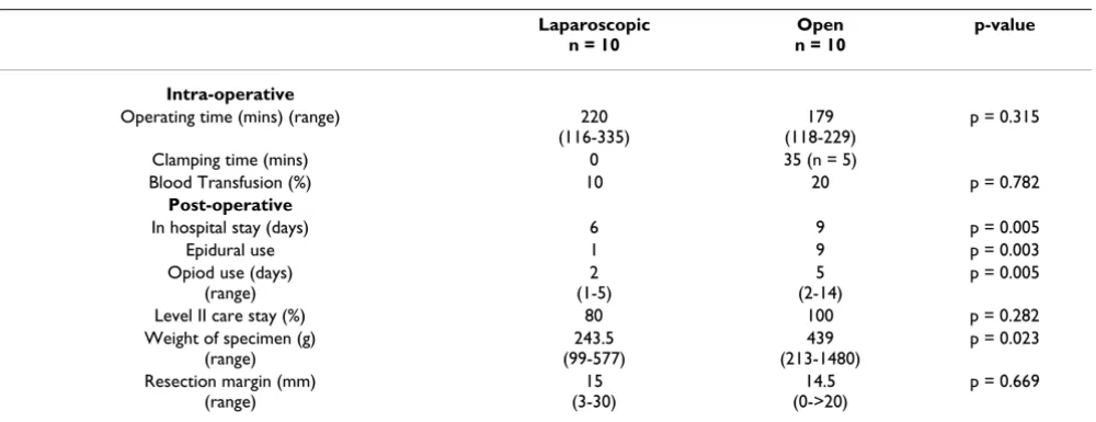
The median operating time for the LG was 220 minutes (116-335 minutes) versus 179 minutes (118-229 min-utes) for the OG. No statistically significant difference was found between the two groups, p = 0.315 (see Table 2). The laparoscopic operating time has reduced over the Table 1: Patient Characteristics
Laparoscopic n = 10 Open n = 10 Median age in years (range) 55
(34 - 72)
56 (25 - 68)
Gender
Male 5 5
Female 5 5
ASA Grade (I:II:III) 1:5:4 2:6:2
Diagnosis
Malignant 6 4
Benign 4 6
Table 2: Intra- and post-operative outcomes
Laparoscopic n = 10
Open n = 10
p-value
Intra-operative
Operating time (mins) (range) 220 (116-335)
179 (118-229)
p = 0.315
Clamping time (mins) 0 35 (n = 5)
Blood Transfusion (%) 10 20 p = 0.782
Post-operative
In hospital stay (days) 6 9 p = 0.005
Epidural use 1 9 p = 0.003
Opiod use (days) (range)
2 (1-5)
5 (2-14)
p = 0.005
Level II care stay (%) 80 100 p = 0.282
Weight of specimen (g) (range)
243.5 (99-577)
439 (213-1480)
p = 0.023
Resection margin (mm) (range)
15 (3-30)
14.5 (0->20)
study period with median operating time of 240 minutes in 2002, reduced to 163 minutes in 2006.
The LG had less analgesic requirements than the OG. This was exemplified by the statistically significant median postoperative opiate use 2 days (1-5) in LG versus 5 days (2-14) in the OG (p = 0.005).
Within the LG 80% (n = 8) of patients required one night in level II care. In the OG 100% of the patients spent one night in level II care (p = 0.282), with one patient requir-ing 4 nights of level II care. This patient was admitted as an emergency with a liver abscess in the left lateral seg-ment of the liver. The prolonged level II care stay was due to multiple factors: poor post-operative analgesic control (unilateral epidural block); ischaemic changes on ECG (Troponin I negative) and a hypoxic episode day 2 post-operatively. A subsequent CT pulmonary angiogram revealed no pulmonary emboli.
The median postoperative in-hospital stay for the LG was 6 days (2-7) compared to 9 days (6-14) for the OG, show-ing a highly significant difference, p = 0.005 (see Table 2).
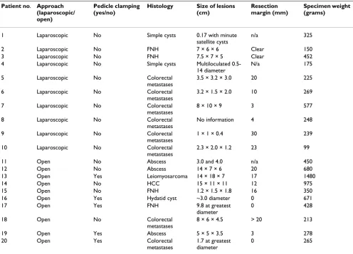
Histology is shown in Table 3. 60% (n = 6) of lesions in the LG and 40% (n = 4) in the open group were malig-nant. The median total weight for the resected specimens in the LG was 243.5 g (99 577 g) versus 439 g (213 g-1480 g) in the OG (p = 0.023). Resection margins for all malignant lesions were clear. In the LG the median resec-tion margin was 15 mm (3-30 mm) with the median in the OG 14.5 mm (< 1 mm ->20 mm)(p = 0.669).
The median follow-up in the LG was 18 months (0-63) and 6 months (0-33) in the OG. Of the malignant cases, post-LLS recurrence in the liver has occurred in 2/6 of the LG (median 14 months) and in 2/4 of the OG (19 months [10-28]). To date no port-site metastases have occurred in these patients.
Table 3: Pathology
Patient no. Approach (laparoscopic/ open)
Pedicle clamping (yes/no)
Histology Size of lesions (cm)
Resection margin (mm)
Specimen weight (grams)
1 Laparoscopic No Simple cysts 0.17 with minute satellite cysts
n/a 325
2 Laparoscopic No FNH 7 × 6 × 6 Clear 150
3 Laparoscopic No FNH 7.5 × 7 × 5 Clear 452
4 Laparoscopic No Simple cysts Multiloculated 0.5-14 diameter
N/a 175
5 Laparoscopic No Colorectal
metastases
3.5 × 3.2 × 3.0 20 225
6 Laparoscopic No Colorectal
metastases
3.2 × 1.5 × 2.0 10 269
7 Laparoscopic No Colorectal
metastases
8 × 10 × 9 3 577
8 Laparoscopic No Colorectal
metastases
No information 4 248
9 Laparoscopic No Colorectal
metastases
1 × 1 × 0.4 30 239
10 Laparoscopic No Colorectal
metastases
2.3 × 2.0 × 1.2 23 99
11 Open No Abscess 3.0 and 4.0 n/a 450
12 Open No Abscess 14 × 7 × 6 20 680
13 Open Yes Leiomyosarcoma 14 × 18 × 7 17 1480
14 Open No HCC 15 × 11 × 11 12 975
15 Open No FNH 1.2 × 1.5 × 1.8 16 350
16 Open Yes Hydatid cyst ~3.0 diameter 0 671
17 Open Yes FNH 9.8 at greatest
diameter
0 428
18 Open No Colorectal
metastases
8 × 6 × 4.5 > 20 213
19 Open Yes Abscess 5 × 5 × 3.5 3 278
20 Open Yes Colorectal
metastases
1.7 at greatest diameter
Discussion
This study echoes the growing body of evidence demon-strating that a laparoscopic approach to liver surgery pro-vides tangible benefits to both patient and hospital. As surgical skill develops it is anticipated that left lateral seg-mentectomy will shift from being a traditionally open procedure to a laparoscopic one. This in turn may benefit an increasing number of patients for whom open surgery could be considered high risk.
The findings of this study are consistent with other pub-lished series showing laparoscopic liver surgery to be fea-sible and safe [2-5,12,20]. A 20% post-operative morbidity rate (< 30 days) was comparable between the open and laparoscopic group. There were no liver related complications, chest or wound infections in either group.
Within the laparoscopic group, conversion to an open procedure occurred in one patient (10%) to ensure nar-rowing of the oesophago-gastric junction did not occur. The need to convert to an open procedure has commonly resulted from uncontrollable bleeding [12,13,18,20]. As surgical techniques improve conversion rates have decreased, with early experiences reporting a 33% sion rate [13] to Chang et al [20] reporting a 2.7% conver-sion rate due to bleeding. In this series no operations were converted to open due to bleeding.
During design of this study we excluded cases in both groups which had undergone previous liver resections, synchronous operations (liver resections, biliary proce-dures, reversal of ileostomy, hernia repairs), polycystic liver disease (inc. fenestration of liver cysts), liver cirrhosis and hand-port assisted procedures. The main objectives of this study were to compare operative time, analgesic requirement and morbidity between the open and lapar-oscopic approach. As such we felt it necessary to control for these variables despite its impact on sample size. This is a retrospective cohort study and as such we recognise the slight disparity in heterogeneity of pathologies included in the final analysis.
Portal clamping was not required in the LG. In the OG 50% (n = 5) of patients underwent clamping for a median duration of 35 mins (11-60 mins). Early experiences in the literature utilised portal clamping to a greater extent with the laparoscopic versus the open approach[18], with a resultant decrease in blood loss. In this series, the open group maintained a low central venous pressure (CVP) and utilised intermittent portal clamping resulting in minimal need for blood transfusion (20% n = 2). Whilst not requiring portal clamping, the laparoscopic technique relied on the positive pressure of the pneumoperitoneum, which in turn minimised potential blood loss, with only one patient (10%) receiving a blood transfusion.
In laparoscopic liver surgery there are long standing con-cerns regarding gas emboli [14] with laparoscopic sur-geons opting for abdominal wall lifting (gasless laparoscopy) or using low CO2 pressures to maintain pneumoperitoneum, to minimise any potential risk. Ani-mal studies have shown an increased risk of cardiac arrhythmias [15] and gas emboli in those with 16 mmHg compared to 8 mmHg (after the left hepatic vein was left open for 3 minutes) [16]. Whilst this implies increased pneumoperitoneal pressures may exacerbate the risk of gas emboli, no human data exists. Potential advantages of increased pneumoperitoneal pressure include reduction in blood loss and improved visualisation of the operative field. In this series pneumoperitoneal pressures were maintained at 15-20 mmHg with no clinical adverse inci-dents however, a prospective study in this field is overdue.
No significant difference was found in the operating time between the two groups (220 vs 179 minutes, p = 0.315), consistent with both Mala et al [21] and Mamada et al [22]. Other groups have shown longer operating times in the LG [18,23]. Of interest, in the LG and the OG the shortest operating times were comparable (116 vs 118 minutes). It must be highlighted that the left lateral seg-mentectomy is considered a training operation. As such, surgeons in training (under the supervision of the Con-sultant) operated on some of the open group (n = 4). These data also includes the first laparoscopic LLS per-formed by the laparoscopic surgeon therefore reflecting the learning curve; the median laparoscopic operating time in 2002 was 240 minutes and by 2006 it was 163 minutes. It is expected that laparoscopic operating time for this procedure will continue to reduce however, these factors may affect comparison of operative time.
administered in the laparoscopic versus open approach following liver resection.
All patients in the OG and 80% of the LG were routinely transferred to level II care with a median stay of one night. With increased confidence and experience this policy has since been modified. Currently patients from both groups no longer routinely require level II care, and the more stringent use of these resources has favourable cost impli-cations for future service development.
In this study the median post-operative in-hospital stay was significantly less in the LG than OG (6 vs 9 days, p = 0.005). In other comparative studies, laparoscopic versus open liver resections for colorectal metastases the median post-operative stay was 4 days vs 8.5 days (p < 0.001) [13], and for hepatic resections 7.8 days vs 11.6 days (p < 0.05) [23] and 10.4 vs 18.0 days (p < 0.05) [21]. This demon-strates reduced post-operative in-patient stay is a repro-ducible, safe benefit of laparoscopic liver resection.
The weights of the resected specimens in the LG were sig-nificantly lower than the OG (p = 0.023). On histological examination of the normal liver in the specimens there was no difference with respect to clamping to account for the weight disparity. Mala et al [21] also noted signifi-cantly lighter specimens resected laparoscopically, along with no significant difference in resection margin involve-ment. This may reflect a slight difference in operative tech-niques between the two approaches. The left hepatic vein is approached and transection of the major vessels occurs intra-hepatically in the laparoscopic group. We speculate that this results in a rounded superior resection, account-ing for this difference.
Oncological integrity is often questioned in laparoscopic liver resections for malignancy. In this study resection margins were clear in all malignant cases (median laparo-scopic 15 mm vs open 14.5 mm). However, early experi-ences of laparoscopic oncological surgery resulted in an increased fear of developing abdominal wall metastases after laparoscopy for hepatic cancer compared with open surgery. Hypotheses suggested the peritoneum may be damaged by the pneumoperitoneum, inducing intra-peri-toneal tumour growth [11]. However this notion is becoming outdated. A short-term animal study by Agnosti et al [27] showed no significant difference in terms of tumour growth, irrespective of gas or pneumoperitoneum pressure used. Jacobi et al [28] suggest intra-peritoneal tumour growth increases for pressures < 10 mmHg and decreases at higher pressures. There is also further evi-dence that reported tumour growth in colonic cancer was significantly reduced after CO2 laparoscopy when com-pared to gasless laparoscopy [29]. A meta-analysis of over 1 500 patients undergoing laparoscopic vs open
colec-tomy for colon cancer found no difference in the 3 year survival rate (82.2% vs 83.3%) [30], concluding that a laparoscopic approach is indeed oncologically safe.
Any potential risk of port-site metastases can be reduced by maintaining an intact surgical specimen and using plastic retrieval devices [20,31], thus minimising contact with the extra-peritoneal structures.
There is evidence that the reduced stress response of lapar-oscopic surgery may be preferential in the malignant cases due to associated lower rates of infection and potential reduction in tumour recurrence [32,33]. Animal data sug-gests that increased surgical stress augments cancer metas-tasis via surgical stress-induced expression of proteinases in the target organ of metastasis [34]. There is also a diminished stress response to the laparoscopic approach versus open liver resection, preserving immune function [32].
An additional benefit of the laparoscopic approach is reduced adhesion formation [32] which may facilitate fur-ther liver resections for metastases. With an increasing trend for non-anatomical and segmental resections with increased parenchymal preservation [35], repeat metastec-tomies (and re-resections) are becoming more common. Petrowsky et al [36] report similar outcomes for patients having either a primary or repeat laparoscopic resection following an initial resection performed laparoscopically. These findings have led to an increased number of patients undergoing further liver metastatectomy, present-ing a further interventional option as multiple staged liver resections become more commonplace.
Conclusion
The laparoscopic approach to left lateral segmentectomy is safe and feasible with reproducible results. In a special-ised unit, it may offer no difference in operating time, morbidity and mortality rates and oncological clearance. Potential benefits include reduced opiate analgesic requirements and shorter hospital stay however, the importance of patient selection cannot be over-empha-sised.
Abbreviations
ASA: American Society of Anaesthesiology; CT: computer-ised tomography; CUSA®: Cavitron Ultrasonic Surgical Aspirator; CVP: central venous pressure; FNH: focal nodu-lar hyperplasia; HCC: hepatocellunodu-lar carcinoma; LG: laparoscopic group; LLS: left lateral segmentectomy; OG: open group.
Completing interests
Authors' contributions
KC conceived of the study, participated in its design, data collection and analysis and drafted the manuscript. FS participated in its design, data analysis and drafting of the manuscript. BM assisted with data collection and drafting of the manuscript. MR and NH participated in study design, carried out some of the operative procedures, assisted with data collection and manuscript revisions. AGP participated in study design and coordination, car-ried out the laparoscopic procedures, assisted with data collection and supervised manuscript revisions.
All authors read and approved the final manuscript.
Acknowledgements
This paper has been presented in part to the Association of Surgeons of Great Britain and Ireland, Edinburgh, UK, May 2006, published in abstract form as BJS 2006; 93(S1): 58, and presented to the 10th World Congress of Endoscopic Surgeons and European Association of Endoscopic Surgeons, Berlin, Germany, September 2006, and published in abstract form as Surg Endosc 2007; 21: S64
References
1. Gagner M, Rheault M, Dubuc J: Laparoscopic partial hepatec-tomy for liver tumour. Surg Endosc 1992, 6:99.
2. Gagner M, Rogula T, Selzer D: Laparoscopic liver resection: ben-efits and controversies. Surg Clin North Am 2004, 84:451-462. 3. Dulucq JL, Wintringer P, Stabilini C, Berticelli J, Mahajna A:
Laparo-scopic liver resections: A single center experience. Surg Endosc 2005, 19:886-891.
4. Cherqui D: Laparoscopic liver resection. Br J Surg 2003,
90:644-646.
5. Mala T, Edwin B, Rosseland AR, Glanhaug I, Fosse E, Mathisen O:
Laparoscopic liver resection: experience of 53 procedures at a single center. J Hepatobiliary Pancreat Surg 2005, 12:298-303. 6. Samama G, Chiche L, Brefort JL, Le Roux Y: Laparoscopic
anatom-ical hepatic resection. Surg Endosc 1998, 12:76-78.
7. Fong Y, Jarnagin W, Conlon K, DeMatteo R, Dougherty E, Blumgart LH: Hand-assisted laparoscopic liver resection: lessons from an initial experience. Arch Surg 2000, 135:854-859.
8. Takiguchi S, Matsuura N, Hamada Y, Taniguchi E, Sekimoto M, Tsuji-naka M, Shiozaki H, Monden M, Ohashi S: Influence of CO2 pneu-moperitoneum during laparoscopic surgery on cancer cell growth. Surg Endosc 2000, 14:41-44.
9. Whelan RL: Laparotomy, laparoscopy, cancer, and beyond.
Surg Endosc 2001, 15:110-115.
10. Gigot JF, Gilneur D, Azagra JS, Goergen M, Ceuterick M, Morino M, Etienne J, Marescaux J, Mutter D, van Krunckelsven L, Descottes B, Valleix D, Lachachi F, Bertrand C, Mansvelt B, Hubens G, Saey J, Schockmel R, under the auspices of the Hepatobiliary and Pancreatic Section of the Royal Belgian Society of Surgery and the Belgian Group for Endoscopic Surgery: Laparoscopic liver resection for malig-nant liver tumours: preliminary results of a multicenter European study. Annals of Surgery 2002, 236(1):90-97.
11. Volz J, Koster S, Spacek Z, Paweletz N: The influence of pneu-moperitoneum used in laparoscopic surgery on an intra abdominal tumour growth. Cancer 1999, 86:770-774.
12. Dagher I, Proske JM, Carloni A, Richa H, Tranchart H, Franco D:
Laparoscopic liver resection: results for 70 patients. Surg Endosc 2007, 21:619-24.
13. Kaneko H, Takagi S, Shiba T: Laparoscopic partial hepatectomy and left lateral segmentectomy: technique and results of a clinical series. Surgery 1996, 120:468-475.
14. Yacoub OF, Cardona I, Coveler LA, Dodson MG: Carbon Dioxide embolism during laparoscopy. Anaesthesiology 1982, 57:533-535. 15. Schmandra TC, Mierdl S, Hollander D, Hanisch E, Gutt C: Risk of gas embolism in hand-assisted versus total laparoscopic hepatic resection. Surg Technol Int 2004, 12:137-143.
16. Eiriksson K, Kylander C, Fors D, Rubertsson S, Arvidsson D: High (16 mmHg) versus low (8 mmHg) pressure pneumoperito-neum in laparoscopic liver resection reduces bleeding but with an increased risk for gas embolism. Presented at the 10th World Congress of Endoscopic Surgery, Berlin 2006:73. Abstract O238 17. Takagi S: Hepatic and portal vein blood flow during carbon
dioxide pneumoperitoneum for laparoscopic hepatectomy.
Surg Endosc 1998, 12:427-431.
18. Lesurtel M, Cherqui D, Laurent A, Tayar C, Fagniez PL: Laparo-scopic versus open left lateral hepatic lobectomy: a case-control study. J Am Coll Surg 2003, 196:236-242.
19. Bismuth H: Surgical anatomy and anatomical surgery of the liver. World J Surg 1982, 6:3-9.
20. Chang S, Laurent A, Tayar C, Karoui M, Cherqui D: Laparoscopy as a routine approach for left lateral sectionectomy. Br J Surg
2007, 94:58-63.
21. Mala T, Edwin B, Gladhaug I, Fosse E, Soreide O, Bergan A, Mathisen O: A comparative study of the short-term outcome following open and laparoscopic liver resection of colorectal metas-tases. Surg Endosc 2002, 16:1059-1063.
22. Mamada Y, Yoshida H, Taniai N, Mizuguchi Y, Kakinuma D, Ishikawa Y, Yokomuro S, Arima Y, Akimaru K, Tajiri T: The usefulness of laparoscopic hepatectomy. J Nippon Med Sch 2007, 74:158-162. 23. Rau HG, Buttler E, Meyer G, Schardey HM, Schildberg FW:
Laparo-scopic liver resection compared with conventional partial hepatectomy - a prospective analysis. Hepatogastroenterology
1998, 45:2333-2338.
24. Moniche S, Jorgensen H, Wetterslev J, Berg Dahl J: Local anaes-thetic infiltration for postoperative pain relief after laparos-copy: a qualitative and quantitative systematic review of intraperitoneal, port-site infiltration and mesosalpinx block.
Anesth Analg 2000, 90:899-912.
25. Veldkamp R, Kuhry E, Hop W, Jeekel J, Kazemier G, Bonjer H, Haglind E, Pahlman L, Cuesta MA, Msika S, Morino M, Lacy AM, Colon cancer Laparoscopic or Open Resection Study Group (COLOR):
Laparoscopic surgery versus open surgery for colon cancer: short-term outcomes of a randomised trial. Lancet Oncol 2005,
6:477-484.
26. Farges O, Jagot P, Kirstetter P, Marty J, Belghiti J: Prospective assessment of the safety and benefit of laparoscopic liver resections. J Hepatobiliary Pancreat Surg 2002, 9:242-8.
27. Agostini A, Robin F, Jais JP, Aggerbeck M, Vilde F, Blanc B, Lecuru F:
Impact of different gases and pneumoperitoneum pressures on tumour growth during laparoscopy in a rat model. Surg Endosc 2002, 16:529-532.
28. Jacobi CA, Wenger FA, Ordemann J, Gutt C, Sabat R, Muller JM:
Experimental study of the effect of intra-abdominal pressure during laparoscopy on tumour growth and port site metas-tasis. Br J Surg 1998, 85:1419-1422.
29. Gutt CN, Riemer V, Kim ZG, Jacobi CA, Paolucci V, Lorenz M:
Impact of laparoscopic colonic resection on tumour growth and spread in an experimental model. Br J Surg 1999,
86:1180-1184.
30. Bonjer HJ, Hop WCJ, Nelson H, Sargent DJ, Lacy AM, Castells A, Guillou PJ, Thorpe H, Brown J, Delgado S, Kuhrij E, Haglind E, Pahl-man L, Transatlantic Laparoscopically Assisted vs Open Colectomy Trials Study Group: Laparoscopically assisted vs open colec-tomy for colon cancer: a meta-analysis. Arch Surg 2007,
142:298-303.
31. Paolucci V, Schaeff B, Schneider M, Gutt C: Tumour seeding fol-lowing laparoscopy: international survey. World J Surg 1999,
23:989-997.
32. Burpee SE, Kurian M, Murakame Y, Benevides S, Gagner M: The metabolic and immune response to laparoscopic vs open liver resection. Surg Endosc 2002, 16:899-904.
33. Lacy AM, Garcia-Valdecasas JC, Delgado S, Castells A, Taura P, Pique JM, Visa J: Laparoscopy-assisted colectomy versus open colec-tomy for treatment of non-metastatic colon cancer: a ran-domised trial. Lancet 2002, 359:2224-2229.
34. Tsuchiya Y, Sawada S, Yoshioka I, Ohashi Y, Matsuo M, Harimaya Y, Tsukada K, Saiki I: Increased surgical stress promotes tumour metastasis. Surgery 2003, 133:547-55.
con-Publish with BioMed Central and every scientist can read your work free of charge "BioMed Central will be the most significant development for disseminating the results of biomedical researc h in our lifetime."
Sir Paul Nurse, Cancer Research UK
Your research papers will be:
available free of charge to the entire biomedical community
peer reviewed and published immediately upon acceptance
cited in PubMed and archived on PubMed Central
yours — you keep the copyright
Submit your manuscript here: BioMedcentral secutive cases over the past decade. Ann Surg 2002,
236:397-407.
36. Petrowsky H, Gonen M, Jarnagin W, Lorenz M, DeMatteo R, Heinrich S, Encke A, Blumgart L, Fong Y: Second liver resections are safe and effective treatment for recurrent hepatic metastases from colorectal cancer: a bi-institutional analysis. Ann Surg
2002, 235:863-871.
Pre-publication history
The pre-publication history for this paper can be accessed here: