R E S E A R C H A R T I C L E
Open Access
Site-dependent differences in the
composite fibers of male pelvic plexus
branches: an immunohistochemical analysis
of donated elderly cadavers
Kuniyasu Muraoka
1,5*, Shuichi Morizane
1, Keisuke Hieda
2, Masashi Honda
1, Takehiro Sejima
1, Gen Murakami
3,
Shin-ichi Abe
4and Atsushi Takenaka
1Abstract
Background:Although the pelvic autonomic plexus branches are considered to be a mixture of sympathetic and parasympathetic nerves, little is known regarding the composite fibers of the pelvic plexus branches. This study aimed to investigate the immunohistochemical features of sympathetic and parasympathetic nerves in the pelvic autonomic plexus branches.
Methods:Using 10 donated elderly male cadavers, the detailed topohistology of nerve fibers at and around the bladder, seminal vesicle, prostate, and rectum was examined. Neuronal nitric oxide synthase (nNOS) and vasoactive intestinal polypeptide (VIP) were used as parasympathetic nerve markers; tyrosine hydroxylase (TH) was used as a sympathetic nerve marker. The myenteric plexus of the colon was utilized as a positive control.
Results:Most nerve fibers in the bladder, seminal vesicle, prostate, and rectum were both nNOS- and TH-positive. Thus, pelvic plexus branches were classified into two types: 1) triple-positive mixed nerves (nNOS+, VIP+, TH+, thick myelinated fibers + or -) and 2) double-positive mixed nerves (nNOS+, VIP-, TH+, thick myelinated fibers + or -). Notably, triple-positive nerves were localized within the posterosuperior part of the plexus (near the rectum) and travelled anteroinferiorly toward the posterolateral corner of the prostate. The posteriorly and inferiorly located nerves were predominantly composed of parasympathetic, rather than sympathetic, fibers. In contrast, nerve fibers within and along the bladder and seminal vesicle contained either no or few VIP-positive nerves. These superiorly located nerves were characterized by clear sympathetic nerve dominance.
Conclusions:The nerves of the pelvic plexus branches were clearly classified into nerves around the bladder and seminal vesicle (VIP-negative) and nerves around the prostate (VIP-positive). Although nNOS- and VIP-positive nerve fibers are candidate cavernous nerves, cavernous nerve identity cannot be definitively concluded for these nerves in the periprostatic region.
Keywords:Pelvic autonomic nerve plexus, Fiber composition, Neuronal nitric oxide synthase, Vasoactive intestinal polypeptide, Tyrosine hydroxylase
* Correspondence:[email protected] 1
Department of Urology, Tottori University Faculty of Medicine, Yonago, Japan
5Division of Urology, Department of Surgery, Tottori University Faculty of
Medicine, 36-1 Nishi-cho, Yonago 683-8504, Japan
Full list of author information is available at the end of the article
Background
Autonomic innervation of the pelvic viscera is formed by sympathetic fibers from the inferior hypogastric plexus and parasympathetic fibers from the pelvic splanchnic nerves [1, 2]. The genital organs and lower urinary tract are controlled by the autonomic nervous system, as well as the somatic nerves. Because functional disruptions, such as urinary incontinence and sexual dysfunction, represent important determinants of quality of life, detailed anatomical studies of the pelvic neuro-anatomy are essential to preserve continence, erection, and bladder function after pelvic surgery [3,4].
Immunohistochemistry is widely applied in pelvic nerve research. However, as evaluation of the peripro-static nerve distribution is typically performed using sur-gically acquired specimens [5–7], the staining of a large area, including adjacent organs, is generally not possible. Therefore, to investigate the anatomy of the pelvic plexus, use of the entire pelvic block of cadavers is ideal. Nitric oxide synthase (nNOS) and vasoactive intestinal polypeptide (VIP) are used as parasympathetic nerve markers, whereas tyrosine hydroxylase (TH) is used as a marker of sympathetic nerves [8]. Recent reports have demonstrated that, in the human pelvic floor, a portion of the pelvic plexus branches contained VIP-, nNOS-, and TH-positive nerve fibers [9–11].
The aim of this study was to investigate the topohistol-ogy of three types of pelvic nerve fibers (nNOS-, VIP-, and TH-positive fibers) by comparison of the peripro-static nerve configuration and distribution with that of adjacent regions.
Methods
This study examined 10 donated male cadavers with a mean age of 73 years (range, 64–82 years). The cause of death was either ischemic heart failure or intracranial bleeding; none of the cadavers had undergone abdom-inal or pelvic surgery, as confirmed by review of patient medical histories, as well as by macroscopic observation of the abdominopelvic cavity. The 10 cadavers were do-nated to Tokyo Dental College for research and educa-tion on human anatomy in accordance with their consent, and their use in research was approved by the Ethics Committee of Tottori University Faculty of Medi-cine. The study was performed in accordance with the provisions of the Declaration of Helsinki 1995, as revised in Edinburgh in 2000.
The donated cadavers had been fixed by arterial perfu-sion of 10%v/v formalin solution, then stored in 50% v/v ethanol solution for > 3 months. From each of the ca-davers, a large tissue block, including the bladder, seminal vesicle, urethra, prostate, and rectum, as well as any con-nective tissue around these viscera, was prepared. After bi-section along the midsagittal line, each of the hemiblocks
was cut into 15-mm thick sections; then, routine proce-dures for paraffin-embedded histology were performed. From each of the macroslices, large horizontal or sagittal sections (70 × 50 mm) were prepared at 2–3-mm inter-vals, then stained with hematoxylin and eosin (H&E). After reviewing the large sections to identify target re-gions, sections (50 × 20 mm) for immunohistochemistry were cut, in close proximity to the former plane. Ultim-ately, we obtained 2–5 large sections and 8–20 standard-sized sections from a single paraffin block con-taining a 15-mm-thick slice.
Most sections were stained with H&E, whereas others were stained via immunohistochemistry and elastic-tissue Masson staining (a variation of Masson-Goldner staining). Primary antibodies for nerve immunohistochemistry were used based on the methods of Hinata et al. [12]; these com-prised mouse monoclonal anti-human S100 protein (1:200 dilution, Dako Z0311; Dako, Glostrup, Denmark), rabbit polyclonal anti-human nNOS (1:200 dilution; Cell Signaling Technology, Beverly, MA, USA), mouse monoclonal anti-human VIP (1:100 dilution, Santa Cruz sc25347; Santa Cruz, CA, USA), and rabbit polyclonal anti-human TH (1:500 dilution, Millipore-Chemicon ab152; Temecula, CA, USA). When possible, the four immunohistochemical stains were applied on adjacent sections; occasionally, non-adjacent sections were sometimes used because of failed immunostaining. As a positive control for immuno-histochemistry, the myenteric plexus of the descending colon was acquired from the 10 specimens; the colic my-enteric nerves invariably exhibit a dense distribution of nNOS-/VIP-coreactive nerve fibers, even in centenarians [13]. The secondary antibody was labeled with horseradish peroxidase (HRP), and antigen-antibody reactions were de-tected via the HRP-catalyzed reaction with diaminobenzi-dine. The immunohistochemistry-labeled samples were counterstained with hematoxylin. Negative controls com-prised samples without primary antibody.
Results
the superior or inferior group of nerves was examined. Figures 1, 2, 3, 4 and 5 were prepared from a combin-ation of observcombin-ations from two specimens.
In all 10 cadavers, the colic myenteric plexus expressed all three markers (nNOS, VIP, and TH; Fig.1), although TH expression was typically weak and found in very few fibers. For descriptive purposes, the pelvic plexus branches to the urogenital organs were divided
into four areas: 1) the middle and superior area between the seminal vesicle and rectum (Fig.1b); 2) the anterosu-perior area between the bladder and seminal vesicle (Fig.1c); 3) the posterosuperior area between the rectum and levator ani muscle (Fig.1d); and, 4) the posteroinfer-ior area at and along the posterolateral corner of the prostate (Fig. 5b). The third group of nerves traveled anteroinferiorly toward the posterolateral corner of the
prostate, such that the third area appeared to be con-nected to the fourth area. All four areas of nerves exhib-ited ganglion cell clusters, but the second and third areas (3–10 clusters per horizontal section) contained greater numbers than the others (1–5 clusters per hori-zontal section). Numbers of cut nerve profiles per mm2 in each horizontal section ranged from 15 to 25 in the first area, 22 to 38 in the second area, 8 to 13 in the
third area, and 5 to 18 in the fourth area. Therefore, nerve density was consistently highest in the first (ante-rosuperior) area of nerves, located between the bladder and seminal vesicle.
More than 70% of nerves in the anterosuperior area were thin (< 0.05-mm diameter), and the density was higher along the seminal vesicle than the bladder (Fig. 1c). In these nerves, TH-positive sympathetic fibers
Fig. 2Immunohistochemistry of nerves in an area between the bladder and seminal vesicle (anterosuperior group of the pelvic plexus branches). Topographical nerve anatomy is shown in Fig.1c. Panelsa,c,e,g, andiexhibit nNOS immunostaining, while panelsb,d,f,andidisplay TH immunostaining. Panelsaandb(oriandj) are adjacent sections, whereas panelscandd(oreandf,gandh) are proximal sections. These nerves did not contain fibers that were reactive for VIP (not shown). In all of these nerves, nNOS- and TH-positive fibers appear to be
were much more abundant than parasympathetic fibers, especially in the thinner nerves (Fig. 2). The proportion of TH-positive fibers in the nerve along the seminal vesicle was much higher than the corresponding propor-tion along the bladder. Sympathetic nerve dominance was also observed in the first (middle and superior) area between the seminal vesicle and rectum (Fig.3). In con-trast to the first and second areas, nerves in the third (posterosuperior) and fourth (posteroinferior) areas
contained VIP-positive fibers, although the number per section was consistently lower than for nNOS-positive nerves (Figs. 4 and 5). Furthermore, in these posterior areas, the number of nNOS-positive fibers was often equal to or greater than the number of TH-positive fi-bers. In addition, with respect to intra-organ nerves, both nNOS-positive fibers and TH-positive fibers were present; these were adjacent to and alongside the glands and mucosa of the prostate, seminal vesicle, and bladder.
Fig. 3Immunohistochemistry of nerves in an area between the rectum and seminal vesicle (middle and superior group of the pelvic plexus branches). Topographical nerve anatomy is shown in Fig.1b. Panelsa,d,f, andhdisplay nNOS immunostaining, while panelsc,e,g, andi exhibit TH immunostaining. Only panelbdisplays VIP immunostaining: the other nerves in this figure did not contain VIP-positive fibers (not shown). Panelsa–c(orfandg;handi) are adjacent sections, whereas panelsdandeare proximal sections. Arrows, arrowheads, and stars in panelsa–cindicate ganglion cells corresponding to each panel: two ganglion cells (stars) appear to be negative for all three markers. All VIP-positive cells (panelb) appear to express nNOS (panela). All TH-positive cells (panelc) do not express either nNOS or VIP. In all of these nerves, nNOS- and TH-positive fibers appear to be intermingled and do not exhibit a clear localization. All panels were prepared at the same
Similarly, the smooth muscles of the prostate and sem-inal vesicle contained a very high proportion of TH-positive fibers, as well as multiple nNOS-positive fi-bers. However, immunoreactive nerves were not found in the bladder detrusor smooth muscles. Other areas of
nerves were characterized according to sympathetic nerve dominance. Of note, in all four areas, the intrapel-vic nerves consistently contained both nNOS-positive and TH-positive fibers. In contrast, the pelvic nerve branches, such as those surrounding the levator ani
muscle, did not contain either nNOS- or VIP-positive fi-bers (data not shown). Ganglion cells, albeit rare, con-tained triple-negative fibers (Figs.2a, b;3a, b, c).
In a ganglion cell cluster (Figs.3a, b, c;5b), nNOS-positive ganglion cells were consistently highest in number, followed by TH-positive cells. Some nNOS-positive cells also
appeared to express VIP when observed in serial sec-tions. The maximum diameter of cell bodies ranged from 15 to 25 μm, irrespective of their sympathetic or parasympathetic function. TH-positive cells were found either intermingled with, or in clusters sepa-rated from, nNOS-positive cells (Fig. 3c).
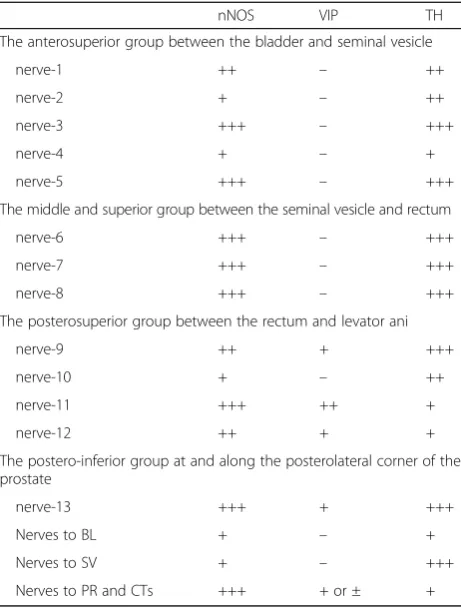
Sympathetic nerve fibers were most frequently ob-served in the superiorly located branches of the pelvic plexus near the bladder and seminal vesicle; in contrast, parasympathetic fibers, a portion of which expressed VIP, were dominant in the inferiorly or posteriorly lo-cated nerves near the prostate. Based on these observa-tions, the pelvic plexus branches were classified into two types: 1) triple-positive mixed nerves (nNOS+, VIP+, TH +, thick myelinated fibers + or -); and, 2) double-positive mixed nerves (nNOS+, VIP-, TH+, thick myelinated fi-bers + or -). Figure6depicts representative pelvic plexus branches, including both triple-positive mixed nerves and double-positive mixed nerves. The composition of nerve fibers shown in Figs. 1, 2, 3, 4 and 5 is summa-rized in Table1.
Discussion
This study focused on the pelvic plexus branches, from the lateral site of the bladder, seminal vesicle, and rectum, to the posteroinferior site at and along the posterolateral cor-ner of the prostate, in elderly male cadavers. Although lim-ited regions of the peripheral pelvic plexus were examined, several patterns were identified in the fiber composition of the pelvic plexus branches. Nerves in the examined region consistently included TH-positive fibers; further, many of the plexus branches were both nNOS- and TH-positive. Notably, the anterosuperior area (between the bladder and seminal vesicle) and the middle and superior area (between the seminal vesicle and rectum) did not contain
VIP-positive nerve fibers. On the contrary, the posterosu-perior area (between the rectum and levator ani muscle) and the posteroinferior area (at and along the posterolateral corner of the prostate) contained VIP-positive nerve fibers. The hypogastric and pelvic splanchnic nerves both contain nNOS-, VIP-, and TH-positive nerves [14, 15]. Kraima et al. [15] reported that VIP-positive fibers were not present in tissues isolated from the lumbar sympathetic chain; thus, areas that do not include VIP-positive nerves may be sup-plied by the lumbar sympathetic chain. In addition, a non-parasympathetic pattern [−, −, +] was not evident in the plexus branches, but was observed both inside and out-side of the levator ani muscle; these nerves appeared to be pudendal nerve branches. Few triple-negative fibers were observed; these are candidate pure sensory nerves [10].
TH is the rate-limiting enzyme in the synthetic path-way of norepinephrine, a neurotransmitter that is found in peripheral sympathetic nerves and their related gan-glia; thus, TH is often used as a marker of sympathetic nerves [10,14–17]. Both nNOS and VIP have been used as parasympathetic nerve markers of the pelvic plexus branches [8–11]; nNOS is found in peripheral parasym-pathetic nerves and catalyzes the formation of nitric oxide [18], whereas, VIP is generally considered to be the primary transmitter released from cholinergic smooth muscle vasodilator and secretomotor fibers [19].
With regard to lower urinary function, the bladder neck is innervated by dense noradrenergic nerves, which have been shown to cause smooth muscle contraction
and subsequent closure of the bladder neck during the storage of urine [20]. During micturition, inhibition of the sympathetic pathway may lead to opening of the bladder neck and prostatic urethra [21]. Additionally, high NOS activity was found in the urethra, whereas intermediate activity was found in the bladder neck, and comparatively low activity was found in the detrusor muscle. VIP-containing nerves form a dense subepithe-lial plexus and project to the detrusor muscle bundles of the bladder [22]. VIP plays an important role in bladder neck opening by promoting relaxation of the smooth muscle [20]. With regard to sexual function, TH- and VIP-positive nerve fibers are very abundant in the hu-man prostate [23, 24] and are considered to play a role in the expulsion of the contents of the prostate gland during seminal emission, as well as during ejaculation, which is largely under adrenergic control [21]. Sympa-thetic innervation mediates corporeal vasoconstriction and corporeal smooth muscle contraction; further, it causes penile detumescence after orgasm, and (in the ab-sence of sexual arousal) maintains the penis in the flac-cid state [25]. The perivascular and trabecular nerve fibers within the corpus cavernosum exhibit positive
immunostaining for both nNOS and VIP [16, 26]. Nitric oxide is released by parasympathetic fibers and is a potent vasodilator associated with the physiology of the male erection. Nitric oxide and VIP participate in the erectile process via activation of the nitric oxide/cGMP and ade-nylyl cyclase/cAMP pathways, respectively [23,24].
The present study demonstrated that nNOS-positive fi-bers are present in the pelvic plexus branches, as well as in the prostate, seminal vesicle, and bladder; further, the number of cut nerves per mm2decreases with proximity to the periphery. Ganzer et al. reported that nerve planim-etry, using a polyclonal antibody against the neural protein S100, revealed that 75% of nerves from the seminal vesi-cles do not reach the striated urethral sphincter level along the prostate [27]. Thus, the nerves around the pros-tatic apex may be the remaining nerves of the pelvic plexus, after distribution to the prostate, seminal vesicle, and bladder. Although nNOS immunoreactivity has been used to identify cavernous nerves, previous data, suggest-ing that nNOS-positive periprostatic nerves are cavernous nerves, may have been overstated.
VIP immunoreactivity is also found in the human penis, where the largest concentrations of VIP are present in the cavernosum body [28]. The present study demonstrated that approximately 10% of nerve fibers progressing toward the prostate and cavernous tissues were VIP-positive. Hinata et al. reported that there were few VIP-positive fibers adjacent to, and posterior to, the rhabdosphincter area [11]. Ehmke et al. reported that > 50% of perivascular nerve fibers and > 90% of trabecular nerve fibers within the corpus cavernosum stained posi-tive for both nNOS and VIP. Furthermore, NOS/VIP im-munoreactivity was reduced (diabetes) or absent (lesion of the cavernous nerve) in penile tissue taken from pa-tients with neurogenic impotence [26]. Although nNOS-and VIP-positive nerve fibers from the prostatic apex to-ward the periphery are regarded as candidate cavernous nerves, there may be considerable interindividual vari-ation in nNOS and VIP immunoreactivity, correspond-ing to erectile function.
Many excellent studies have been published regarding the macro- and microscopic anatomy of the pelvic auto-nomic nerve plexus and its branches. Takenaka et al. re-ported that the main route of the cavernous nerve branches from a location near the root of the pelvic splanchnic nerves, then joins in a spray-shaped distribu-tion to the central area of the neurovascular bundle, travelling along the distal side of the pelvic plexus [2]. The distal pelvic plexus, including the cavernous nerves, passes through the rectourethral muscle [29,30]. Clinic-ally, even after non-nerve sparing prostatectomy, erectile function may be maintained [31,32], which implies that the cavernous nerves along the posterior side of the prostate and urethra are preserved, in some cases, after
Table 1Summary of composite nerve fibers of the pelvic plexus branches
nNOS VIP TH
The anterosuperior group between the bladder and seminal vesicle
nerve-1 ++ – ++
nerve-2 + – ++
nerve-3 +++ – +++
nerve-4 + – +
nerve-5 +++ – +++
The middle and superior group between the seminal vesicle and rectum
nerve-6 +++ – +++
nerve-7 +++ – +++
nerve-8 +++ – +++
The posterosuperior group between the rectum and levator ani
nerve-9 ++ + +++
nerve-10 + – ++
nerve-11 +++ ++ +
nerve-12 ++ + +
The postero-inferior group at and along the posterolateral corner of the prostate
nerve-13 +++ + +++
Nerves to BL + – +
Nerves to SV + – +++
Nerves to PR and CTs +++ + or ± +
+, > 10 positive nerve fibers were seen in the nerve; ++, positive nerves occupied 30–70% of a cross-sectional area of the nerve; +++, positive fibers occupied nearly all parts of the nerve with a high density
resecting the so-called neurovascular bundle. Further-more, differences in the postoperative recovery of erect-ile function depend on the quantity of damaged cavernous nerves. Therefore, athermal dissection and re-duced traction may lead to the preservation of function, even when non-nerve-sparing procedures are used.
There are several limitations to the present study. Be-cause of the lack of immunohistochemical analysis of the hypogastric and pelvic splanchnic nerves, it was not possible to propose a complete scheme from a pregangli-onic fiber, via a ganglion cell and postganglipregangli-onic fibers, to the target organs. Additionally, nNOS immunohisto-chemistry is difficult to successfully perform in cadaveric specimens that have undergone long periods of preser-vation [9]. Because the quality of nNOS immunohisto-chemistry varies among individuals, we did not perform a quantitative evaluation, which might have included a determination of the percentages of each nerve type.
Conclusions
Because the dominance of VIP-positive nerve fibers var-ied by site, nerves of the pelvic plexus branches were clearly categorized as those nerves around the bladder and seminal vesicle (VIP-negative) and those nerves around the prostate (VIP-positive). Furthermore, the re-sults confirmed that nNOS expression is a general char-acteristic of the pelvic plexus branches, rather than a specific characteristic of the cavernous nerve. Although nNOS- and VIP-positive nerve fibers are candidate cav-ernous nerves, the periprostatic nNOS-positive fibers may not be cavernous nerves, even when they are ob-served alongside VIP-positive fibers.
Abbreviations
H&E:hematoxylin and eosin; HRP: horseradish peroxidase; nNOS: nitric oxide synthase; TH: tyrosine hydroxylase; VIP: vasoactive intestinal polypeptide
Acknowledgments
We are grateful to the individuals who donated their bodies to Tokyo Dental College for research and education in human anatomy without any economic benefit. We also thank their families for agreeing to the donation, as well as for their patience in waiting for return of the bodies after completion of the study.
Availability of data and materials
The datasets used and/or analyzed during the current study are available from the corresponding author on reasonable request.
Authors’contributions
Conception and design: KM, SM, GM, SA, and AT. Acquisition of data: KM, SM, KH, and MH. Analysis and interpretation of data: KM, SM, and TS. Drafting of the manuscript: KM, GM, and AT. Critical review of the manuscript for important intellectual content: KM and AT. Statistical analysis: KM and AT. Administrative, technical, or material support: GM and SA. All authors read and approved the final manuscript.
Ethics approval and consent to participate
All cadavers had been donated to Tokyo Dental College for research and education on human anatomy in accordance with their consent, and their use in research was approved by the Ethics Committee of Tottori University Faculty of Medicine (the reference number 17A144).
Competing interests
The authors declare that they have no competing interests.
Publisher’s Note
Springer Nature remains neutral with regard to jurisdictional claims in published maps and institutional affiliations.
Author details
1Department of Urology, Tottori University Faculty of Medicine, Yonago,
Japan.2Department of Urology, Hiroshima University Faculty of Medicine, Hiroshima, Japan.3Division of Internal Medicine, Iwamizawa Kojin-kai Hospital, Iwamizawa, Japan.4Department of Anatomy, Tokyo Dental College, Tokyo, Japan.5Division of Urology, Department of Surgery, Tottori University Faculty of Medicine, 36-1 Nishi-cho, Yonago 683-8504, Japan.
Received: 4 April 2017 Accepted: 16 May 2018
References
1. Mauroy B, Demondion X, Drizenko A, Goullet E, Bonnal JL, Biserte J, Abbou C. The inferior hypogastric plexus (pelvic plexus): its importance in neural preservation techniques. Surg Radiol Anat. 2003;25(1):6–15.
2. Takenaka A, Murakami G, Soga H, Han SH, Arai Y, Fujisawa M. Anatomical analysis of the neurovascular bundle supplying penile cavernous tissue to ensure a reliable nerve graft after radical prostatectomy. J Urol. 2004;172(3):1032–5.
3. Sanda MG, Dunn RL, Michalski J, Sandler HM, Northouse L, Hembroff L, Lin X, Greenfield TK, Litwin MS, Saigal CS, et al. Quality of life and satisfaction with outcome among prostate-cancer survivors. N Engl J Med. 2008;358(12): 1250–61.
4. Beveridge TS, Johnson M, Power A, Power NE, Allman BL. Anatomy of the nerves and ganglia of the aortic plexus in males. J Anat. 2015;226(1):93–103. 5. Kiyoshima K, Yokomizo A, Yoshida T, Tomita K, Yonemasu H, Nakamura M,
Oda Y, Naito S, Hasegawa Y. Anatomical features of periprostatic tissue and its surroundings: a histological analysis of 79 radical retropubic
prostatectomy specimens. Jpn J Clin Oncol. 2004;34(8):463–8. 6. Lee SB, Hong SK, Choe G, Lee SE. Periprostatic distribution of nerves in
specimens from non-nerve-sparing radical retropubic prostatectomy. Urology. 2008;72(4):878–81.
7. Hisasue S, Hashimoto K, Kobayashi K, Takeuchi M, Kyoda Y, Sato S, Masumori N, Tsukamoto T. Baseline erectile function alters the cavernous nerve quantity and distribution around the prostate. J Urol. 2010;184(5):2062–7.
8. Butler-Manuel SA, Buttery LD, A'Hern RP, Polak JM, Barton DP. Pelvic nerve plexus trauma at radical and simple hysterectomy: a quantitative study of nerve types in the uterine supporting ligaments. J Soc Gynecol Investig. 2002;9(1):47–56.
9. Hieda K, Cho KH, Arakawa T, Fujimiya M, Murakami G, Matsubara A. Nerves in the intersphincteric space of the human anal canal with special reference to their continuation to the enteric nerve plexus of the rectum. Clin Anat. 2013;26(7):843–54.
10. Hinata N, Hieda K, Sasaki H, Murakami G, Abe S, Matsubara A, Miyake H, Fujisawa M. Topohistology of sympathetic and parasympathetic nerve fibers in branches of the pelvic plexus: an immunohistochemical study using donated elderly cadavers. Anat Cell Biol. 2014;47(1):55–65.
11. Hinata N, Murakami G, Miyake H, Abe S, Fujisawa M. Histological study of the cavernous nerve mesh outside the periprostatic region: anatomical basis for erectile function after nonnerve sparing radical prostatectomy. J Urol. 2015;193(3):1052–9.
12. Hinata N, Hieda K, Sasaki H, Kurokawa T, Miyake H, Fujisawa M, Murakami G, Fujimiya M. Nerves and fasciae in and around the paracolpium or paravaginal tissue: an immunohistochemical study using elderly donated cadavers. Anat Cell Biol. 2014;47(1):44–54.
13. Bernard CE, Gibbons SJ, Gomez-Pinilla PJ, Lurken MS, Schmalz PF, Roeder JL, Linden D, Cima RR, Dozois EJ, Larson DW, et al. Effect of age on the enteric nervous system of the human colon. Neurogastroenterol Motil. 2009;21(7): 746–e746.
15. Kraima AC, van Schaik J, Susan S, van de Velde CJ, Hamming JF, Lakke EA, DeRuiter MC. New insights in the neuroanatomy of the human adult superior hypogastric plexus and hypogastric nerves. Auton Neurosci. 2015;189:60–7. 16. Tamura M, Kagawa S, Kimura K, Kawanishi Y, Tsuruo Y, Ishimura K.
Coexistence of nitric oxide synthase, tyrosine hydroxylase and vasoactive intestinal polypeptide in human penile tissue–a triple histochemical and immunohistochemical study. J Urol. 1995;153(2):530–4.
17. Takenaka A, Kawada M, Murakami G, Hisasue S, Tsukamoto T, Fujisawa M. Interindividual variation in distribution of extramural ganglion cells in the male pelvis: a semi-quantitative and immunohistochemical study concerning nerve-sparing pelvic surgery. Eur Urol. 2005;48(1):46–52. discussion 52
18. Stanarius A, Uckert S, Machtens SA, Stief CG, Wolf G, Jonas U. Immunocytochemical distribution of nitric oxide synthase in the human corpus cavernosum: an electron microscopical study using the tyramide signal amplification technique. Urol Res. 2001;29(3):168–72.
19. Lundberg JM. Evidence for coexistence of vasoactive intestinal polypeptide (VIP) and acetylcholine in neurons of cat exocrine glands. Morphological, biochemical and functional studies. Acta Physiol Scand Suppl. 1981;496:1–57. 20. Gosling JA, Dixon JS, Jen PY. The distribution of noradrenergic nerves in the
human lower urinary tract. A review. Eur Urol. 1999;36(Suppl 1):23–30. 21. Iwata T, Ukimura O, Inaba M, Kojima M, Kumamoto K, Ozawa H, Kawata M,
Miki T. Immunohistochemical studies on the distribution of nerve fibers in the human prostate with special reference to the anterior fibromuscular stroma. Prostate. 2001;48(4):242–7.
22. Smet PJ, Moore KH, Jonavicius J. Distribution and colocalization of calcitonin gene-related peptide, tachykinins, and vasoactive intestinal peptide in normal and idiopathic unstable human urinary bladder. Lab Investig. 1997;77(1):37–49.
23. Gonzalez-Cadavid NF, Ignarro LJ, Rajfer J. Nitric oxide and the cyclic GMP system in the penis. Mol Urol. 1999;3(2):51–9.
24. Andersson KE. Mechanisms of penile erection and basis for pharmacological treatment of erectile dysfunction. Pharmacol Rev. 2011;63(4):811–59. 25. Kandeel FR, Koussa VK, Swerdloff RS. Male sexual function and its disorders:
physiology, pathophysiology, clinical investigation, and treatment. Endocr Rev. 2001;22(3):342–88.
26. Ehmke H, Junemann KP, Mayer B, Kummer W. Nitric oxide synthase and vasoactive intestinal polypeptide colocalization in neurons innervating the human penile circulation. Int J Impot Res. 1995;7(3):147–56.
27. Ganzer R, Stolzenburg JU, Neuhaus J, Weber F, Fuchshofer R, Burger M, Bründl J. Anatomical study of pelvic nerves in relation to seminal vesicles, prostate and urethral sphincter: Immunohistochemical staining, computerized Planimetry and 3-dimensional reconstruction. J Urol. 2015; 193(4):1205–12.https://doi.org/10.1016/j.juro.2014.10.001. Epub 2014 Oct 6. 28. Polak JM, Bloom SR. Localisation and measurement of VIP in the
genitourinary system of man and animals. Peptides. 1984;5(2):225–30. 29. Takenaka A, Murakami G, Matsubara A, Han SH, Fujisawa M. Variation in
course of cavernous nerve with special reference to details of topographic relationships near prostatic apex: histologic study using male cadavers. Urology. 2005;65(1):136–42.
30. Takenaka A, Leung RA, Fujisawa M, Tewari AK. Anatomy of autonomic nerve component in the male pelvis: the new concept from a perspective for robotic nerve sparing radical prostatectomy. World J Urol. 2006;24(2):136–43. 31. Tewari AK, Srivastava A, Huang MW, Robinson BD, Shevchuk MM, Durand M,
Sooriakumaran P, Grover S, Yadav R, Mishra N, et al. Anatomical grades of nerve sparing: a risk-stratified approach to neural-hammock sparing during robot-assisted radical prostatectomy (RARP). BJU Int. 2011;108(6 Pt 2):984–92. 32. Moskovic DJ, Alphs H, Nelson CJ, Rabbani F, Eastham J, Touijer K,