R E S E A R C H
Open Access
PRMT1 expression in renal cell
tumors-application in differential diagnosis and
prognostic relevance
Jelena Filipovi
ć
1,2*, Martina Bosi
ć
1, Sanja
Ć
irovi
ć
1, Maja
Ž
ivoti
ć
1, Du
š
ko Dun
đ
erovi
ć
1, Dejan
Đ
or
đ
evi
ć
2,
Sne
ž
ana
Ž
ivkovi
ć
-Peri
š
i
ć
3, Aleksandar Lipkovski
4and Jasmina Markovi
ć
-Lipkovski
1,2Abstract
Background:Protein arginine methyltransferase-1 (PRMT1) is associated with the progression of various tumor types and the process of epithelial to mesenchymal transition (EMT). However, the expression of PRMT1 in renal cell tumors (RCT) is unknown.
Methods:We evaluated PRMT1 immunohistochemical (IHC) expression on tissue microarray (TMA) of 208 specimens of RCT, including clear cell renal cell carcinomas (ccRCC), papillary RCC type I and II (pRCC I and II), chromophobe RCC (chRCC), renal oncocytomas (RO), collecting duct carcinomas - Bellini (CDC) and multilocular cystic renal cell neoplasms of low malignant potential (MLCRN-LMP). Moreover, a subset of ccRCC, pRCC, chRCC, RO were also studied using conventional sections. PRMT1 expression in tumor tissue was compared to the IHC
expression of EMT-related transcription factors (ZEB1, RUNX1, and TWIST1) and cell surface markers (ß-catenin, N-and E-cadherin). Additionally, qRT-PCR expression of PRMT1 in ccRCC, pRCC, N-and chRCC was evaluated N-and the results were compared to the mRNA PRMT1 transcript profiling data in The Cancer Genome Atlas (TCGA) and Genotype-Tissue Expression (GTEx) cohort.
Results:PRMT1 immunoreactivity was observed in the majority of ccRCC, RO, all MLCRN-LMP, but in a minority of chRCC (p= 0.044), and it was associated with low grade and low stage ccRCC (p= 0.014; p = 0.044, respectively). ZEB1 immunoreactivity was noted in all RO, in minority of chRCC and neither of MLCRN-LMP (p< 0.001). The majority of PRMT1-negative ccRCC was negative to ZEB1 and showed cytoplasmic expression of TWIST1 (p= 0.028;p< 0.001, respectively). PRMT1 positive ccRCC mostly expressed RUNX1 (p= 0.019). PRMT1 and ZEB1 expression were associated with better cancer-specific survival in patients with ccRCC (p= 0.029;p= 0.009, respectively). In multivariate analysis, ZEB1 expression was an independent prognostic factor for cancer-specific survival (hazard ratio [HR], 0.367;p= 0.026). Significant IHC heterogeneity was observed in PRMT1, ZEB1 and TWIST1 expression (p< 0.001). Homogenous loss of PRMT1 was associated with high grade and high stage ccRCC, while the homogenous loss of PRMT1 and ZEB1 was more frequent in patients who died of ccRCC (p= 0.017;p= 0.040;p= 0.044;p= 0.009, respectively). Relative
mRNA-PRMT1 expression in both cohorts was down-regulated in tumor tissue compared to non-tumor parenchyma (p=
0.009). Unlike in our samples, mRNA-PRMT1 expression in the TCGA cohort was not correlated to ccRCC tumor stage or grade. PRMT1, ZEB1, and TWIST1 expression were not associated with EMT related aberrant ß-catenin expression, a gain of N-cadherin or loss of E-cadherin expression. Only RUNX1 was associated with a gain of N-cadherin (p= 0.003).
(Continued on next page)
© The Author(s). 2019Open AccessThis article is distributed under the terms of the Creative Commons Attribution 4.0 International License (http://creativecommons.org/licenses/by/4.0/), which permits unrestricted use, distribution, and reproduction in any medium, provided you give appropriate credit to the original author(s) and the source, provide a link to the Creative Commons license, and indicate if changes were made. The Creative Commons Public Domain Dedication waiver (http://creativecommons.org/publicdomain/zero/1.0/) applies to the data made available in this article, unless otherwise stated.
* Correspondence:[email protected]
1
Faculty of Medicine, Institute of Pathology, University of Belgrade, Dr. Subotića 1, Belgrade, Serbia
2Clinic for Urology, Clinical Center of Serbia, Faculty of Medicine, University
of Belgrade, Belgrade, Serbia
(Continued from previous page)
Conclusions:IHC expression of PRMT1 may be characteristic for low grade and low stage ccRCC, while the homogenous loss of PRMT1 may be significant for high grade and high stage ccRCC. Both, PRMT1 and/or ZEB1 expression, could be associated with better survival of the patients with ccRCC.
Keywords:PRMT1, EMT, Renal cell tumors, TCGA, GTEx
Background
Renal cell tumors (RCT) comprise 80% of primary kidney tumors. Among RCT malignant types, clear cell RCC (ccRCC) accounts for 65–70%, papillary RCC (pRCC) 18.5%, chromophobe RCC (chRCC) 5–7%, and collecting duct carcinomas-Bellini (CDC) 1–2%. Renal oncocytoma (RO), as a benign tumor, comprises 5–9%, while MLCRN-LMP, as a borderline lesion, presents in less than 1% [1].
Deregulated gene expression, especially that related to epigenetic control mechanisms clearly contributes to the development of renal tumors [2,3]. Epigenetic gene con-trol includes methylation of DNA and/or histone proteins and regulation of miRNA signaling, without a change in DNA sequence [4]. Protein arginine methylation is crucial in the epigenetic regulation of gene transcription, mRNA splicing, DNA repair, protein cellular localization, and sig-naling processes [4]. Protein arginine methyltransferase 1 (PRMT1) is the predominant type I arginine methyltrans-ferase in mammals, normally expressed in various tissues [4, 5]. Significant changes in expression of PRMT1 have been observed in various cancer types, such as lung car-cinoma, colon cancer, breast carcar-cinoma, prostate and bladder cancer, gastric carcinomas, and gliomas [6–14]. An interesting finding documented in previous studies is that PRMT1 can induce epithelial to mesenchymal transi-tion (EMT), cellular senescence and apoptosis in various cancer cell types [15,16]. EMT represents a switch from an epithelial phenotype to a mesenchymal phenotype, which results in increased cellular motility, decreased cel-lular interactions, and non-polarized cell organization. EMT is partially regulated by Wnt/β-catenin signaling pathway, whose hallmark is membrane dissociation of ß-catenin and its translocation into the cytoplasm or nucleus, which then leads to N-cadherin overexpression and reduced E-cadherin expression [17]. The function of PRMT1 in modulating EMT was suggested through the regulation of Zinc Finger E-Box Binding Homeo-box 1 (ZEB1) in breast carcinoma and Twist Family BHLH Transcription Factor 1 (TWIST1) in non-small lung carcinoma (10). Furthermore, it was shown that PRMT1 directly methylates Runt-related transcription factor 1 (RUNX1) and functions as a co-activator for RUNX1-dependent transcriptional activation in differ-ent hematopoietic cell lineages [18].
The present study was designed to investigate PRMT1 expression in the renal parenchyma, to determine the diagnostic and prognostic significance of PRMT1 expres-sion in various RCT types and to correlate its expresexpres-sion
with EMT-related transcription factors: ZEB1 and
TWIST1, EMT regulator RUNX1 and Wnt/β-catenin EMT signaling pathway-associated markers: ß-catenin, E-cadherin and N-E-cadherin.
Methods
Patient characteristics and tumor samples
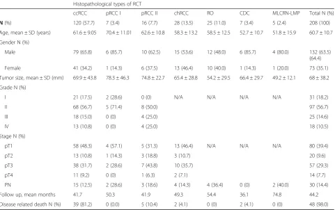
A total of 208 patients with different types of RCT who underwent radical or partial nephrectomy between January 1st, 2010 and December 31st, 2016 at Clinic for Urology, Clinical Center of Serbia, were included in this retrospect-ive study. Medical archival records were retrieved to obtain clinicopathological parameters including age, gender and tumor size (maximum tumor diameter). Descriptive charac-teristics of all 208 patients and histological characcharac-teristics are listed in Table 1. Most of the study group was com-posed of elderly male patients. Among all 208 patients, 178 (85.6%) underwent radical nephrectomy and 30 (14.4%) underwent partial nephrectomy. Tumor size ranged from 10 mm to 34 cm in the largest diameter, both in patients with ccRCC. Sarcomatoid change was observed in 2 ccRCC with nuclear grade IV. Information about patients’ out-comes, including the time between nephrectomy and cancer-related death or last follow-up (if death did not occur), was also recorded. The median follow-up time was 44.2 months, ranging from 1 month in a patient with chRCC to 94 months in a patient with RO. For 14 (6.7%) patients with RO, survival data wasn’t available.
Pathological evaluation criteria
ISUP, 2016 [1]. Since there are not clear recommenda-tions for grading chRCC and CDCs, they were not graded [19]. Tumor staging was evaluated according to the updated American Joint Committee on Cancer (AJCC) tumor–node–metastasis (TNM) classification 8th edition, by reviewing the gross description and rep-resentative slides [20].
TMA
TMA was constructed manually. As TMA only contains a limited amount of tissue, we were aware of the possible variation of immunostaining due to tissue het-erogeneity. To provide the high accuracy of our results in the representation of the whole section, we sampled three different cores 1.2 mm from each specimen, as it was is recommended previously [21]. The hematoxylin-eosin (H&E) stained slides were examined and the three different the most high-grade areas (subcapsular, middle and periphery of the tumor), were marked by patholo-gists. Microarray samples were punched out from the selected regions of each donor block and transferred into a new recipient paraffin block using a hollow needle with an inner diameter of 1,2 mm. An additional core of
normal-appearing kidney cortex and medulla was taken randomly for each case. Five-micrometer sections were cut from completed TMA blocks and transferred to ad-hesive slides to be used for immunohistochemical stain-ing. Thus, 5μm thickness sections of each tissue array block were stained by H&E to confirm the grade and histological type of each tissue core spot.
Conventional sections
In order to validate our methodology and to increase the confidence in our results, we used the corresponding whole-mount sections in a subset of 45 cases used for TMA. The selected cohort included: 15 ccRCC, 3 pRCC I, 7 pRCC II, 10 chRCC, and 10 RO. ccRCC and pRCC were chosen according to different nuclear grades. Seven cases of ccRCC were in low grade, eight cases were in high grade, including one with sarcomatoid features. In addition, we selected five cases from each of low- and high-grade pRCC. ChRCC and RO were taken randomly. Five-micrometer sections were cut from Formalin-Fixed Paraffin-Embedded (FFPE) tissue blocks of each case and transferred to adhesive slides for immunohisto-chemical staining.
Table 1Patients’demographics and tumor characteristics
Histopathological types of RCT
ccRCC pRCC I pRCC II chRCC RO CDC MLCRN-LMP Total N (%)
N(%) 120 (57.7) 7 (3.4) 16 (7.7) 28 (13.5) 25 (11.0) 7 (3.4) 5 (2.4) 208 (100)
Age, mean ± SD (years) 61.6 ± 9.05 70.4 ± 11.01 62.6 ± 10.8 58.3 ± 13.2 58.5 ± 12.5 52.7 ± 10.7 51.8 ± 15.9 60.7 ± 10.7
Gender N (%)
Male 79 (65.8) 6 (85.7) 10 (62.5) 15 (53.6) 12 (48.0) 6 (85.7) 4 (80.0) 132 (63.5)
(64.4)
Female 41 (34.2) 1 (14.3) 6 (37.5) 13 (46.4) 10 (40.0) 1 (14.3) 1 (20.0) 73 (35.1)
Tumor size, mean ± SD (mm) 69.9 ± 43.8 78.3 ± 46.3 74.8 ± 22.7 65.4 ± 28.8 54.2 ± 29.5 66.4 ± 29.7 49.2 ± 12.1 68 ± 38.2
Grade N (%)
I 21 (17.5) 2 (28.6) 0 (0) N/A N/A N/A N/A 31 (18.2)
II 68 (56.7) 5 (71.4) 8 (50.0) 97 (56.7)
III 18 (15.0) 0 (0) 4 (25.0) 25 (14.6)
IV 13 (10.8) 0 (0) 4 (25.0) 18 (10.5)
Stage N (%)
pT1 58 (48.3) 4 (57.1) 5 (31.3) 13 (46.4) N/A N/A N/A 80 (39.4)
pT2 13 (10.8) 1 (14.3) 3 (18.8) 3 (10.7) 20 (9.6)
pT3 38 (31.7) 2 (28.6) 7 (43.8) 10 (35.7) 57 (29.3)
pT4 11 (9.2) 0 (0) 1 (6.3) 2 (7.1) 14 (7.7)
PN 15 (12.5) 2 (28.6) 3 (18.6) 4 (14.3) 4 (36.4) 0 (0) 2 (40.0) 30 (14.4)
Follow up, mean months 41.7 50.3 41.9 49.3 54.4 36.1 74.8 44.2
Disease related death N (%) 39 (81.2) 0 (0.0) 5 (10.4) 2 (4.1) 0 (0) 2 (4.1) 0 (0) 48 (98.0)
Immunohistochemistry
All TMA and conventional sections were deparaffinized at 60 °C for 20 min and rehydrated in graded ethanols. Antigen retrieval was performed by immersing the tis-sues in citrate buffer (pH = 6.0) for 20 min in a water bath. Endogenous peroxidase and non-specific staining
were blocked with 3% H2O2 for 20 min at room
temperature. The TMA sections were incubated for 1 hour at room temperature with the following primary antibodies: PRMT1 (ab92299, Abcam, dilution: 1/200), ZEB1 (HPA027524, Sigma Aldrich, dilution: 1/500), RUNX1 (sc-365,644, Santa Cruz, dilution: 1/50), TWIST1 (ABD29 Merck Millipore, dilution: 1/100), E-cadherin (spm471, Santa Cruz, dilution: 1/100), N-cadherin (6D11, DAKO, dilution: 1/100), ß-catenin (ß-catenin-1, Dako, di-lution: 1/100). Whole-mount sections were incubated with the PRMT1 (ab92299, Abcam, dilution: 1/200) and ZEB1 (HPA027524, Sigma Aldrich, dilution: 1/500). All slides were then incubated with EnvisioninFLEX/HRP (K4063, Dako, Denmark) for 30 min. Staining patterns were visual-ized by exposure to 3,3′-diaminobenzidine (DAB) and counterstained with Mayer’s hematoxylin. Finally, the slides were dehydrated in ethanol, cleared in xylene and mounted for examination. Human kidney tissue was used as a positive control of staining and replacement of the primary antibody with Tris-buffered saline was used as a negative control.
Evaluation of immunostaining
Immunostaining of all markers was independently evaluated by three pathologists (J.F. M.B. M.Ž.), who were blinded to the patient outcome and pathological information. Valid immunoreactivity was considered as following: nuclear staining of PRMT1, ZEB1 and RUNX1, cytoplasmic and/or nuclear for TWIST1, mem-branous for N- and E-cadherin, while for ß-catenin, membranous staining was considered to be normal and nuclear and/or cytoplasmic immunopositivity was aber-rant. Tumors were considered positive when more than 5% of tumor cells expressed positive staining to all ana-lyzed markers, with at least moderate intensity, and at least in one core. In addition, the analysis of IHC hetero-geneity was performed. If the staining pattern was equal among the cores, it was grouped as a homogenous negative or homogenous positive. The group with differ-ent immunopositivity among the cores was considered as heterogenous. In addition, PRMT1 expression in nor-mal renal parenchyma was examined. Moreover, PRMT1 and ZEB1 expression in a subset of TMA cases were compared to corresponding whole-mount sections. We analyzed whether homogenous negative, homogenous positive or heterogeneous staining on TMA corresponds to the same staining pattern in subcapsular, middle or peripheral areas on whole-mount sections. A consensus
was achieved for all samples. The slides were evaluated using the light microscope BX53 with DP12-CCD cam-era (Olympus, Germany).
RNA isolation and qRT-PCR analysis of mRNA PRMT1 Tissues were collected on surgery and kept in liquid ni-trogen. Fresh samples of 5 ccRCC, 2 pRCC and 1 chRCC with adjacent normal renal tissue were analyzed in tripli-cates. Tissue lysis was obtained in lysis buffer in a Tissue Lyser LT Adapter (Qiagen, United Kingdom) for 5 minutes. A TRIzol Reagent (Takara, Shiga, Japan) and Minipipet angio Kit (Isto Takara) were used for total RNA extraction in accordance with the manufacturer’s protocol. The quantity of the isolated RNA was assessed using NanoDrop 2000 spectrophotometer (Thermo Scientific, Germany). 100 ng of total RNA was digested with DNaseI (Sigma Aldrich, United Kingdom) and used for cDNA synthesis using Super Script II Reverse Transcriptase (Life Technologies, Germany). For quanti-tative real-time reverse transcription PCR (qRT-PCR) analysis, diluted cDNA (1/10) was used as a template in a Fast SYBR Green Master Mix (Life Technologies, Germany) and run on Step One Plus Real-Time PCR System (Applied Biosystems) in a total reaction volume of 20μl. Primers for PRMT1 gene (forward, 5′-ATCTCG CACCACACCTTCTA-3′; reverse, 5′-CGGTATAGAT
GTCCACCTCCTTTATG −3′), were designed and
pur-chased from Primer Design.
TCGA and GTEx cohort
mRNA PRMT1 transcript profiling data in Fragment Per Kilo Base Million (FPKM) values of 877 tumor samples in The Cancer Genome Atlas (TCGA) cohorts and 32 samples of normal tissue in Genotype-Tissue Expression (GTEx) cohort were downloaded from the Human Protein Atlas data portal (https://www.proteinatlas. org/) in April 2019. Patients clinicopathological data are collected from the TCGA resource network, as in [22]. Merged data are available in Additional file2: Table S1. Pa-tient characteristics are summarized in Additional file 3: Table S2. Overall, 60,1% of patients (n= 528) were diag-nosed as ccRCC; 23,5% (n= 285) were classified as pRCC and 7,3% (n= 64), were classified as chRCC. Approximately 61% of the patients were male. mRNA PRMT1 expression level was analyzed in cancerous and normal renal paren-chyma. Furthermore, mRNA PRMT1 expression level in ccRCC, pRCC and chRCC was compared to tumor stage, while tumor grade was available only for ccRCC.
Statistical analysis
Kaplan Meier and Cox regression were used for sur-vival analysis. All pvalues less than 0.05 were consid-ered significant. All data were analyzed using SPSS 20.0 (IBM corp.) statistical software.
Results
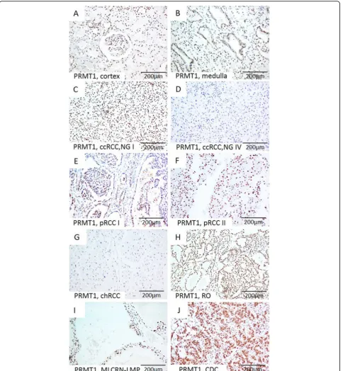
Expression of PRMT 1 in normal kidney parenchyma We observed nuclear PRMT1 IHC expression in all sam-ples of the renal parenchyma used as a positive control.
PRMT1 was expressed in various cortical structures, such as epithelial cells of proximal and distal tubules, glomeru-lar mesangial cells and parietal cells of Bowman’s capsule. In the medulla, PRMT1 was observed in epithelial cells of collecting ducts (Fig.1a-b).
PRMT 1, ZEB 1, RUNX 1, and TWIST 1 expression in RCT types
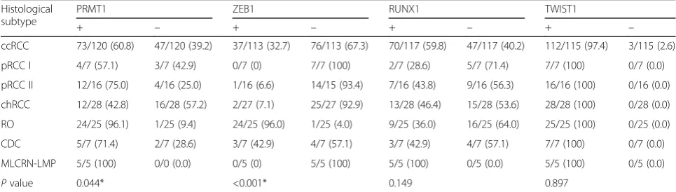
Immunohistochemical expression of PRMT1, ZEB1, RUNX1, and TWIST1 in different tumor types is sum-marized in Table 2. Expression patterns of PRMT1 and ZEB1 immunopositivity were associated with different RCT types (p values 0.044 and < 0.001, respectively) (Table 2). PRMT1 expression was observed in all ana-lyzed tumor types, notably in the majority of ccRCC and pRCC, almost all RO, all MLCRN-LMP, and in minority of chRCC (Fig.1c-j). ZEB1 was negative in a majority of ccRCC and pRCC and in all MLCRN-LMP (Fig. 2 a-d.) ZEB1 was negative in a majority of chRCC, but positive in almost all RO cases (Fig.2e-f.). RUNX1 immunoposi-tivity was noted in all tumor types, notably in a majority of ccRCC and in all MLCRN-LMP, however, without statistical significance (Fig. 2 g-h). TWIST1 was also observed in all tumor types, mostly with cytoplasmic immunopositivity and occasionally with nuclear expres-sion, but without statistical significance (Fig. 3 a-h). Statistically significant differences in co-expression of PRMT1 with ZEB1, RUNX1 and TWIST1 were observed only in ccRCC, but not in other analyzed tumor types
(Table 3). The majority of PRMT1 negative ccRCC
showed mutual loss of ZEB1 (p= 0.028). On the other hand, the majority of PRMT1 positive cases expressed RUNX1 (p= 0.019). Also, the majority of PRMT1 nega-tive ccRCC showed solely cytoplasmic expression of TWIST1 without nuclear positivity (p< 0.001).
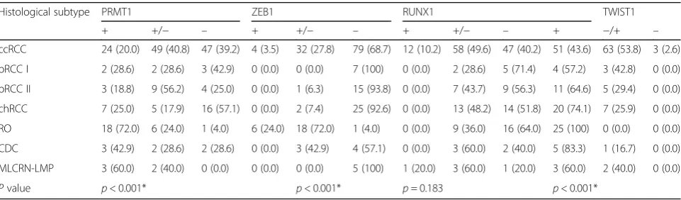
The heterogeneity of PRMT1 and ZEB1 IHC expression in RCT types
The significant heterogeneity of PRMT1, ZEB1, and TWIST1 immunostaining was observed among analyzed tumor types (p< 0.001) (Table 4). Thus, PRMT1 expres-sion among ccRCC was mostly heterogenous, while ZEB1 was mainly homogenous negative (Additional file1: Figure S1A-F). PRMT1 and ZEB1 were more frequently homogenous negative in chRCC (Additional file 1: Fig-ure S1G-L). RO the most frequently expressed PRMT1
homogenously, while ZEB1 positivity was mostly
heterogenous (Additional file1: Figure S1M-R). TWIST1
in the majority of cases was heterogenous or
homogenous positive (Table4).
PRMT1 and ZEB1 IHC expression on conventional sections Homogenous positive or homogenous negative PRMT1 and ZEB1 expression on TMA, corresponded to the same staining pattern on whole-mount section, regard-less of the tumor type or nuclear grade. Heterogenous PRMT1 and ZEB1 expression on TMA corresponded to the same subcapsular and occasional peripheral and/or middle areas on analyzed conventional sections. One case of ccRCC with sarcomatoid features showed mutual loss of PRMT1 and ZEB1 on TMA and whole-mount section (Additional file4: Table S3).
Association of PRMT1, RUNX1, ZEB1 and TWIST 1 with clinicopathological parameters in various types of RCT As clinical behavior of tumor stage pT1 is similar to pT2, and pT3 to pT4, we analyzed the significance of PRMT1, RUNX1, ZEB1 and TWIST 1 expression in pT1/pT2 versus pT3/pT4 stage group. Likewise, tumor grades were grouped as low grade (I/II) and high grade (III/IV) [23]. Analysis of expression profiles showed as-sociation only of PRMT1 expression with
clinic-Table 2Immunohistochemical positivity of PRMT1, ZEB1, RUNX1 and TWIST1 among different tumor types
Histological subtype
PRMT1 ZEB1 RUNX1 TWIST1
+ – + – + – + –
ccRCC 73/120 (60.8) 47/120 (39.2) 37/113 (32.7) 76/113 (67.3) 70/117 (59.8) 47/117 (40.2) 112/115 (97.4) 3/115 (2.6)
pRCC I 4/7 (57.1) 3/7 (42.9) 0/7 (0) 7/7 (100) 2/7 (28.6) 5/7 (71.4) 7/7 (100) 0/7 (0.0)
pRCC II 12/16 (75.0) 4/16 (25.0) 1/16 (6.6) 14/15 (93.4) 7/16 (43.8) 9/16 (56.3) 16/16 (100) 0/16 (0.0)
chRCC 12/28 (42.8) 16/28 (57.2) 2/27 (7.1) 25/27 (92.9) 13/28 (46.4) 15/28 (53.6) 28/28 (100) 0/28 (0.0)
RO 24/25 (96.1) 1/25 (9.4) 24/25 (96.0) 1/25 (4.0) 9/25 (36.0) 16/25 (64.0) 25/25 (100) 0/25 (0.0)
CDC 5/7 (71.4) 2/7 (28.6) 3/7 (42.9) 4/7 (57.1) 3/7 (42.9) 4/7 (57.1) 7/7 (100) 0/7 (0.0)
MLCRN-LMP 5/5 (100) 0/0 (0.0) 0/5 (0) 5/5 (100) 5/5 (100) 0/5 (0.0) 5/5 (100) 0/5 (0.0)
Pvalue 0.044* <0.001* 0.149 0.897
Abbreviations: PRMT1, protein arginine methyltransferase 1; ZEB1, Zinc Finger E-Box Binding Homeobox 1; RUNX1, Runt-related transcription factor 1; TWIST1, Twist Family BHLH Transcription Factor 1; ccRCC, clear cell renal cell carcinomas; pRCC I, papillary renal cell carcinoma type I; pRCC II, papillary renal cell carcinoma type I; chRCC, chromophobe renal cell carcinoma;CDC, collecting duct carcinomas-Bellini; MLCRN-LMP, multilocular cystic renal neoplasm of low malignant potential; +, positive;−, negative
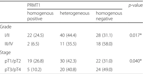
pathological parameters in ccRCC patients (Table 5). PRMT1 expression was often present in low-grade ccRCC and low stage ccRCC (p= 0.014, p= 0.044, re-spectively). Moreover, homogenous loss of PRMT1 was observed in 58% high grade and in 49% of high stage ccRCCs (p= 0.017, p= 0.040, respectively) (Table 6). Interestingly, two cases of ccRCC with sarcomatoid change showed homogenous loss to PRMT1 and ZEB1, heterogenous positivity to RUNX1 and cytoplasmic TWIST1 expression. Gender and age of ccRCC patients were not associated with PRMT1 expression. Further, RUNX1, ZEB1 or TWIST1 expression didn’t show
association with clinicopathological features in any of the analyzed tumor types.
Association of PRMT1, RUNX1, ZEB1 and TWIST 1 with markers of EMT
Among analyzed RCT types, aberrant ß-catenin cyto-plasmic immunoreactivity was observed in 147 (71.7%) of cases, without nuclear positivity. N-cadherin expres-sion was observed in 123 (59.1%) cases, while the loss of E-cadherin immunopositivity was detected in 41 (19.7%) cases. PRMT1 expression was associated neither with
aberrant ß-catenin expression nor with N- cadherin gain or E-cadherin loss in analyzed tumor types. However, the association between RUNX1, N-and E-cadherin ex-pression was significant only in ccRCC (Table7). The ma-jority of RUNX1 positive ccRCC expressed N-cadherin (p= 0.003). E-cadherin loss was less frequent in RUNX1 positive than in RUNX1 negative ccRCC (p= 0.019).
Association of PRMT1, Zeb1, RUNX 1 and TWIST 1 expression with prognosis
During the follow-up time, disease-related death was documented in 48 (23.1%) patients. Diagnosed RCC types in patients who died of cancer related disease were
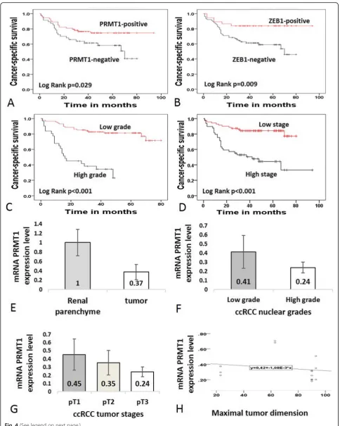
as following: 39 (80%) ccRCC, 1(2%) pRCC I, 4(8.2%) pRCC II, 2 (4%) chRCC and 2 (4%) CDCs. One patient with oncocytoma died of an unrelated cause. Kaplan– Meier survival analyses revealed that PRMT1, ZEB1, nuclear grade, and tumor stage were significant prognos-tic factors in ccRCC. In parprognos-ticular, the expression of PRMT1 and ZEB1 in ccRCC, as well as low-nuclear tumor grade and low-tumor stage were significantly as-sociated with better cancer-specific survival (p= 0.029, p= 0.009,p< 0.001 and p< 0.001, respectively) (Fig.4 a-d; Table 8). Moreover, patients who died of ccRCC showed homogenous loss of PRMT1 in 44.7%, while 41.8% had homogenous loss of ZEB1 (p= 0.044, p=
0.009, respectively) (Table 9). RUNX1 and TWIST1 and patients’ gender didn’t show a correlation with cancer-specific survival (Table8).
qRT-PCR PRMT1 expression in cRCC, chRCC, and pRCC A decrease of relative PRMT1 mRNA expression level was detected in tumor tissue compared to adjacent renal parenchyma (Fig. 4 e). Furthermore, relative PRMT1 mRNA expression level was lower in ccRCC than in chRCC and pRCC. However, ccRCC samples with nu-clear grade II and lower tumor stages (I and II), had higher PRMT1 mRNA expression level (Fig. 4 f-g). In addition, we observed a tendency to decrease in PRMT1 mRNA expression level along with the increase of tumor size (Fig.4h). In these samples, PRMT1 IHC expression was observed in all three analyzed RCC types. In two cases of ccRCC with the highest grade, stage and with
the largest tumor dimension relative mRNA PRMT1 ex-pression was the lowest and PRMT1 IHC exex-pression was homogenous negative and heterogenous. Interest-ingly, both cases of pRCC, type I expressed PRMT1 homogenously, although the relative PRMT1 mRNA level was different (Additional file5: Table S4).
mRNA PRMT1 expression in TCGA and GTEx cohort The results of mRNA PRMT1 expression analysis in the
TCGA cohort are presented in Fig. 5. mRNA PRMT1
expression level in tumor tissue was significantly lower than that in renal parenchyma (p= 0.009; Fig. 5a). In tumor tissue, mRNA PRMT1 expression level was sig-nificantly lower in ccRCC, when compared to pRCC and chRCC (p< 0.001; Fig.5b). However, mRNA PRMT1 ex-pression level in ccRCC was not associated with tumor stage or grade, neither was associated with tumor stage of pRCC (p= 0.136; p= 0.094; p= 0.355, respectively (Fig. 5 c-e). On the other hand, mRNA PRMT1 expres-sion level in chRCC was significantly higher in stage IV (p< 0.001; Fig.5f).
Discussion
RCT is a heterogeneous group of neoplasms, with differ-ent biological behavior and prognosis [1]. Thus, persist-ent efforts to predict outcome in patipersist-ents with renal neoplasms and to diagnose RCT precisely, have led sci-entists to investigate various biological markers. Further-more, it has been widely accepted that epigenetic regulation plays an important role in many biological processes, including tumorigenesis [2].
PRMT1 is usually considered as an epigenetic molecu-lar marker, expressed in many non-tumorous and tu-morous tissues. PRMT1 immunopositivity in our study was equally observed in cortex and medulla of all cases used as a positive control. To the best of our knowledge, only one study reported IHC PRMT1 expression in renal
Table 3Evaluation of co-expression patterns of PRMT1 and other transcription factors ZEB1, RUNX1, and TWIST1 in ccRCC
IHC marker (total cases) PRMT1 Pvalue
positive negative
ZEB1 (113)
positive 27 (40.9) 10 (21.3) 0.028*
negative 39 (59.1) 37 (78.7)
RUNX1 (117)
positive 48 (68.6) 22 (46.8) 0.019*
negative 22 (31.4) 25 (53.2)
TWIST1 (115)
nuclear ± cytoplasmic 37 (53.6) 10 (21.7) <0.001*
cytoplasmic 32 (46.4) 36 (78.3)
Abbreviations: PRMT1, protein arginine methyltransferase 1; ZEB1, Zinc Finger E-Box Binding Homeobox 1; RUNX1, Runt-related transcription factor 1; TWIST1, Twist Family BHLH Transcription Factor 1; ccRCC, clear cell renal cell carcinomas
*Statistically significantp-value
Table 4Heterogeneity of PRMT1, ZEB1, RUNX1 and TWIST1 immunohistochemical expression among different tumor types
Histological subtype PRMT1 ZEB1 RUNX1 TWIST1
+ +/− – + +/− – + +/− – + −/+ –
ccRCC 24 (20.0) 49 (40.8) 47 (39.2) 4 (3.5) 32 (27.8) 79 (68.7) 12 (10.2) 58 (49.6) 47 (40.2) 51 (43.6) 63 (53.8) 3 (2.6)
pRCC I 2 (28.6) 2 (28.6) 3 (42.9) 0 (0.0) 0 (0.0) 7 (100) 0 (0.0) 2 (28.6) 5 (71.4) 4 (57.2) 3 (42.8) 0 (0.0)
pRCC II 3 (18.8) 9 (56.2) 4 (25.0) 0 (0.0) 1 (6.3) 15 (93.8) 0 (0.0) 7 (43.7) 9 (56.3) 11 (64.6) 5 (29.4) 0 (0.0)
chRCC 7 (25.0) 5 (17.9) 16 (57.1) 0 (0.0) 2 (7.4) 25 (92.6) 0 (0.0) 13 (48.2) 14 (51.8) 20 (74.1) 7 (25.9) 0 (0.0)
RO 18 (72.0) 6 (24.0) 1 (4.0) 6 (24.0) 18 (72.0) 1 (4.0) 0 (0.0) 9 (36.0) 16 (64.0) 25 (100) 0 (0.0) 0 (0.0)
CDC 3 (42.9) 2 (28.6) 2 (28.6) 0 (0.0) 3 (42.9) 4 (57.1) 0 (0.0) 3 (60.0) 2 (40.0) 5 (83.3) 1 (16.7) 0 (0.0)
MLCRN-LMP 3 (60.0) 2 (40.0) 0 (0.0) 0 (0.0) 0 (0.0) 5 (100) 1 (20.0) 3 (60.0) 1 (20.0) 3 (60.0) 2 (40.0) 0 (0.0)
Pvalue p< 0.001* p< 0.001* p= 0.183 p< 0.001*
Abbreviations: PRMT1, protein arginine methyltransferase 1; ZEB1, Zinc Finger E-Box Binding Homeobox 1; RUNX1, Runt-related transcription factor 1; TWIST1, Twist Family BHLH Transcription Factor 1; ccRCC, clear cell renal cell carcinomas; pRCC I, papillary renal cell carcinoma type I; pRCC II, papillary renal cell carcinoma type I; chRCC, chromophobe renal cell carcinoma;CDC, collecting duct carcinomas-Bellini; MLCRN-LMP, multilocular cystic renal neoplasm of low malignant potential; +, homogenous positive; +/−heterogenous;-, homogenous negative
tubules, without PRMT1 expression in glomerular struc-tures, which could possibly be explained due to the use of different antibody PRMT1 clone [5].
The role of PRMT1 in tumorigenesis is still controver-sial and seems to depend on tumor type. Usually, it is overexpressed in tumors with high-grade morphology and high stage of the disease [14,16]. Nevertheless, the protective effect of PRMT1 has been suggested in pa-tients with gastric carcinomas, positively affecting sur-vival and recurrence rates, especially in patients treated with adjuvant therapy [9]. Similarly, more frequent PRMT1 expression was observed in less advanced ccRCC, with low grade and low stage of the disease, while homogenous PRMT1 loss was detected in high grade and
high stage ccRCC. Additionally, we found PRMT1 expres-sion as more common in benign tumors, such as oncocy-toma. Heterogeneous PRMT1, ZEB1, and TWIST1 expression are presumably related to ITH [24–26]. ITH is extensively studied in many tumor types, especially in ccRCC [25–29]. The significance of ITH is still arguable. Although it has been linked to aggressive tumor behavior, it seems that ITH appears early in carcinogenesis and therefore may not be necessarily associated with enhanced tumor malignancy [26, 28, 29]. Thus, heterogenous PRMT1 IHC expression in ccRCC may emphasize early acquired epigenetic diversity, which deserves further analyses.
Previously, other authors reported mRNA-PRMT1 level overexpression in cancerous tissue of the bladder, lung, breast, and glioma, when compared to non-tumor tissue. However, renal tumor and renal non-tumor tis-sues were not examined in their studies [5, 10, 13, 14]. Therefore, we analyzed mRNA PRMT1 expression level in TCGA/GTEx group in relation to our small sample. In both cohorts, mRNA PRMT1 expression level was downregulated in tumor tissue, when compared to coun-terparts, while the lowest level was observed in ccRCC type. Unlike in our study, mRNA PRMT1 expression level in ccRCC of TCGA cohort was not associated with tumor stage or tumor grade. Additionally, mRNA PRMT1 expression level up-regulation was associated only with high stage chRCC in the TCGA cohort. The observed differences in mRNA expression may be re-lated to our small sample available for qRT-PCR ana-lysis. Furthermore, different results obtained by IHC
Table 5The comparative analysis of PRMT1, ZEB1, RUNX1 and TWIST1 immunopositivity with clinic-pathological features of the patients with ccRCC
PRMT1 p-value ZEB1 p-value RUNX p-value TWIST1 p-value
+ – + – + – + –
Grade
I/II 62 (68.8) 28 (31.2) 0.009* 38 (35.7) 54 (64.3) 0.093 51 (59.3) 35 (40.7) 0.846 84 (97.7) 2 (2.3) 1
III/IV 13 (41.9) 18 (58.1) 6 (19.4) 25 (80.6) 19 (61.3) 12 (38.7) 30 (96.8) 1 (3.2)
Stage
pT1/pT2 49 (69.0) 22 (31.0) 0.046* 23 (34.3) 44 (65.7) 0.409 37 (54.4) 31 (45.6) 0.159 67 (97.1) 2 (2.9)
pT3/pT4 25 (51.0) 24 (49.0) 13 (27.1) 35 (72.9) 33 (67.3) 16 (32.7) 47 (97.9) 1 (2.9) 1
Gender
Male 44 (55.7) 35 (44.3) 0.110 23 (30.7) 52 (69.3) 0.509 46 (60.5) 30 (39.5) 0.834 76 (98.8) 1 (1.2) 0.276
Female 29 (70.7) 12 (29.3) 14 (36.8) 24 (63.2) 24 (58.5) 17 (41.5) 39 (95.1) 2 (4.9)
Age, mean ± SD (years)
61 ± 9.45 62.6 ± 8.38 0.346 63 ± 8.75 61.2 ± 9 0.378 62 ± 8.9 61 ± 8.7 0.704 61.9 ± 8.8 59 ± 5.03 0.536
Tumour size, mean ± SD (mm)
67 ± 48.75 74.5 ± 32.3 0.362 63 ± 35.1 74.1 ± 45.7 0.181 70 ± 50 71.7 ± 32.8 0.830 70.6 ± 43.7 65 ± 18.03 0.823
Abbreviations: PRMT1, protein arginine methyltransferase 1; ZEB1, Zinc Finger E-Box Binding Homeobox 1; RUNX1, Runt-related transcription factor 1; TWIST1, Twist Family BHLH Transcription Factor 1; ccRCC-clear cell renal cell carcinomas; SD, standard deviation;
ISUP (International Society of Urological Pathology) nuclear grading system and updated American Joint Committee on Cancer (AJCC) tumor–node–metastasis (TNM) classification 8th edition for tumor staging were used
*Statistically significantp-value
Table 6Immunohistochemical heterogeneity of PRMT1 in ccRCC with different nuclear grade and stage
PRMT1 p-value
homogenous positive
heterogeneous homogenous negative
Grade
I/II 22 (24.5) 40 (44.4) 28 (31.1) 0.017*
III/IV 2 (6.5) 11 (35.5) 18 (58.0)
Stage
pT1/pT2 19 (26.8) 30 (42.3) 22 (31.0) 0.040*
pT3/pT4 5 (10.2) 20 (40.8) 24 (49.0)
Abbreviation: PRMT1, protein arginine methyltransferase 1
ISUP (International Society of Urological Pathology) nuclear grading system and updated American Joint Committee on Cancer (AJCC) tumor–node–
analysis in our cohort and mRNA expression analysis in TCGA group may be due to the variations in post-transcriptional mechanisms involved in mRNA PRMT1 translation into protein, as already suggested [30,31]. In addition, gene expression variations in individual case within the same cancer subtype should be taken into ac-count [32].
Our results imply PRMT1 as a ubiquitously expressed marker in the elements of the renal parenchyma. There-fore, PRMT1 may be one of “housekeeping” genes in renal tissue, required for the epigenetic regulation of normal kidney development [33]. Thus, according to re-sults, we may speculate that any change in PRMT1 ex-pression could lead to carcinogenesis and unfavorable prognosis of the patients.
EMT is considered responsible for renal tumorigenesis and progression, which is characterized by TWIST1 cytoplasmic localization, overexpression of N-cadherin, and loss of E-cadherin expression. In addition, ß-catenin dissociation from the membrane and its translocation to the cytoplasm or nucleus is the main feature of Wnt/ß-catenin EMT signaling pathway [17, 34–37]. EMT has been regulated by ZEB1, RUNX1 and TWIST1 tran-scription factors [11,16,38–40]. Furthermore, as an epi-genetic regulator, PRMT1 can act as an EMT inducer or repressor in various cancers. Thus, in breast carcinoma, PRMT1 methylates ZEB1 promoter, which then induces EMT and therefore implies ZEB1 as a negative prognos-tic parameter [16]. On the other hand, PRMT1 activates RUNX1, that prevents EMT and therefore, RUNX1 serves as a tumor suppressor in various cancers [18,39]. Finally, in non-small lung carcinoma, PRMT1 methylates
TWIST1 and induces EMT related aggressive cancer be-havior [11]. Following this, in more aggressive types of RCC and high-grade and/or high-stage tumors we ex-pected frequent ZEB1 expression and co-expression with PRMT1. Unlike PRMT1, ZEB1 was not related to any of the analyzed clinicopathological parameters within dif-ferent RCC types, but survival analysis revealed ZEB1 as an independent prognostic parameter of better survival of the patients with ccRCC. PRMT1 loss was often ac-companied by ZEB1 loss in ccRCC. Thus, we assume that ZEB1 loss in ccRCC may be dependent on PRMT1 loss. This finding is different than those obtained in breast carcinoma and may be related to the different molecular mechanism of ZEB1 activity and specific PRMT1/ZEB1 interaction in RCT which haven’t been investigated so far. In this study, frequent homogenous PRMT1 and heterogenous ZEB1 IHC expression were observed in RO, considered to have benign behavior, while the majority of chRCC and RO was homogenously negative to ZEB1. This observation was confirmed on our small cohort of corresponding whole-mount
sec-tions. This may suggest that PRMT1/ZEB1
co-expression may be significant in RO tumorigenesis and may serve as a helpful IHC tool in the diagnosis of RO. However, this finding requires further investigation and confirmation on larger sample size, including conven-tional sections analysis. On the other hand, in less advanced carcinomas, we expected frequent RUNX1 expression and co-expression with PRMT1. Therefore, we may speculate that RUNX1/PRMT1 co-expression may be significant for the favorable behavior of renal neoplasms. Finally, in advanced carcinomas, we expected
Table 7Association of PRMT1, ZEB1, RUNX1, and TWIST1 with WNT-β-catenin/EMT hallmarks (β-catenin, N- and E-cadherin) in ccRCC
Transcription factor
Wnt-β-catenin/EMT hallmarks
Aberrantβ-catenin Pvalue N-cadherin+ Pvalue E-cadherin- Pvalue
PRMT1
+ 37/37 (100) 1 58/69 (84.1) 0.878 20/66 (30.3) 0.468
- 36/36 (100) 40/47 (85.1) 11/46 (24.0)
ZEB1
+ 21/21 (100) 1 33/37 (89.2) 0.380 12/36 (33.3) 0.280
- 52/52 (100) 63/76 (83.0) 17/73 (23.6)
RUNX1
+ 46/46 (100) 1 65/70 (92.8) 0.003* 14/70 (20.0) 0.019*
- 26/26 (100) 32/45 (71.1) 17/42 (40.5)
TWIST1
n+/−c 20/20 (100) 1 38/46 (82.6) 0.563 14/46 (30.4) 0.528
c 52/52 (100) 58/67 (86.6) 16/64 (25.0)
- 1/1 (100) 2/3 (66.5) 1/2 (50.0)
frequent TWIST1 cytoplasmic expression and co-expression with PRMT1. In ccRCC, loss of PRMT1 was associated with cytoplasmic TWIST1 expression, charac-teristic for aggressive ccRCC [36]. Therefore, we suggest that loss of PRMT1 in ccRCC could lead to TWIST1 cytoplasmic expression and more aggressive behavior. Moreover, two cases of high-grade ccRCC with sarcoma-toid features showed homogenous PRMT1/ZEB1 loss
and TWIST1 localization in the cytoplasm. This obser-vation may be of great importance, that should be evalu-ated on larger cohort of RCT with sarcomatoid and/or rhabdoid change and compared to other high-grade RCC. EMT Wnt/ß-catenin pathway markers were not associated with PRMT1, ZEB1 or TWIST1 expression. Only RUNX1 was associated with N-cadherin overex-pression. Our results suggest, that retention of PRMT1
Table 8Survival analysis. Mean survival of the patients within all types (n= 194) and ccRCC (n= 120); univariate and multivariate cancer-specific survival in the patients with ccRCC according to analyzed immunohistochemical markers and clinic-pathologic parameters
Prognostic factors
Cancer-specific survival (months)
Univariate Multivariate
All types mean (95% CI) p-value ccRCC mean (95% CI) p-value Hazard ratio (95% CI) p-value Hazard ratio (95%CI) p-value
PRMT1
positive 78 (73–84) 0.009* 74 (66–82) 0.029* 0.504 (0.269–0.947) Ref.
0.033* 0.622 (0.326–1.188) 0.150
negative 56 (48–63) 51 (42–61)
ZEB1
positive 83 (76–91) 0.011* 80 (70–90) 0.009* 0.344 (0.140–0.801) 0.014* 0.367 (0.152–0.889) 0.026
negative 68 (61–74) 52 (45–60)
RUNX1
positive 75 (68–82) 0.937 67 (60–74) 0.506 0.872 (0.463–1.644) 0.673 N/A
negative 69 (61–78) 57 (48–66)
TWIST1
positive 73 (68–78) 0.886 67 (60–74) 0.878 1.168 (0.159–8.553) 0.879 N/A
negative 54 (34–75) 54 (34–75)
Grade N/A
Low 67 (62–72) <0.001* 5.802 (3.000–11.222) <0.001* 6.284 (3.175–12.438) <0.001*
High 25 (18–32)
Stage N/A
Low 70 (63–76) <0.001* 1.062 (0.849–1.330) 0.597 1.187 (0.912–1.547) 0.203
High 48 (36–60)
PN 63 (51–74)
Gender
Male 75 (66–83) 0.686 58 (51–65) 0.736 1.123 (0.569–2.218) 0.737 N/A
Female 72 (66–78) 70 (58–81)
Abbreviations:PRMT1 protein arginine methyltransferase 1,ZEB1 Zinc Finger E-Box Binding Homeobox 1,RUNX1 Runt-related transcription factor 1,TWIST1 Twist Family BHLH Transcription Factor 1,CIconfidence interval,PNpartial nephrectomy,ref. referent value. *Statistically significantp-value
(See figure on previous page.)
expression may be a significant feature of less advanced ccRCC, while PRMT1 homogenous loss could be spe-cific for high grade and high stage ccRCC. The role of PRMT1 in RCT, as an epigenetic marker, is barely re-lated to WNT/ß-catenin-mediated EMT processes, since neither of EMT related cadherins or ß-catenin were as-sociated with PRMT1 expression. Another possibility to explain the role of PRMT1 in various RCT emerge from the previous suggestions that PRMT1 indirectly, through different transcription factors, may affect tumor cell growth or cell apoptosis. Overall, this experimental study represents the first step of the future research that will evaluate the role of PRMT1, as a marker of favorable prognosis in ccRCC and its role in carcinogenesis of RCT.
Conclusions
IHC expression of PRMT1 may be a valuable marker of low grade and low stage ccRCC, while homogenous PRMT1 loss may be significant for high grade and high stage ccRCC. Heterogenous PRMT1 IHC expression may emphasize intratumor heterogeneity which should be extensively examined in further studies. Loss of PRMT1 and/or ZEB1 IHC expression could be an indi-cator of poor prognosis in patients with ccRCC. As an epigenetic regulator, PRMT1 may be involved in other mechanisms of RCT development unrelated to EMT, such as regulation of the cell cycle or apoptosis, which remain to be clarified in the future.
Limitations of this current study
Our study has several innate limitations to note. First, our small sample size of non-ccRCC tumor types with their retrospective nature was a major limitation. Other RCT types are relatively rare compared to ccRCC, espe-cially MLCRN-LMP and CDC. Therefore, the main dis-cussion of our paper relied on ccRCC, RO and chRCC. However, this is an experimental study, and the results of the current analysis on TMA should be confirmed in a larger multi-institutional study and using whole-mount sections. Furthermore, quantification of mRNA expres-sion level could not be performed for all tumor types
Fig. 5mRNA PRMT1 transcript profiling data (FPKM values) for TCGA cohorts relative to tumor tissue and tumor stage.amRNA PRMT1 expression level was decreased in tumor tissue related to non-tumor parenchyma; (b) In tumor tissue, mRNA PRMT1 expression level was significantly lower in ccRCC, when compared to pRCC and chRCC; (c-d) However, mRNA PRMT1 expression level in ccRCC was not associated with tumor stage, neither with nuclear grade; (e) Likewise, mRNA PRMT1 expression level in pRCC was not associated with tumor stage; (f) On the other hand, mRNA PRMT1 expression increase was observed in stage IV of chRCC; mRNA expression levels are presented with Fragments Per Kilobase Million values (FPKM) (+/−1 SD). Statistically significant differencep< 0.05
Table 9The comparation of PRMT1 and ZEB1 expression heterogeneity and outcome of the patients with ccRCC
Prognostic marker PRMT1 p value ZEB1 pvalue
Dead Dead
homogenous positive 4/24 (16.7) 0.044* 0/4 (0.0) 0.009*
heterogenous 14/49 (28.6) 5/32 (15.6)
homogenous negative 21/47 (44.7) 33/79 (41.8)
and for all markers, because of the lack of tissues and re-agents for qRT-PCR analysis. Our hypothesis should, therefore, be further confirmed by obtaining tissue for other tumor types.
Supplementary information
Supplementary informationaccompanies this paper athttps://doi.org/10. 1186/s13000-019-0901-6.
Additional file 1: Figure S1.Representative TMA microscopic photographs of PRMT1 and ZEB1 expression in ccRCC, chRCC and RO. (A-C) Strong heterogenous nuclear expression of PRMT1 and (D-F) homogenous negative ZEB1 staining in ccRCC, (G-L) Homogenous negative PRMT1 and ZEB1 in chRCC, (M-R) Strong homogenous positive nuclear PRMT1 expression and heterogenous nuclear ZEB1 expression in RO. Original magnification, × 40. Abbreviation: TMA, tissue microarray
Additional file 2: Table S1.mRNA PRMT1 expression level (FPKM) and clinical characteristics for all patients and their tumors
Additional file 3: Table S2.Patients characteristics in analyzed TCGA cohort
Additional file 4: Table S3.PRMT1 and ZEB1 IHC expression on TMA and corresponding whole-mount sections
Additional file 5: Table S4.Relative mRNA PRMT level and IHC PRMT1 expression in analyzed RCC
Abbreviations
AJCC:American Joint Committee on Cancer; CDC: collecting duct carcinomas–Bellini; cDNA: complementary DNA; chRCC: chromophobe renal renal cell carcinoma; DAP: diaminobenzidine; DNA: Deoxyribonucleic acid; EMT: epithelial to mesenchymal transition; FFPE: Formalin-Fixed Paraffin-Embedded; FPKM: Fragment Per Kilo Base Million; GTEx: Genotype-Tissue Expression; H&E: hematoxylin and eosin; IHC: immunohistochemical; ISUP: International Society of Urological Pathology; miRNA: microRNA; MLCRN-LMP: multilocular cystic renal cell neoplasms of low malignant potential; pRCC I: papillary RCC type I; pRCC II: papillary renal cell carcinoma type II; PRMT1: Protein arginine methyltransferase-1; qRT-PCR: Real-Time Quantitative Reverse Transcription Polymerase Chain Reaction; RCC: renal cell carcinomas; RCT: renal cell tumors; RNA: ribonucleic acid; RO: renal oncocytomas; RUNT1: Runt-related transcription factor 1; TCGA: The Cancer Genome Atlas; TMA: tissue microarray; TNM: tumor–node–metastasis; TWIST1: Twist Family BHLH Transcription Factor 1; ZEB1: Zinc Finger E-Box Binding Homeobox 1
Acknowledgments
Not applicable.
Authors’contributions
JF and MB designed the study, analyzed histological slides, interpreted the results and drafted the manuscript. SĆperformed immunohistochemical staining, analyzed stained slides and interpreted the results. MŽperformed qRT-PCR, interpreted the results, reviewed and edited the manuscript. DD constructed TMA, analyzed histological slides and interpreted the results. DĐ collected surgical material and interpreted the results. SŽP performed survival analysis and interpreted the results. AL performed the statistical analysis, interpreted the results and made language corrections. JML and MB reviewed the literature, reviewed and edited the manuscript. All authors read and approved the final manuscript.
Funding
This research was supported by Project of Ministry of Education, Science and Technological Development, Belgrade Serbia (number OI175047).
Availability of data and materials
The most data generated or analyzed during this study are included in this article and its supplementary information files.
Ethics approval and consent to participate
This study was approved by the Institutional Review Board of School of Medicine, University of Belgrade, Serbia.
Consent for publication
Not applicable.
Competing interests
The authors declare that they have no competing interests.
Author details
1Faculty of Medicine, Institute of Pathology, University of Belgrade, Dr.
Subotića 1, Belgrade, Serbia.2Clinic for Urology, Clinical Center of Serbia,
Faculty of Medicine, University of Belgrade, Belgrade, Serbia.3Institute of Public Health“Dr. Milan JovanovićBatut”, Belgrade, Serbia.4Faculty of
Mathematics, University of Belgrade, Belgrade, Serbia.
Received: 6 February 2019 Accepted: 10 October 2019
References
1. Moch H, Humphrey PA, Ulbright TM RV, editor. WHO Classification of Tumors of the Urinary System and Male Genital Organs. 4th ed. Lyon, France: International Agency for Research on Cancer (IARC); 2016. 14–76 p. 2. Morris MR, Latif F. The epigenetic landscape of renal cancer. Nat Rev
Nephrol. 2017;13:47–60.
3. Cancer T, Atlas G. Comprehensive molecular characterization of clear cell renal cell carcinoma. Nature. 2013;24:3–9.
4. Nicholson TB, Chen T, Richard S. The physiological and pathophysiological role of PRMT1-mediated protein arginine methylation. Pharmacol Res. 2009; 60:466–74.
5. Vezzalini M, Aletta JM, Beghelli S, Moratti E, Della Peruta M, Mafficini A, et al. Immunohistochemical detection of arginine methylated proteins (MeRP) in archival tissues. Histopathology. 2010;57:725–33.
6. Yang Y, Bedford MT. Protein arginine methyltransferases and cancer. Nat Rev Cancer. 2013;13:37–50.
7. Zhang R, Li X, Liang Z, Zhu K, Lu J, Kong X, et al. Theoretical insights into catalytic mechanism of protein arginine methyltransferase 1. PLoS One. 2013;8:1–11.
8. Madreiter-Sokolowski CT, Győrffy B, Klec C, Sokolowski AA, Rost R, Waldeck-Weiermair M, et al. UCP2 and PRMT1 are key prognostic markers for lung carcinoma patients. Oncotarget. 2017;8:80278–85.
9. Altan B, Yokobori T, Ide M, Mochiki E, Toyomasu Y, Kogure N, et al. Nuclear PRMT1 expression is associated with poor prognosis and chemosensitivity in gastric cancer patients. Gastric Cancer. 2016;19:789–97.
10. Wang S, Tan X, Yang B, Yin B, Yuan J, Qiang B, et al. The role of protein arginine-methyltransferase 1 in gliomagenesis. BMB Rep. 2012;45:470–5. 11. Avasarala S, Van Scoyk M, Rathinam MKK, Zerayesus S, Zhao X, Zhang W,
et al. PRMT1 is a novel regulator of epithelial-mesenchymal-transition in non-small cell lung cancer. J Biol Chem. 2015;290:13479–89.
12. Mathioudaki K, Papadokostopoulou A, Scorilas A, Xynopoulos D, Agnanti N, Talieri M. The PRMT1 gene expression pattern in colon cancer. Br J Cancer. 2008;99:2094–9.
13. Mathioudaki K, Scorilas A, Ardavanis A, Lymberi P, Tsiambas E, Devetzi M, et al. Clinical evaluation of PRMT1 gene expression in breast cancer. Tumor Biol. 2011;32:575–82.
14. Elakoum R, Gauchotte G, Oussalah A, Wissler MP, Clément-Duchêne C, Vignaud JM, et al. CARM1 and PRMT1 are dysregulated in lung cancer without hierarchical features. Biochimie. 2014;97:210–8.
15. Liu C, Tao TAO, Xu BIN, Lu KAI, Zhang LEI, Jiang L, et al. BTG1 potentiates apoptosis and suppresses proliferation in renal cell carcinoma by interacting with PRMT1. Oncol Lett. 2015;10:619–24.
16. Gao Y, Zhao Y, Zhang J, Lu Y, Liu X, Geng P, et al. The dual function of PRMT1 in modulating epithelial-mesenchymal transition and cellular senescence in breast cancer cells through regulation of ZEB1. Sci Rep. 2016; 6:19874.
17. Kalluri R, Weinberg RA. Review series the basics of epithelial-mesenchymal transition. J Clin Invest. 2009;119:1420–8.
19. Paner GP, Amin MB, Alvarado-Cabrero I, Young AN, Sticker HJ, Moch H, et al. A novel tumor grading scheme for Chromophobe renal cell carcinoma. Am J Surg Pathol. 2010;34:1233–40.
20. Mahul B. Amin, Edge SB, Frederick L. Gfeene. David R. Byrd, Robert K, Brookland JEG et al, editors. American Joint Committee on Cancer AJCC Cancer Staging Manual. 8th ed. Springer International Publishing; 2017. 739–748 p.
21. Kampf C, Olsson I, Ryberg U, Sjöstedt E, Pontén F. Production of tissue microarrays, immunohistochemistry staining and digitalization within the human protein Atlas. J Vis Exp. 2012;63:1–8.
22. Ricketts CJ, Cubas AA De, Fan H, Spellman PT, Rathmell WK, Linehan WM, et al. Molecular Characterization of Renal Cell Carcinoma The Cancer Genome Atlas Comprehensive Molecular Characterization of Renal Cell Carcinoma 2018;313–26.
23. Park JH, Lee C, Suh JH, Chae JY, Kim HW, Moon KC. Decreased ARID1A expression correlates with poor prognosis of clear cell renal cell carcinoma. Pathol Res Pract. 2015;211:824–8.
24. Zaldumbide L, Erramuzpe A, Guarch R, Pulido R, Cortés JM, López JI. Snail heterogeneity in clear cell renal cell carcinoma. BMC Cancer. 2016;16(194):1–8. 25. Guarch R, Lawrie CH, Larrinaga G, Angulo JC, Pulido R. High levels of
intratumor heterogeneity characterize the expression of epithelial-mesenchymal transition markers in high-grade clear cell renal cell carcinoma. Ann Diagn Pathol. 2018;34:27–30.
26. Zaldumbide L, Erramuzpe A, Guarch R, Cortes M J, Lopez JI. Large ( > 3 . 8 cm ) clear cell renal cell carcinomas are morphologically and
immunohistochemically heterogeneous. Virchows Arch 2015;466:61–66. 27. Nassar A, Radhakrishnan A, Cabrero IA, Cotsonis GACC. Intratumoral
heterogeneity of Immunohistochemical marker expression in breast carcinoma. Appl Immunohistochem Mol Morphol. 2010;18:433–41. 28. Cortés JM, Petris DP LJ. Detection of intratumor Heterogeneity in Modern
Pathology : A Multisite tumor sampling Perspective. Front Med. 2017;4(25). 29. Renovanz M, Kim EL. Intratumoral heterogeneity, its contribution to therapy
resistance and methodological caveats to assessment. Front Oncol. 2014;4:142. 30. Rudolph B, Lichtinghagen R, Musholt PB, Lein M, Ro A, Kristiansen G, et al.
European Urology Different mRNA and Protein Expression of Matrix Metalloproteinases 2 and 9 and Tissue Inhibitor of Metalloproteinases 1 in Benign and Malignant ProstateTissue. Eur Urol. 2002;42:398–406. 31. Greenbaum D, Colangelo C, Williams K, Gerstein M. Comparing protein
abundance and mRNA expression levels on a genomic scale. Genome Biol. 2003;4:117.
32. Uhlen M, Zhang C, Sunjae Lee S, Sjöstedt E, Fagerberg L, Bidkhori GBR, et al. A pathology atlas of the human cancer transcriptome. Science. 2017;357: eaan2507.
33. Uhlen M, Fagerberg L, Uhlén M, Fagerberg L, Hallström BM, Lindskog C, et al. Tissue-based map of the human proteome. Science. 2015;347:6220. 34. Zhao Z, Rahman MA, Chen ZG, Dong SM. Multiple biological functions of
Twist1 in various cancers. Oncotarget. 2017;8:20380–93. 35. Ohba K, Miyata Y, Matsuo T, Asai A, Mitsunari K, Shida Y, et al. High
expression of Twist is associated with tumor aggressiveness and poor prognosis in patients with renal cell carcinoma. Int J Clin Exp Pathol. 2014;7: 3158–65.
36. Rasti A, Madjd Z, Abolhasani M, Mehrazma M, Janani L, Saeednejad Zanjani L, et al. Cytoplasmic expression of Twist 1, an EMT - related transcription factor, is associated with higher grades renal cell carcinomas and worse progression - free survival in clear cell renal cell carcinoma. Clin Exp Med. 2018;18:177–90.
37. Yang J, Mani SA, Donaher JL, Ramaswamy S, Itzykson RA, Come C, et al. Twist, a Master Regulator of Morphogenesis, Plays an Essential Role in Tumor Metastasis. Cell. 2004;117:927–39.
38. Chimge NO, Little GH, Baniwal SK, Adisetiyo H, Xie Y, Zhang T, et al. RUNX1 prevents estrogen-mediated AXIN1 suppression andβ-catenin activation in ER-positive breast cancer. Nat Commun. 2016;7:10751.
39. Hong D, Messier TL, Tye CE, Dobson JR, Fritz AJ, Sikora KR, et al. Runx1 stabilizes the mammary epithelial cell phenotype and prevents epithelial to mesenchymal transition. Oncotarget. 2017;8:17610–27.
40. Tanas Isikci O, He H, Grossmann P, Alaghehbandan R, Ulamec MMK, et al. Low-grade spindle cell proliferation in clear cell renal cell carcinoma is unlikely to be an initial step in sarcomatoid differentiation. Histopathology. 2018;72(5):804–013.
Publisher’s Note