Open Access
Research article
Predictors of diagnostic yield in bronchoscopy: a retrospective
cohort study comparing different combinations of sampling
techniques
Kjetil Roth*
1,2, Jon A Hardie
1, Alf H Andreassen
1, Friedemann Leh
3and
Tomas ML Eagan
1Address: 1Dept. of Thoracic Medicine, Haukeland University Hospital, Bergen, Norway, 2Dept. of Internal Medicine, Aalesund Hospital, Aalesund, Norway and 3Dept. of Pathology, Haukeland University Hospital, Bergen, Norway
Email: Kjetil Roth* - [email protected]; Jon A Hardie - [email protected]; Alf H Andreassen - [email protected]; Friedemann Leh - [email protected]; Tomas ML Eagan - [email protected]
* Corresponding author
Abstract
Background: The reported diagnostic yield from bronchoscopies in patients with lung cancer varies greatly. The optimal combination of sampling techniques has not been finally established.
The objectives of this study were to find the predictors of diagnostic yield in bronchoscopy and to evaluate different combinations of sampling techniques.
Methods: All bronchoscopies performed on suspicion of lung malignancy in 2003 and 2004 were reviewed, and 363 patients with proven malignant lung disease were included in the study. Sampling techniques performed were biopsy, transbronchial needle aspiration (TBNA), brushing, small volume lavage (SVL), and aspiration of fluid from the entire procedure. Logistic regression analyses were adjusted for sex, age, endobronchial visibility, localization (lobe), distance from carina, and tumor size.
Results: The adjusted odds ratios (OR) with 95% confidence intervals (CI) for a positive diagnostic yield through all procedures were 17.0 (8.5–34.0) for endobronchial lesions, and 2.6 (1.3–5.2) for constriction/compression, compared to non-visible lesions; 3.8 (1.3–10.7) for lesions > 4 cm, 6.7 (2.1–21.8) for lesions 3–4 cm, and 2.5 (0.8–7.9) for lesions 2–3 cm compared with lesions <= 2 cm. The combined diagnostic yield of biopsy and TBNA was 83.7% for endobronchial lesions and 54.2% for the combined group without visible lesions. This was superior to either technique alone, whereas additional brushing, SVL, and aspiration did not significantly increase the diagnostic yield.
Conclusion: In patients with malignant lung disease, visible lesions and larger tumor size were significant predictors of higher diagnostic yield, after adjustment for sex, age, distance from carina, side and lobe. The combined diagnostic yield of biopsy and TBNA was significant higher than with either technique alone.
Published: 26 January 2008
BMC Pulmonary Medicine 2008, 8:2 doi:10.1186/1471-2466-8-2
Received: 24 August 2007 Accepted: 26 January 2008
This article is available from: http://www.biomedcentral.com/1471-2466/8/2 © 2008 Roth et al; licensee BioMed Central Ltd.
Background
The incidence of lung cancer in Norway increased from 21.1/100 000 person years in 1967–1971 to 36.1/100 000 in 2000–2004 for men and from 4.5/100 000 to 21.1/100 000 for women [1]. Bronchoscopy is the main diagnostic procedure in patients with endoscopic visible lesions. British Thoracic Society (BTS) guidelines from 2001 rec-ommend biopsies, brushings, and washings for sampling from visible lesions. The diagnostic yield for visible lesions should be at least 80% [2]. Transbronchial needle aspiration (TBNA) was not included in the recommenda-tion, and the optimal combination of sampling tech-niques in peripheral lesions was not settled [2].
Computer tomography (CT) guided sampling techniques have a diagnostic yield of approximately 90% in periph-eral lung cancer [3], with the disadvantage of a high inci-dence of pneumothorax [4]. Previous studies of bronchoscopy in peripheral lesions have shown a great variability in the diagnostic yield, with sensitivity for can-cer between 20% and 86% [5-9]. The reported predictors of positive samples have been size [6,9-16], location [10,13,14,17], visible lesion, compression or constriction [12,18,19], CT bronchus sign [13,17,20], fuzzy or sharp border [11], the use of a C-arm fluoroscope [8,21], and sampling technique [11,14,15,18,22-29]. Many studies were based on bronchoscopies performed by selected investigators in highly specialized centres [5,8,10,18].
The great variability in the previous studies makes it diffi-cult to know if the real life situation in a clinical practice is comparable with the reported results. The choice of sampling techniques is often left to the physician who performs the bronchoscopy, and it is not known if a standardised approach gives better results.
The aims of this study were to evaluate the sensitivity of bronchoscopy for detecting malignant disease in clinical practice, identify predictors of a high diagnostic yield, and to evaluate different pairs of sampling techniques.
Methods
All 1438 bronchoscopies performed between January 2003 and December 2004 at Haukeland University Hos-pital, Bergen, Norway, were retrospectively reviewed. All procedures where the indication for bronchoscopy was to obtain samples from a lesion suspicious of malignancy and where the final diagnosis obtained through all possi-ble methods was malignant lung disease were eligipossi-ble for inclusion in the study. If a patient had repeated broncho-scopies, only the first bronchoscopy was included.
Of 493 patients with a lesion suspicious of malignancy, 367 patients were investigated with bronchoscopy of later proven malignant disease. Three patients were excluded
because no samplings had been performed during bron-choscopy, and one patient was excluded because it was not possible to perform the bronchoscopy. Thus, 363 patients were included in the study sample.
Twenty three medical doctors; nine pulmonologists and fourteen trainees (pulmonary residents and fellows) formed the bronchoscopies. The investigations were per-formed with Olympus BF 1T 160 bronchoscopes, using Boston "Radial Jaw3" for biopsies, Boston 21 Gauche "Stifcor" transbronchial aspiration needle for TBNA, and Boston "Cellebrity" for brushings. TBNA was taken directly from endobronchial lesions, under visual control from constriction and compression, and blind or under fluoroscopic guidance from peripheral lesions. The proce-dures were performed transorally without an endotra-cheal tube. Patients were semi-sedated with pethidine hydrochloride 25–75 mg or midazolam 2.5–5 mg. Biop-sies and small volume lavage (SVL), a bronchial washing with 10–20 ml saline were fixated in formalin. TBNA and brushings were alcohol fixed on a glass slide. In addition, aspiration from the whole bronchoscopy was collected and a sample of 10–20 ml was fixated in formalin.
Two of the authors (KR and TME) registered endobron-chial visibility and indications for bronchoscopy based on a review of the bronchoscopy reports. Endobronchial vis-ibility was categorized into 1) visible lesion, 2) constriction, compression or suspected submucosal changes, or 3) non-visible lesion. The largest size of the lesion was measured from the CT scan in all but five cases, in which size was estimated from the chest radiograph. The distance from carina to the lesion was measured on a posterior-anterior chest radio-graph or on a reconstruction from the CT scan. In 40 cases the distance from carina was impossible to measure. The localization (side and lobe) of the lesion was registered from the CT scan. In cases of multiple lesions, the sam-pled lesions were registered. The cases were categorized as indeterminate when it was impossible to decide which lesion that had been sampled.
later proven malignant disease: 1) A computer based search through patient journals for a later malignant diag-nosis. 2) A review of the journals of all patients who died before November 2005. 3) Follow up until November 2005 of all patients discharged with a diagnosis of an uncertain pulmonary lesion.
The statistical analyses were performed in SPSS, using Chi square tests for univariate analyses, multivariate logistic regression to estimate the odds ratios and adjust for con-founding, and McNemar's test to compare different sam-pling techniques. The Regional Norwegian Ethical Committee and the Norwegian Social Science Data Serv-ice approved the study.
Results
The baseline characteristics of the patients are displayed in Table 1. The first bronchoscopy provided a conclusive diagnosis of malignant disease in 161 of the 363 patients (44%). Two patients had cytological specimens
suspi-cious of cancer in the first bronchoscopy, with no other sampling techniques to confirm the diagnosis. Almost 40% of the patients diagnosed with cancer these two years were women.
The final diagnostic method and pathological classifica-tion is shown in Table 2. Transthoracic sampling tech-niques provided the diagnosis in 105/363 patients. Of the transthoracic samples, 87 were obtained by CT guided sampling, 12 by ultrasound guided sampling, two were pleural biopsies, and four were pleural effusions (Table 2).
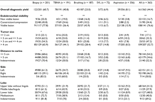
Table 3 presents the diagnostic yield of the different bron-choscopic sampling techniques. The sampling techniques performed were biopsy (201/363), TBNA (191/363), brushing (187/363), SVL (72/363), and aspiration of fluid from the entire procedure (356/363). Biopsy consist-ently gave the highest diagnostic yield with the possible exception in the case of lesions smaller than 2 cm. In uni-variate analyses endobronchial visibility, tumor size, and distance from carina were predictors of a higher diagnos-tic yield (Table 3). The overall sensitivity for cancer increased from 16.7% in non-visible lesions, to 34.4% for compression, constriction or submucosal disease, and fur-ther to 76.6% in visible lesions (χ2: p < 0.001).
In non visible lesions the diagnostic yield using a C-arm fluoroscope was 17/48 (35.4%) compared to 4/83 (4.8%)
Table 2: The diagnostic method and final diagnosis of 363 cases with malignant lung disease.
n %
Final diagnostic method
First bronchoscopy* 163 44.9
Repeated bronchoscopy 28 7.7
Transthoracic sampling 105 28.9
Mediastinoscopy 2 0.6
Operation, autopsy and open lung biopsy 11 3.0 Sampling from other organs than the lung 15 4.1 Clinical diagnosis of cancer 17 4.7 Malignant diagnosis obtained before bronchoscopy 22 6.1 Pathology
Small cell lung cancer 53 14.6
Adenocarcinoma 100 27.5
Squamous cell carcinoma 66 18.2
Large cell carcinoma 10 2.8
Non classifiable non small cell lung cancer. 82 22.6
Metastasis to the lung 21 5.8
Other cancer in the lung# 14 3.9
Only clinical diagnosis 17 4.7
* 2 patients diagnosed with uncertain pathology.
#Other cancer in the lung: Carcinoid tumor:6, Lymphoma:5, Mesothelioma:2,
Neuroendocrine tumor:1.
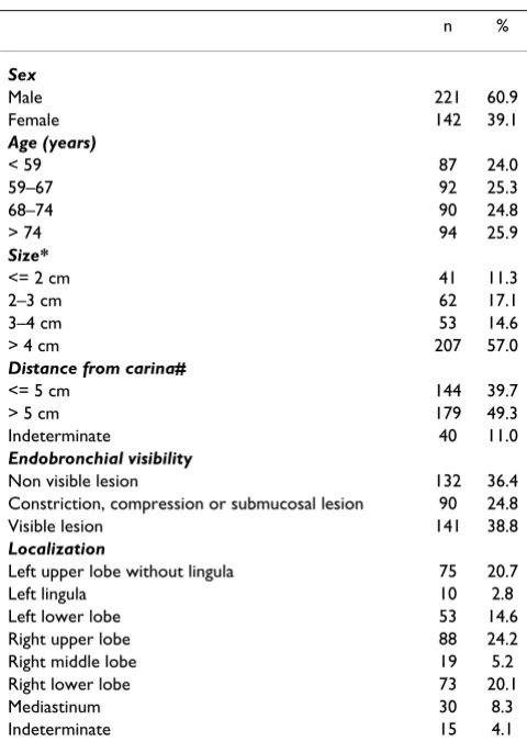
Table 1: Baseline characteristics of 363 cases
n %
Sex
Male 221 60.9
Female 142 39.1
Age (years)
< 59 87 24.0
59–67 92 25.3
68–74 90 24.8
> 74 94 25.9
Size*
<= 2 cm 41 11.3
2–3 cm 62 17.1
3–4 cm 53 14.6
> 4 cm 207 57.0
Distance from carina#
<= 5 cm 144 39.7
> 5 cm 179 49.3
Indeterminate 40 11.0
Endobronchial visibility
Non visible lesion 132 36.4
Constriction, compression or submucosal lesion 90 24.8
Visible lesion 141 38.8
Localization
Left upper lobe without lingula 75 20.7
Left lingula 10 2.8
Left lower lobe 53 14.6
Right upper lobe 88 24.2
Right middle lobe 19 5.2
Right lower lobe 73 20.1
Mediastinum 30 8.3
Indeterminate 15 4.1
*Size was measured on axial Ct thorax, only chest radiograph available in 5 cases.
without a C-arm fluoroscope (χ2: p < 0.001), data not
shown in Table 3.
Table 4 presents predictors of diagnostic yield for each sampling technique in a multivariate analysis after adjust-ment for age, sex, lobe, endobronchial visibility, size and distance from carina. The adjusted odds ratio (OR) for a
positive diagnostic result, was 2.6 (1.3–5.2) for constric-tion, compression or suspected submucosal changes and 17.0 (8.5–34.0) for visible lesions, compared to non visi-ble lesions. Endobronchial visibility was a significant pre-dictor for a higher diagnostic yield in all sampling techniques. Larger tumor size predicted an overall higher diagnostic yield, but was statistically significant only for
Table 4: Predictors of a higher diagnostic yield. Odds ratio (95%CI) in multivariate analysis.
Biopsy (n = 201) TBNA (n = 191) Brushing (n = 187) Aspiration (n = 356) All (n = 363)
Endobronchial visibility ** ** * ** **
Non-visible lesion 1 1 1 1 1
Compression/constriction 2.4 (0.8–7.0) 1.2 (0.3–4.4) 0.4 (0.1–1.5) 0.8 (0.1–4.6) 2.6 (1.3–5.2) Visible lesion 10.8 (3.8–30.7) 5.0 (1.4–17.5) 3.1 (1.1–8.3) 6.4 (1.9–20.9) 17.0 (8.5–34.0)
Tumor size * *
<= 2 cm 1 1 1 1 1
2–3 cm 7.9 (1.4–45.1) 0.8 (0.2–4.0) 1.1 (0.2–7.3) 2.2 (0.4–12.7) 2.5 (0.8–7.9) 3–4 cm 9.4 (1.7–52.2) 3.1 (0.6–15.3) 2.8 (0.5–17.0) 2.1 (0.3–13.6) 6.7 (2.1–21.8) > 4 cm 6.7 (1.5–30.6) 2.0 (0.5–7.6) 2.0 (0.4–10.9) 1.0 (0.2–5.4) 3.8 (1.3–10.7)
Odds ratio adjusted for age, sex, lobe and distance from carina. TBNA = Transbronchial needle aspiration. * Likelihood ratio: p < 0.05. ** Likelihood ratio: p < 0.001.
Table 3: Diagnostic yield of different sampling techniques
Biopsy (n = 201) TBNA (n = 191) Brushing (n = 187) SVL (n = 72) Aspiration (n = 356) All (n = 363)
Overall diagnostic yield 122/201 (60.7) 78/191 (40.8) 43/187 (23.0) 5/72 (6.9) 29/356 (8.1) 161/363 (44.4)
Endobronchial visibility ** ** ** ** **
Non visible lesion 9/36 (25.0) 4/21 (19.0) 13/68 (16.0) 3/46 (6.5) 5/130 (3.8) 22/132 (16.7) Constriction/compression 22/48 (45.8) 17/69 (24.6) 5/49 (10.2) 1/11 (9.1) 2/88 (2.3) 31/90 (34.4) Visible lesion 91/117 (77.8) 57/101 (56.4) 25/57 (43.9) 1/15 (6.7) 22/138 (15.9) 108/141 (76.6)
Tumor size * **
<= 2 cm 3/13 (23.1) 4/16 (25.0) 2/19 (10.5) 0/13 (0.0) 2/41 (4.9) 7/41 (17.1) > 2 cm and <= 3 cm 15/24 (62.5) 6/18 (25.0) 4/35 (11.4) 0/19 (0.0) 6/59 (10.2) 20/62 (32.3) > 3 cm and <= 4 cm 19/35 (54.3) 12/24 (50.0) 8/31 (25.8) 1/13 (7.7) 4/51 (7.8) 25/53 (47.2) > 4 cm 85/129 (65.9) 56/127 (44.1) 29/102 (28.4) 4/27 (14.8) 17/205 (8.3) 109/207 (52.7)
Distance to carina * * *
<= 5 cm 59/86 (68.6) 40/93 (43.0) 23/68 (33.8) 0/12 (0.0) 13/142 (9.2) 78/144 (54.2) > 5 cm 44/88 (50.0) 26/74 (35.1) 17/102 (16.7) 3/52 (5.8) 12/165 (6.8) 62/179 (34.6) Indeterminate 19/27 (70.4) 12/24 (50.0) 3/17 (17.6) 2/8 (25.0) 4/37 (10.8) 21/40 (52.5)
Side
Left 49/80 (61.3) 26/75 (34.7) 20/80 (25.0) 4/27 (14.8) 14/147 (9.5) 62/151 (41.1) Right 68/115 (59.1) 46/106 (43.4) 22/103 (21.4) 1/42 (2.4) 14/195 (7.2) 92/198 (46.5) Indeterminate 5/6 (83.3) 6/10 (60.0) 1/4 (25.0) 0/3 (0.0) 1/14 (7.1) 7/14 (50.0)
Lobe
Upper lobe without lingula 47/92 (51.1) 29/91 (31.9) 20/92 (21.7) 3/27 (11.1) 15/162 (9.3) 68/164 (41.5) Middle lobe/lingula 8/13 (61.5) 6/14 (42.9) 6/18 (33.3) 0/9 (0.0) 0/27 (0.0) 11/29 (37.9) Lower lobe 50/74 (67.6) 29/58 (50.0) 13/60 (21.7) 2/30 (6.7) 11/124 (8.9) 61/127 (48.0) Mediastinum 8/11 (72.7) 7/18 (38.9) 2/13 (15.4) 0/5 (0.0) 0/30 (0.0) 12/30 (40.0) Indeterminate 9/11 (81.8) 7/10 (70) 2/4 (50.0) 0/1 (0.0) 3/13 (23.1) 9/13 (69.2)
*p < 0.05. **p < 0.001
biopsies. In the multivariate analyses, distance from car-ina and localization of the lesion did not predict the diag-nostic yield.
Different pairs of sampling techniques were compared (Table 5). The sample size was not large enough to com-pare endobronchial visibility in three categories. There-fore non-visible lesions, compression, constriction, or suspected submucosal changes were combined to one cat-egory. In 86 patients with visible lesions, biopsy and TBNA were performed with a combined diagnostic yield of 83.7% (72/86, Table 5). In 38 of the 86 procedures, brushing was performed in addition to biopsy and TBNA, and in 85 of the procedures aspirations were also exam-ined. Cytological examination of the brushings and aspi-rations provided an increased diagnostic yield of one case (NS). Compared to the combination of biopsy and TBNA in these 86 procedures, seven cases would have been missed without TBNA (p = 0.02), and 22 cases would have been missed without biopsy (p < 0.001). The result in the group with non-visible lesions, compression, constriction or suspected submucosal changes was similar. In this group, biopsy and TBNA was performed in 48 patients with a combined diagnostic yield of 54.2% (Table 5). In these procedures additional 47 aspirations and 25 brush-ings were performed, which increased the diagnostic yield by only one case (NS). The diagnostic yield with a combi-nation of biopsy and TBNA was significantly higher than with biopsy or TBNA alone.
Discussion
Endobronchial visibility and tumor size were the predic-tors of diagnostic yield in bronchoscopies of patients with malignant disease in this study. The diagnostic yield was 16.7% in procedures with non-visible lesions, 34.4% in procedures with compression, constriction or suspected submucosal changes, and 76.6% in procedures with
endobronchial visible lesions. The combination of biopsy and TBNA had the highest diagnostic yield both in visible lesions and in the combined group of non-visible lesions, constriction, compression, or suspected submucosal changes. Biopsy and TBNA together was significant better than either technique alone.
There are some methodological issues to consider.
The main strength of this study is that it reflects the diag-nostic value of bronchoscopy in a regular clinical practice. For many operative procedures, an important factor is the skill of the operator. The current study did not have the power to examine diagnostic yield by operator. However, the large number of operators partly reflects the large number of patients seen with lung cancer, at our centre. The results of this study should be comparable to centres that include trainees and where each doctor performs approximately thirty bronchoscopies per year.
Patient selection bias is a problem in studies of diagnostic yield in bronchoscopy. Haukeland University Hospital is the only centre for diagnosing lung cancer in Hordaland County and some smaller surrounding municipalities. Thus all patients from surrounding area would be included. The follow up time to December 2005 with inclusion of clinical cancer were important factors to ensure inclusion of all cancers present among those exam-ined in 2003/04.
Another selection bias pertains to the choice of sampling methods. In our centre, the choice of sampling method is left to the judgement of the examiner. Thus, although in most bronchoscopies more than one sampling method was employed, for example biopsy and TBNA, rarely all five available techniques were used. In many procedures there is an urge to use the least amount of material and
Table 5: Diagnostic yield of different combinations of sampling techniques
The result of the first two sampling techniques in the
combination
The result of all sampling techniques performed
The result of the first sampling technique (1)
The result of the second sampling technique (2)
DY (%) DY (%) p DY (%) p DY (%) p
Non-visible lesion/compression or constriction:
Biopsy(1) and TBNA(2) 26/48 (54.2) 27/48 (56.3) NS 20/48 (41.7) 0.03 16/48 (33.3) 0.002 Biopsy(1) and Brushing(2) 18/42 (42.9) 20/42 (47.6) NS 14/42 (33.3) NS 9/42 (21.4) 0.004 TBNA(1) and Brushing(2) 16/51 (31.4) 21/51 (41.2) 0.06 11/51 (21.6) 0.06 10/51 (19.6) 0.03 Endobronchial visible lesion:
Biopsy(1) and TBNA(2) 72/86 (83.7) 73/86 (84.9) NS 65/86 (75.6) 0.02 50/86 (58.1) < 0.001 Biopsy(1) and Brushing(2) 37/46 (80.4) 39/46 (84.8) NS 34/46 (73.9) NS 23/46 (50.0) < 0.001 TBNA(1) and Brushing(2) 30/47 (63.8) 39/47 (83.0) 0.004 27/47 (57.4) NS 22/47 (46.8) 0.008
time necessary, in order to avoid complications and dis-comfort for the patients. Thus, an operator who feels to have obtained an adequate sample may terminate the pro-cedure without all techniques being employed. On the other hand, some sampling techniques may be dropped if complications arise, or the tumor seems impossible to reach. Clearly this is a weakness in the current study, when comparing the different sampling methods. However, the current study reflects how bronchoscopies are normally performed. In studies where all possible techniques are used, there may be a selection bias in that patients where the procedure is terminated prior to full sampling, for instance due to complications, are excluded from the study. This bias would likely inflate the results from stud-ies in which only patients having undergone all proce-dures are included.
Previous studies have categorized "compression, constric-tion or suspected submucosal changes" either with visible lesions or with non-visible lesions. Some studies have included these as non specific findings [12,30], thus increasing the diagnostic yield of peripheral lesions. Other studies have classified the findings as submucosal-peribronchial disease [31,32], decreasing the diagnostic yield of visible lesions. We have classified this group as a separate category. The finding that these lesions show a diagnostic yield intermediate between visible and non-visible supports our choice of classification. However, for the analyses in table 5, sample size dictated that non-visi-ble lesions be grouped with "compression, constriction or suspected submucosal changes". As TBNA and biopsy were more often used for the later category, and brushings more often for non-visible lesions, there could be a ten-dency for the results of TBNA to appear better in the com-bined category.
The overall diagnostic yield of 77% in visible lesions, is similar to previously reported results [7,21,25,30,31]. The overall diagnostic yield of 17% in non visible lesions is lower than in some studies [3,5-15], but higher than the yield reported in a Scottish multi-centre study (9%) [30].
The individual diagnostic yield of biopsy, brushing or TBNA from visible lesions were similar to previous reported studies [18,21,28,29]. SVL and aspiration had lower diagnostic yields for visible and non visible lesions, compared with other studies [21,25,33]. Studies with bronchoalveolar lavage have had a higher diagnostic yield, possibly due to higher fluid volume [22,23]. The low diagnostic yield might also be explained by the proce-dure of taking a small sample of 10–20 ml from the fluid aspirated and the lack of wedging the bronchoscope into the affected bronchus. Although there are relatively few prospective trials, they tend to report a higher diagnostic yield than retrospective trials, which may be due to the
benefits of planning [20,28]. Studies from highly special-ized centres or from selected procedures with all sampling techniques, have a higher diagnostic yield compared to the current study [5,8,13].
The C-arm fluoroscope increased the diagnostic yield from 5% to 35%. The rate of fluoroscopy in the current study was low, mainly because the C-arm fluoroscope was operated by radiographers who were not always available. The current study was not powered to examine differences in diagnostic yield between procedures with and without the C-arm fluoroscope. However, this study suggests a sig-nificant benefit from using the fluoroscope.
Several studies have examined predictors for a higher diagnostic yield, but only in univariate analysis [9-11,13-16,18,20]. Significant predictors found have been size [10,14-16,20], location [9-11], and endobronchial visibil-ity [18]. In the current study, endobronchial visibilvisibil-ity and tumor size prevailed as significant predictors when several potential predictors were examined together. Stratified on each sampling technique, the effect of size was significant only for biopsy. Both brushing and TBNA gave better results in larger lesions, but the results were not statisti-cally significant. This could indicate that sample size was too small to show the effect of size on these sampling methods. However, brushings might be sampled from a wider area than biopsy, and the size of the lesion might be less important for this method.
Conclusion
This study evaluated predictors of diagnostic yield of bronchoscopy reflecting clinical real life. Endobronchial visible lesion and larger tumor size predicted a higher diagnostic yield. The diagnostic yield was comparable with previous studies for visible lesions but was lower than in many of the previous studies for non-visible lesions.
BTS guideline recommended biopsy, brushing and wash-ing for visible lesions. This study has shown that biopsy and TBNA might be better. In the combined group of compression, constriction, suspected submucosal disease and non visible lesions, biopsy and TBNA was better than each sampling technique alone, however this study did not have sufficient power to determine whether brushing should be performed or not. These groups should be fur-ther investigated to find the optimal combination of sam-pling techniques and a cost effectiveness analysis could be performed. Washings were performed in almost all proce-dures and did not increase the diagnostic yield signifi-cantly in any groups.
Abbreviations
BTS = British Thoracic Society, CI = confidence interval, CT = computer tomography, OR = odds ratio, SD = stand-ard deviation, SNOMED = systemized nomenclature of medicine, SVL = small volume lavage, TBNA = transbron-chial needle aspiration.
Competing interests
The author(s) declare that they have no competing inter-ests.
Authors' contributions
TME and KR read the bronchoscopy reports and registered the information provided, viewed the chest radiographs and the CT scans, implemented the information from the pathological department, performed the statistical analy-sis, and wrote the manuscript.
JH and AHA participated in the design of the study, and reviewed the manuscript thoroughly.
FL provided all the information from the pathological department, was part of the planning of the study, and reviewed the manuscript thoroughly.
Acknowledgements
The study was founded by a one year grant for KR from Helse Vest and by a grant from Helse Sunnmore.
References
1. Cancer registry of Norway [http://www.kreftregisteret.no]. (Nov 2006).
2. Honeybourne D, Babb J, Bowie P, Brewin A, Fraise A, Garrard C, Harvey J, Lewis R, Neumann C, Wathen CG, Williams T: British
Thoracic Society guidelines on diagnostic flexible bronchos-copy. Thorax 2001, 56:I1-I21.
3. Schreiber G, McCrory DC: Performance characteristics of dif-ferent modalities for diagnosis of suspected lung cancer: Summary of published evidence. Chest 2003, 123:115S-128S. 4. Charig MJ, Phillips AJ: CT-guided cutting needle biopsy of lung
lesions – Safety and efficacy of an out-patient service. Clinical Radiology 2000, 55:964-969.
5. Gasparini S, Ferretti M, Secchi EB, Baldelli S, Zuccatosta L, Gusella P:
Integration of transbronchial and percutaneous approach in the diagnosis of peripheral pulmonary nodules or masses. Experience with 1,027 consecutive cases. Chest 1995,
108:131-137.
6. Hattori S, Matsuda M, NISHIHAR H, Horai T: Early diagnosis of small peripheral lung cancer – Cytologic diagnosis of very fresh cancer cells obtained by TV-brushing technique. Acta Cytologica 1971, 15:460-467.
7. Lam WK, So SY, Hsu C: Fiberoptic bronchoscopy in the diagno-sis of bronchial cancer: Comparison of washings, brushings and biopsies in central and peripheral tumors. Clinical Oncology
1983, 9:35-42.
8. Torrington KG, Kern JD: The utility of fiberoptic bronchoscopy in the evaluation of the solitary pulmonary nodule. Chest
1993, 104:1021-1024.
9. Wallace JM, Deutsch AL: Flexible fiberoptic bronchoscopy and percutaneous needle lung aspiration for evaluating the soli-tary pulmonary nodule. Chest 1982, 81:665-671.
10. Baaklini WA, Reinoso MA, Gorin AB, Sharafkanch A, Manian P: Diag-nostic yield of fiberoptic bronchoscopy in evaluating solitary pulmonary nodules. Chest 2000, 117:1049-1054.
11. Chechani V: Bronchoscopic diagnosis of solitary pulmonary nodules and lung masses in the absence of endobronchial abnormality. Chest 1996, 109:620-625.
12. Estarriol MH, Goday MR, Sanchez MV, Padro XB, Sot MTC, Quetglas FS: Bronchoscopic lung biopsy with fluoroscopy to study 164 localized pulmonary lesions. Archivos de Bronconeumologia 2004,
40:483-488.
13. Naidich DP, Sussman R, Kutcher WL, Aranda CP, Garay SM, Ettenger NA: Solitary pulmonary nodules. CT -bronchoscopic correla-tion. Chest 1988, 93:595-598.
14. Radke JR, Conway WA, Eyler WR, Kvale PA: Diagnostic accuracy in peripheral lung lesions. Factors predicting success with flexible bronchoscopy. Chest 1979, 76:176-179.
15. Reichenberger F, Weber J, Tamm M, Bolliger CT, Dalquen P, Perru-choud AP, Soler M: The value of transbronchial needle aspira-tion in the diagnosis of peripheral pulmonary lesions. Chest
1999, 116:704-708.
16. Stringfield JT, Markowitz DJ, Bentz RR, Welch MH, Weg JG: Effect of tumor size and location on diagnosis by fiberoptic bron-choscopy. Chest 1977, 72:474-476.
17. Aristizabal JF, Young KR, Nath H: Can chest CT decrease the use of preoperative bronchoscopy in the evaluation of suspected bronchogenic carcinoma? Chest 1998, 113:1244-1249. 18. Castella J, Buj J, Puzo C, Anton PA, Burgues C: Diagnosis and
stag-ing of bronchogenic carcinoma by transtracheal and trans-bronchial needle aspiration. Annals of Oncology 1995, 6(Suppl 3):s21-s24.
19. Pilotti S, Rilke F, Gribaudi G, Spinelli P: Cytologic diagnosis of pul-monary carcinoma on bronchoscopic brushing material. Acta Cytologica 1982, 26:655-660.
20. Bilaceroglu S, Kumcuoglu Z, Alper H, Osma E, Cagirici U, Gunel O, Bayol U, Celikten E, Perim K, Kose T: CT bronchus sign-guided bronchoscopic multiple diagnostic: Procedures in carcino-matous solitary pulmonary nodules and masses. Respiration
1998, 65:49-55.
21. Kvale PA, Bode FR, Kini S: Diagnostic accuracy in lung cancer; Comparison of techniques used in association with flexible fiberoptic bronchoscopy. Chest 1976, 69:752-757.
22. Debeljak A, Mermolja M, Sorli J, Zupancic M, Zorman M, Remskar J:
Bronchoalveolar lavage in the diagnosis of peripheral pri-mary and secondary malignant lung tumors. Respiration 1994,
61:226-230.
Publish with BioMed Central and every scientist can read your work free of charge "BioMed Central will be the most significant development for disseminating the results of biomedical researc h in our lifetime."
Sir Paul Nurse, Cancer Research UK
Your research papers will be:
available free of charge to the entire biomedical community
peer reviewed and published immediately upon acceptance
cited in PubMed and archived on PubMed Central
yours — you keep the copyright
Submit your manuscript here:
http://www.biomedcentral.com/info/publishing_adv.asp
BioMedcentral
24. Gay PC, Brutinel WM: Transbronchial needle aspiration in the practice of bronchoscopy. Mayo Clinic Proceedings 1989,
64:158-162.
25. Mak VHF, Johnston IDA, Hetzel MR, Grubb C: Value of washings and brushings at fiberoptic bronchoscopy in the diagnosis of lung cancer. Thorax 1990, 45:373-376.
26. Mori K, Yanase N, Kaneko M, Ono R, Ikeda S: Diagnosis of periph-eral lung cancer in cases of tumors 2 cm or less in size. Chest
1989, 95:304-308.
27. Pirozynski M: Bronchoalveolar lavage in the diagnosis of peripheral, primary lung cancer. Chest 1992, 102:372-374. 28. Popp W, Rauscher H, Ritschka L, Redtenbacher S, Zwick H, Dutz W:
Diagnostic sensitivity of different techniques in the diagnosis of lung tumors with flexible fiberoptic bronchoscope. Com-parison of brush biopsy, imprint cytology of forceps biopsy, and histology of forceps biopsy. Cancer 1991, 67:72-75. 29. Zavala DC: Diagnostic fiberoptic bronchoscopy: Techniques
and results of biopsy in 600 patients. Chest 1975, 68:12-19. 30. Mclean AN, Semple PD, Franklin DH, Petrie G, Millar EA, Douglas JG:
The Scottish multi-centre prospective study of bronchos-copy for bronchial carcinoma and suggested audit standards.
Respiratory Medicine 1998, 92:1110-1115.
31. Kacar N, Tuksavul F, Edipoglu O, Ermete S, Guclu SZ: Effectiveness of transbronchial needle aspiration in the diagnosis of exo-phytic endobronchial lesions and submucosal/peribronchial diseases of the lung. Lung Cancer 2005, 50:221-226.
32. Govert JA, Kopita JM, Matchar D, Kussin PS, Samuelson WM: Cost-effectiveness of collecting routine cytologic specimens dur-ing fiberoptic bronchoscopy for endoscopically visible lung tumor. Chest 1996, 109:451-456.
33. van der Drift MA, van der Wilt GJ, Thunnissen FBJM, Janssen JP: A prospective study of the timing and cost-effectiveness of bronchial washing during bronchoscopy for pulmonary malignant tumors. Chest 2005, 128:394-400.
34. Karahalli E, Yilmaz A, Turker H, Ozvaran K: Usefulness of various diagnostic techniques during fiberoptic bronchoscopy for endoscopically visible lung cancer: Should cytologic exami-nations be performed routinely? Respiration 2001, 68:611-614.
Pre-publication history
The pre-publication history for this paper can be accessed here: