R E S E A R C H
Open Access
TGR5 expression in normal kidney and
renal neoplasms
Chaohui Lisa Zhao
1, Ali Amin
1, Yiang Hui
1, Dongfang Yang
1and Weibiao Cao
1,2*Abstract
Background:The G protein-coupled bile acid receptor (TGR5) is a cell surface receptor which induces the production of intracellular cAMP and promotes epithelial-mesenchymal transition in gastric cancer cell lines. TGR5 is found in a wide variety of tissues including the kidney. However, the patterns of TGR5 expression have not been well characterized in physiologic kidney or renal neoplasms. We explore the expression of TGR5 in benign renal tissue and renal neoplasms and assess its utility as a diagnostic marker.
Methods:Sixty-one renal cortical neoplasms from 2000 to 2014 were retrieved. TGR5 protein expression was examined by immunohistochemistry. TGR5 mRNA was also measured by real-time PCR.
Results:In normal renal tissue, TGR5 was strongly positive in collecting ducts, distal convoluted tubules and thin loop of Henle. Proximal convoluted tubules showed absent or focal weak staining. In clear cell renal cell carcinomas (RCCs), 25 of 27 cases (92%) were negative for TGR5 (p< 0.001). TGR5 mRNA was also significantly decreased in clear cell RCCs, suggesting that decreased TGR5 protein expression may be attributable to the downregulation of TGR5 mRNA in these tumors. All 11 papillary RCCs expressed TGR5 with 45% (5/11) exhibiting moderate to strong staining. All chromophobe RCCs and oncocytomas were positive for TGR5 with weak to moderate staining. TGR5 mRNA expression in these tumors was similar to normal kidney. All urothelial carcinomas of the renal pelvis strongly expressed TGR5 including a poorly differentiated urothelial carcinoma with sarcomatoid features. Conclusion:TGR5 is strongly expressed in collecting ducts, distal convoluted tubules and thin loop of Henle. TGR5 protein and mRNA expression were notably decreased in clear cell RCCs and may be helpful in differentiating these tumors from other RCCs.
Keywords:G protein-coupled bile acid receptor, TGR5, Renal cell carcinoma, Urothelial cell carcinoma, Oncocytoma
Background
Renal malignancies are the 7th most common cancer in men in the US with approximately 14,000 attributable deaths annually [1]. Malignant renal cell carcinomas (RCCs) include clear cell RCCs, papillary RCCs (type 1 and type 2), chromophobe RCCs, collecting duct carcinomas, clear cell papillary RCCs, and others [2]. Benign renal epithelial neoplasms include papillary adenomas, oncocytomas and metanephric adenomas [2]. Overall, RCCs constitute 80–85% of primary renal neoplasms. Urothelial cell car-cinoma of the renal pelvis is the second most common
malignant neoplasm in the kidney [1]. Morphologic similarities among these tumors may present difficulties in accurately classifying these lesions which follow dif-ferent clinical outcomes.
The G protein-coupled bile acid receptor (TGR5) is a cell-surface receptor mediating bile acid effects [3]. Both primary and secondary bile acids bind to TGR5 to in-duce cyclic AMP (cAMP) production [3]. In turn, TGR5 has been shown to activate a MAP kinase signaling pathway [4] and to be linked to an increase in the intra-cellular ATP/ADP ratio [5]. It plays an important role in energy homeostasis, bile acid-regulated lipid metabol-ism, and glucose metabolism [6,7] . TGR5 is also impli-cated in the mitochondrial response to inflammation and the development of fibrosis in the kidney [8, 9]. TGR5 expression is present in a variety of tissues including * Correspondence:Weibiao_Cao@brown.edu
1
Department of Pathology, Rhode Island Hospital and the Warren Alpert Medical School of Brown University, 593 Eddy Street, APC 12, Providence, RI 02903, USA
2Department of Medicine, Rhode Island Hospital and the Warren Alpert
Medical School of Brown University, Providence, RI 02903, USA
kidney [9–11]. Some studies have reported strong TGR5 expression in renal tubular cells [10, 11] as well as the mesangial cells of rat kidney [10]. However, its distribution of expression has not been well characterized. Renal tu-mors have also not been interrogated for TGR5. In this study, we showed that the TGR5 receptor was strongly expressed in distal convoluted tubules, thin loop of Henle and collecting ducts and that TGR5 lost expres-sion in clear cell RCCs.
Methods Case selection
With Institutional Review Board approval at Rhode Is-land Hospital, sixty-one renal cortical neoplasms were identified from the archives of the Department of Path-ology between 2000 and 2014. These included 27 clear cell RCCs and 11 papillary RCCs, 8 of which were type 1 and 3 of which were type 2. The remainder of the cases consisted of 8 papillary urothelial carcinomas in-cluding one poorly differentiated case involving the cor-tex, 6 chromophobe RCCs, 5 oncocytomas, 2 clear cell papillary RCCs, 1 metanephric adenoma, and 1 poorly differentiated RCC with sarcomatoid features. Formalin-fixed paraffin-embedded tissue sections and blocks were retrieved. The corresponding hematoxylin-eosin slides were reviewed for confirmation of diagnosis and adequacy of material by a specialist in genitourinary pathology (AA). Multiple representative sections of each case were examined. Corresponding normal kidney control sections separate from the tumors were also reviewed for all cases. Snap-frozen tissue from normal kidney, clear cell RCCs, papillary RCCs, chromophobe RCC and oncocytomas was obtained from the Rhode Island Hospital tissue bank.
Tissue microarray construction
Paraffin blocks containing areas of carcinomas were identified on the hematoxylin and eosin-stained sections. Areas of the tumor were identified and marked on the source block. The source block was cored using a 1-mm needle and the tissue was transferred to a recipient“master block” using a Beecher Tissue Microarrayer (Beecher In-struments, Silver Spring, MD). Three to six cores of each tumor were arrayed per specimen. Additionally, a core of normal kidney tissue was also sampled when present [12].
Immunohistochemistry
Immunohistochemistry for TGR5 was performed on 4-μm paraffin sections of the tissue microarray or whole tissue sections. Slides were stained with TGR5 antibody (rabbit anti-human, polyclonal, 1:1000; Sigma-Aldrich, St. Louis, MO) using the DAKO Envision + Dual Link System and the DAKO Liquid 3,3′-Diaminobenzidine Substrate Chromogen System (DAKO North America, Carpinteria, CA). Bile ducts from liver tissue were used
as positive controls. Negative controls were included by replacement of the primary antibody with non-reacting antibodies derived from the same species. The specifi-city of TGR5 antibody has previously been confirmed by Western blot analysis in our lab [12].
Tissue microarray was employed to study normal kid-ney and 44 cases of renal neoplasms, including all clear cell RCCs, 1 metanephric adenoma, 8 papillary urothelial carcinomas, 4 type 1 papillary RCCs, 3 oncocytomas, and 1 chromophobe RCC.
Immunohistochemistry was also performed on whole tissue sections for 17 cases including 4 papillary RCC (4 type 1 papillary RCC and 3 type 2 papillary RCC), 5 chromophobe RCC, 2 oncocytomas, 2 clear cell papillary RCC, and 1 poorly differentiated RCC with some sarco-matoid features.
Immunohistochemistry assessment
Immunohistochemical results were evaluated semi-quantitatively within neoplastic tissue. Cells displaying strong intensity staining for TGR5 were scored as 3+, moderate staining as 2+, and weak staining as 1+. The extent of staining was scored as follows: 0 (< 5%), 1+ (5–25%), 2+ (26–50%), 3+ (51–100%). A combined score of intensity and extent was calculated and assigned as fol-lows: weak staining 1–2, moderate staining 3–4, and strong staining 5–6. At least three cores were scored per case. Analysis of three cores per case in this fashion has been shown to be comparable to whole tissue sections [13]. All sections were independently scored by two pa-thologists (WC and CZ) blinded to clinicopathological features and outcome.
Quantitative real time PCR
TGR5 mRNA was measured using real-time PCR analysis as we have previously described [14]. The primers used were as follows: TGR5 sense (5′- CTGGCCCTGGCAAG CCTCAT-3’), TGR5 antisense (5′- CTGCCATGTAGCG
CTCCCCGT-3’), 18S sense (5’-CGGACAGGATTGACA
City, CA). The transcript level of each specific gene was normalized to 18S amplification.
Statistical analysis
A Chi-square test was utilized to compare groups where appropriate. Data were expressed as mean ± SEM. Statis-tical differences between two groups were determined by Student’s t-test. Differences among multiple groups were tested using analysis of variance (ANOVA) and checked for significance using Fisher’s protected least significant difference test. Ap-value of 0.05 or less was considered statistically significant.
Results
TGR5 protein expression
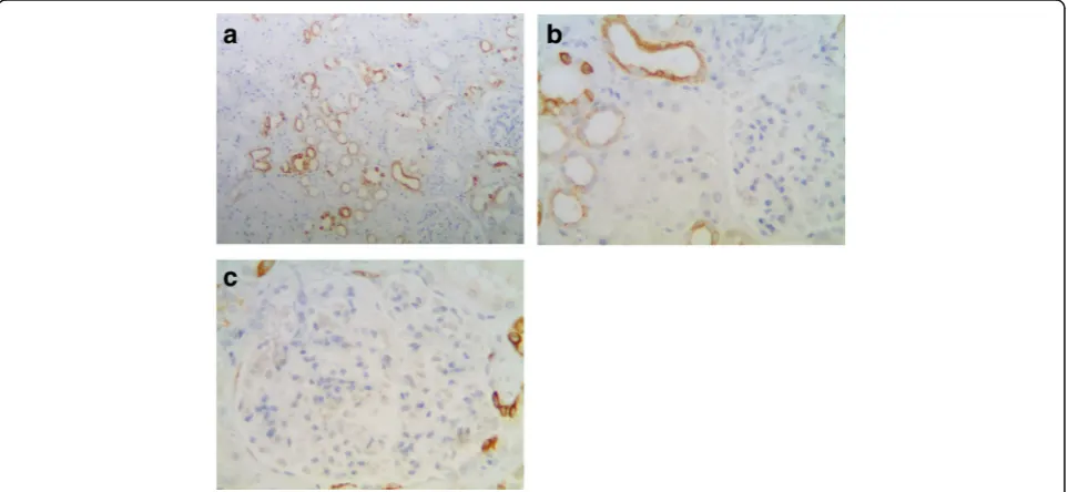
In normal kidney, the proximal tubular cells are more columnar and eosinophilic than cells of the distal tubules on H&E slides. The ascending limb has no brush border and the cells are more cuboidal than adjacent proximal tubular cells [15]. According to this criterion, TGR5 was strongly expressed in the distal convoluted tubules, thin loop of Henle, and collecting ducts (Fig. 1a). In contrast, the proximal convoluted tubular cells showed absent or focal weak staining (Fig. 1b). TGR5 expression was not observed in the glomerular tuft, but focal immunoreactiv-ity was identified in parietal epithelial cells (Fig.1c).
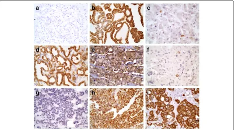
The expression of TGR5 in renal cell neoplasms is summarized in Table1. Notably, among 25 cases of clear cell RCCs, 92% (25/27) of cases were negative for TGR5 staining (p< 0.001, Fig. 2a). The remaining 2 cases of
clear cell RCCs (2/27) exhibited TGR5 focal staining in areas with papillary features. One of these two TGR5-positive cases also exhibited sarcomatoid differentiation. Both cases of clear cell papillary RCCs (2/2) displayed strong TGR5 staining (Fig.2d).
All papillary RCCs (11/11) demonstrated TGR5 staining. The TGR5 staining in Type 1 papillary RCCs (8 cases) were variable from weak to strong staining (Fig.2b). All 3 cases of type 2 papillary RCCs showed only focal weak staining (Fig.2c). There were no statistically significant differences between type 1 and type 2 papillary RCCs (p= 0.152).
All chromophobe RCCs (6/6) exhibited weak to mod-erate TGR5 staining (Fig.2e). The oncocytomas all (5/5) showed weak TGR5 expression (Fig.2f). No statistically significant differences between chromophobe RCCs and oncocytomas (p= 0.082) were identified. The metaneph-ric adenoma (0/1; Fig.2g) and poorly differentiated RCC with sarcomatoid features (0/1) were negative for TGR5 staining.
Urothelial carcinomas all uniformly (8/8) expressed TGR5 (Fig. 2h) including the case of poorly differenti-ated sarcomatoid case (Fig.2i).
TGR5 mRNA expression
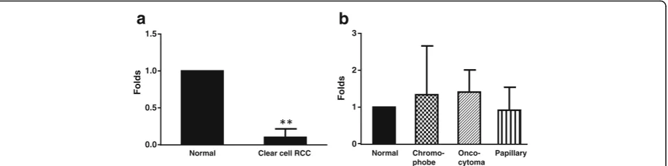
TGR5 mRNA was significantly diminished in clear cell RCC (N= 5, p< 0.001, fig. 3a) in comparison to normal non-neoplastic renal tissue. TGR5 mRNA expression in papillary RCCs, chromophobe RCCs, and oncocytomas did not significantly differ from normal renal tissue (fig.3b).
Fig. 1TGR5 staining in normal renal cortex.aTGR5 is strongly expressed in the distal convoluted tubule, thin loop of Henle, and collecting duct
(100X).bAbsent or only minimal focal weak staining is observed in the proximal convoluted tubular cells (arrow, 400X).cIn the glomeruli, TGR5
Discussion
The distal convoluted tubules play a key role in regulat-ing extracellular fluid volume and electrolyte homeosta-sis. Distal tubular cells are rich in mitochondria and biochemical studies reveal that distal tubular cells have a higher level of Na+/K+-ATPase activity than any other tubular segment [16–18]. Recently, TGR5 has been identified as a cell surface receptor, which can induce the production of intracellular cAMP and activation of a MAP kinase signaling pathway [4]. TGR5 has also been linked to an increase in the intracellular ATP/ADP ratio [5]. TGR5 agonist may be useful in the treatment of kidney injury and
various metabolic diseases including diabetes and obesity [9]. Our data reveal that TGR5 is strongly positive in normal distal convoluted tubules, the thin loop of Henle, and collecting ducts. In comparing the glomeruli and proximal tubular cells, increased expression in the distal tubular cells corresponds to elevated Na+/K+-ATPase activity in the distal tubular cells. This might account for high TGR5 expression in the distal tubular cells in the kidney.
Immunohistochemical staining demonstrated no expres-sion of TGR5 in the glomeruli with the exception of some parietal cells within Bowman’s capsule. This is consistent
Table 1The expression of TGR5 in RCCs
Case number (N) Negative Weak Moderate Strong
Clear cell RCC 27 25 (92%) 0 1 (4%) 1 (4%)
Papillary RCC 11 0 6 (55%) 4 (36%) 1 (9%)
Type 1 8 0 3 (37%) 4 (50%) 1 (13%)
Type 2 3 0 3 (100%) 0 0
Papillary urothelial carcinoma 8 0 1 (12%) 0 7 (88%)
Clear cell papillary RCCs 2 0 0 0 2 (100%)
Chromophobe RCC 6 0 2 (33%) 4 (67%) 0
Oncocytoma 5 0 5 (100%) 0 0
Fig. 2TGR5 staining -aClear cell RCC: negative for TGR5 (400X).bType 1 papillary RCC: strongly positive for TGR5 (400X).cType 2 papillary RCC:
weakly positive with focal cells expressing TGR5 (400X).dClear cell papillary RCC: strongly positive for TGR5 (400X).eChromophobe RCC: moderate
expression of TGR5 (400X).fOncocytoma: weakly positive with focal cells expressing TGR5 (400X).gMetanephric adenoma: negative for TGR5 (400X).
hUrothelial carcinoma of the renal pelvis: strongly positive for TGR5 (400X).iPoorly differentiated urothelial carcinoma with sarcomatoid features:
with prior studies in the literature [19]. Xiong et al. previ-ously described TGR5 expression in rat mesangial cells [10]. This discrepancy may be due to one of three scenar-ios. First, there may be intrinsic differences in TGR5 ex-pression between species. Second, the primary polyclonal antibody employed in the prior study was obtained from a different manufacturer which may have resulted in dis-crepant immunoreactivity. Third, TGR5 may be expressed in low levels in these cells which are undetectable by immunohistochemistry.
Strong expression of TGR5 in distal tubular cells and collecting ducts lends credence to the theory that some renal tumors such as papillary RCCs, chromophobe RCCs, and oncocytomas likely derive from distal convoluted tubules or collecting ducts as suggested by some studies [20, 21]. Electron microscopic and immunohistochemi-cal data implicate the interimmunohistochemi-calated cells of the collecting duct as the cell of origin for chromophobe RCCs [22] and oncocytomas [23]. These tumors variably express TGR5. In contrast, clear cell RCCs are suggested to ori-ginate from proximal tubular cells [20] which are nega-tive or focal very weakly posinega-tive for TGR5. TGR5 mRNA is significantly decreased in clear cell RCC corresponding to the diminished TGR5 protein expression, suggesting that decreased TGR5 protein expression may be attributable to the downregulation of TGR5 mRNA in these tumors.
Clear cell RCCs comprise 70% to 80% of all RCCs [24]. Clear cell papillary RCCs constitute a diagnostic challenge given their morphologic similarities to clear cell RCCs [25]. Carbonic anhydrase 9 (CA-IX) is expressed in both clear cell and clear cell papillary RCCs [25]. CA-IX is expressed in approximately 85% of clear cell RCCs [25]. Since the ex-pression TGR5 is negative in up to 92% of clear cell RCCs, this marker may be useful in the diagnostic work-up of these tumors. Additional studies directly comparing TGR5 and CA-IX are warranted to better understand the utility of TGR5 in this context. Notably, TGR5 was strongly
expressed in clear cell papillary RCCs although only 2 cases were included in our cohort.
Papillary RCCs are the second most common histo-logic subtype and comprise 7–15% of all RCCs [25]. Immunohistochemical markers such as CK7, alpha me-thyl acyl coenzyme A racemase (AMACR), and c-kit (CD117), have been leveraged to differentiate these tu-mors from clear cell RCCs. CK7 is expressed in 80– 87% papillary RCCs and is variably positive in 0–37% of clear cell RCCs [24]. AMACR is positive in 80–100% of papillary RCCs and variably positive in 4–68% of clear cell RCCs [24]. C-kit (CD117) expression is variable in clear cell RCCs (0–5%), papillary RCCs (0–13%), chro-mophobe RCCs (82–100%), and oncocytomas (58–100%) [24]. We found that TGR5 is positive in all papillary RCCs including both type 1 and type 2. Oncocytomas and chro-mophobe RCCs also all expressed TGR5. However, only 8% of clear cell RCCs were positive. This supports the potential usefulness of the inclusion of TGR5 into the diagnostic work-up of these tumors.
Urothelial carcinomas of the renal pelvis including sar-comatoid type of poorly differentiated urothelial carcin-omas strongly expressed TGR5. Although only one case poorly differentiated RCC with sarcomatoid features was included in our study, this case was negative for TGR5. This may suggest that TGR5 could be used to differen-tiate poorly differendifferen-tiated urothelial carcinomas from poorly differentiated RCCs. Further studies with a lar-ger cohort are necessary to establish the utility of TGR5 for this application.
Our cases suggest that TGR5 would have overall lim-ited diagnostic utility in differentiating among non-clear cell RCCs. All papillary RCCs showed TGR5 positive staining. No significant differences in TGR5 expression between type 1 and type 2 papillary RCCs were detected. Similarly, TGR5 staining may not be useful to differenti-ate chromophobe RCCs from oncocytomas.
Normal Chromo-phobe
Onco-cytoma
Papillary
0 1 2 3
Folds
Normal Clear cell RCC
0.0 0.5 1.0 1.5
Folds
a
b
Fig. 3TGR5 mRNA expression. A. TGR5 mRNA was significantly decreased in clear cell RCC (N= 5, un-paired t test), when compared with normal renal tissue. The data suggest that decreased TGR5 protein expression may be due to downregulation of TGR5 mRNA in clear cell RCC. B. TGR5 mRNA
Conclusion
In summary, we employed immunohistochemistry to es-tablish that TGR5 expression is robust in the distal con-voluted tubules, thin loop of Henle, and collecting ducts. Among renal neoplasms, papillary RCCs, clear cell papil-lary RCCs, chromophobe RCCs and oncocytomas show various degrees of TGR5 expression. UCCs of the renal pelvis also strongly express TGR5. We demonstrate that the majority of clear cell RCCs, particularly cases without papillary features, are negative attributable to their cur-tailed expression of TGR5 mRNA. Overall, our data sug-gest that TGR5 staining may contribute to the diagnostic workup in distinguishing clear cell RCC from other renal tumors.
Abbreviations
AMACR:Alpha methyl acyl coenzyme A racemase; CA-IX: Carbonic Anhydrase 9; RCC: Renal cell carcinoma; TGR5: G protein-coupled bile acid receptor
Acknowledgements
None.
Funding
None.
Availability of data and materials
Please contact the corresponding author for data requests.
Authors’contributions
CZ performing the histological examination, analyzing data and writing the manuscript. AA performing the histological examination. YH writing the manuscript. DY performing immunohistochemical staining. WC performing real-time PCR and histological examination, analyzing data and writing the manuscript. All authors have read and approved the final manuscript.
Ethics approval and consent to participate
This study was approved by the Institutional Review Board at the Rhode
Island Hospital (the IRB committee number 0258–02). Additional informed
consent was not required by the IRB for this retrospective study since our hospital standard consent form for surgical or other procedure specifically states that" I understand that the Hospital may use for teaching or scientific purposes, or may otherwise dispose of, tissues, fluids or organs removed during the procedure".
Consent for publication
Not applicable
Competing interests
The authors declare that they have no competing interests.
Publisher’s Note
Springer Nature remains neutral with regard to jurisdictional claims in published maps and institutional affiliations.
Received: 3 October 2017 Accepted: 20 March 2018
References
1. Siegel RL, Miller KD, Jemal A. Cancer statistics, 2015. CA Cancer J Clin. 2015;65:5–29.
2. Kuroda N, Tanaka A. Recent classification of renal epithelial tumors. Med
Mol Morphol. 2014;47:68–75.
3. Kawamata Y, Fujii R, Hosoya M, Harada M, Yoshida H, Miwa M, Fukusumi S,
Habata Y, Itoh T, Shintani Y, et al. A G protein-coupled receptor responsive
to bile acids. J Biol Chem. 2003;278:9435–40.
4. Masyuk AI, Huang BQ, Radtke BN, Gajdos GB, Splinter PL, Masyuk TV,
Gradilone SA, LaRusso NF. Ciliary subcellular localization of TGR5 determines
the cholangiocyte functional response to bile acid signaling. Am J Physiol
Gastrointest Liver Physiol. 2013;304:G1013–24.
5. Thomas C, Gioiello A, Noriega L, Strehle A, Oury J, Rizzo G, Macchiarulo A,
Yamamoto H, Mataki C, Pruzanski M, et al. TGR5-mediated bile acid sensing
controls glucose homeostasis. Cell Metab. 2009;10:167–77.
6. Katsuma S, Hirasawa A, Tsujimoto G. Bile acids promote glucagon-like
peptide-1 secretion through TGR5 in a murine enteroendocrine cell line
STC-1. Biochem Biophys Res Commun. 2005;329:386–90.
7. Watanabe M, Houten SM, Mataki C, Christoffolete MA, Kim BW, Sato H,
Messaddeq N, Harney JW, Ezaki O, Kodama T, et al. Bile acids induce energy expenditure by promoting intracellular thyroid hormone activation. Nature.
2006;439:484–9.
8. Carino A, Graziosi L, D'Amore C, Cipriani S, Marchiano S, Marino E, Zampella A,
Rende M, Mosci P, Distrutti E, et al. The bile acid receptor GPBAR1 (TGR5) is expressed in human gastric cancers and promotes epithelial-mesenchymal
transition in gastric cancer cell lines. Oncotarget. 2016;7(38):61021–35.
9. Herman-Edelstein M, Weinstein T, Levi M. Bile acid receptors and the kidney.
Curr Opin Nephrol Hypertens. 2018;27:56–62.
10. Xiong F, Li X, Yang Z, Wang Y, Gong W, Huang J, Chen C, Liu P, Huang H.
TGR5 suppresses high glucose-induced upregulation of fibronectin and transforming growth factor-beta1 in rat glomerular mesangial cells by
inhibiting RhoA/ROCK signaling. Endocrine. 2016;54:657–70.
11. Yang Z, Xiong F, Wang Y, Gong W, Huang J, Chen C, Liu P, Huang H. TGR5
activation suppressed S1P/S1P2 signaling and resisted high glucose-induced
fibrosis in glomerular mesangial cells. Pharmacol Res. 2016;111:226–36.
12. Cao W, Tian W, Hong J, Li D, Tavares R, Noble L, Moss SF, Resnick MB.
Expression of bile acid receptor TGR5 in gastric adenocarcinoma. Am J
Physiol Gastrointest Liver Physiol. 2013;304:G322–7.
13. Jourdan F, Sebbagh N, Comperat E, Mourra N, Flahault A, Olschwang S,
Duval A, Hamelin R, Flejou JF. Tissue microarray technology: validation in colorectal carcinoma and analysis of p53, hMLH1, and hMSH2
immunohistochemical expression. Virchows Arch. 2003;443:115–21.
14. Li D, Cao W. Bile acid receptor TGR5, NADPH oxidase NOX5-S and CREB
mediate bile acid-induced DNA damage in Barrett's esophageal adenocarcinoma cells. Sci Rep. 2016;6:31538.
15. Valtin H. Structural and functional heterogeneity of mammalian nephrons.
Am J Phys. 1977;233:F491–501.
16. Snarski J, Snarski A, Bachelet M, Bader C, Ulmann A. (Na-K)ATPase activity
along the nephrons in normal and adrenalectomized rats measured by
quantitative cytochemistry. Cell Biochem Funct. 1985;3:127–32.
17. Garg LC, Narang N. Ouabain-insensitive K-adenosine triphosphatase in distal
nephron segments of the rabbit. J Clin Invest. 1988;81:1204–8.
18. McCormick JA, Ellison DH. Distal convoluted tubule. Compr Physiol. 2015;5:45–98.
19. Wang XX, Edelstein MH, Gafter U, Qiu L, Luo Y, Dobrinskikh E, Lucia S,
Adorini L, D'Agati VD, Levi J, et al. G protein-coupled bile acid receptor TGR5 activation inhibits kidney disease in obesity and diabetes. J Am Soc Nephrol.
2016;27:1362–78.
20. Axelson H, Johansson ME. Renal stem cells and their implications for kidney
cancer. Semin Cancer Biol. 2013;23:56–61.
21. Fleming S. Distal nephron neoplasms. Semin Diagn Pathol. 2015;32:114–23.
22. Nagashima Y. Chromophobe renal cell carcinoma: clinical, pathological and
molecular biological aspects. Pathol Int. 2000;50:872–8.
23. Valladares Ayerbes M, Aparicio Gallego G, Diaz Prado S, Jimenez Fonseca P,
Garcia Campelo R, Anton Aparicio LM. Origin of renal cell carcinomas. Clin
Transl Oncol. 2008;10:697–712.
24. Truong LD, Shen SS. Immunohistochemical diagnosis of renal neoplasms.
Arch Pathol Lab Med. 2011;135:92–109.
25. Alshenawy HA. Immunohistochemical panel for differentiating renal cell