REVIEW
Stem cells as a potential therapy for diabetes
mellitus: a call-to-action in Latin America
Mairim Alexandra Solis
1*, Ilais Moreno Velásquez
1, Ricardo Correa
2,3and Lynn L. H. Huang
4,5,6Abstract
Latin America is a fast-growing region that currently faces unique challenges in the treatment of all forms of diabetes mellitus. The burden of this disease will be even greater in the coming years due, in part, to the large proportion of young adults living in urban areas and engaging in unhealthy lifestyles. Unfortunately, the national health systems in Latin-American countries are unprepared and urgently need to reorganize their health care services to achieve dia-betic therapeutic goals. Stem cell research is attracting increasing attention as a promising and fast-growing field in Latin America. As future healthcare systems will include the development of regenerative medicine through stem cell research, Latin America is urged to issue a call-to-action on stem cell research. Increased efforts are required in studies focused on stem cells for the treatment of diabetes. In this review, we aim to inform physicians, researchers, patients and funding sources about the advances in stem cell research for possible future applications in diabetes mellitus. Emerging studies are demonstrating the potential of stem cells for β cell differentiation and pancreatic regeneration. The major economic burden implicated in patients with diabetes complications suggests that stem cell research may relieve diabetic complications. Closer attention should be paid to stem cell research in the future as an alternative treatment for diabetes mellitus.
Keywords: Diabetes mellitus, Stem cells, Latin America, Stem cell differentiation, Regenerative medicine
© The Author(s) 2019. This article is distributed under the terms of the Creative Commons Attribution 4.0 International License (http://creat iveco mmons .org/licen ses/by/4.0/), which permits unrestricted use, distribution, and reproduction in any medium, provided you give appropriate credit to the original author(s) and the source, provide a link to the Creative Commons license, and indicate if changes were made. The Creative Commons Public Domain Dedication waiver (http://creat iveco mmons .org/ publi cdoma in/zero/1.0/) applies to the data made available in this article, unless otherwise stated.
Background
Diabetes mellitus (DM), in all its forms, is a metabolic disorder that occurs due to deficient production of insulin by the pancreas. Physiological control of blood glucose levels can be restored in a number of ways: exog-enous administration of insulin, medications that stimu-late insulin, medications that decrease insulin resistance and/or replace the β cell mass (the producers of insu-lin) [1]. Pancreatic regeneration of the lost functional β cell mass is an attractive strategy for recovery from the disease. Current approaches utilized for pancreatic replacement of damaged β cells include cadaveric islet transplantation, induction of endogenous regeneration and administration of stem cell-derived β cells [2]. Trans-plantation of pancreatic islets has proven to be success-ful for functional replenishment of damaged islets [3,
4]. However, to achieve sustained metabolic control for 1 year, at least 2 million β cells per kg body weight need to be transplanted [5], resulting in a limited availability of healthy islets for this application. The increasing suc-cess rate of deriving glucose responsive β-like cells from human stem cells encouraged a new era of β cell replace-ment therapy, as stem cell therapy could potentially deliver 100–200 million β cells per graft. The current epidemiologic burden of diabetes, globally and especially in Latin America, urges the scientific community to tar-get the key influencers of endogenous β cell regeneration that may be applied to increase the successful rate of dif-ferentiated functional β cells, with the hope that in the near future, the load of diabetic patients will be amelio-rated through stem cell therapy. Latin America, although at a slow pace, has made important advances in stem cell research. The epidemiology and costs incurred by DM in the region are motivating local stem cell researchers to focus their efforts in developing optimal strategies for obtaining stem cell-derived β cells, which will be the topic of this review.
Open Access
*Correspondence: msolis@gorgas.gob.pa
1 Gorgas Memorial Institute for Health Studies, Panama City, Republic of Panama
Epidemiology and costs of diabetes mellitus: Latin‑American perspective
Globally, more than 415 million people were living with DM in 2015 [6]. A 31.1% increase in diabetes-related deaths was reported from 2006 to 2016, leading to 1.43 million deaths in 2016 [7]. In addition, between these years, disability-adjusted life years and years of life lost increased by 24.4 and 25.3%, respectively, reflecting a true global pandemic [8, 9].
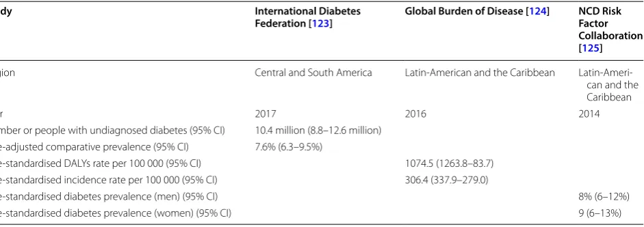
According to estimates from the Global Burden of Dis-ease, the burden of DM is greater than expected in Latin America and the Caribbean region [9]. This disease is estimated to affect 10–15% of the adult population in the Caribbean [10] and 9.4% in South and Central-American regions [6]. Recent estimates for DM in Latin America are summarized in Table 1. Furthermore, a strong rela-tionship between socioeconomic deprivation and DM has been consistently reported in several studies, mir-roring the social determinants of health in this metabolic disease [9, 11–14]. Recently, a population-based cross sectional study from the Southern Cone of Latin America reported prevalence estimates of DM between 8.4 and 14.3%, with 20% of these cases going undiagnosed [15].
Diabetes is a chronic disease with one of the highest costs to the healthcare system due to its multiple health hazards, high incidence of cardio-metabolic comorbidi-ties, and disabilities that impair individual productiv-ity [16, 17]. Approximately 7% of patients living with DM face costly long-term complications, many of which can be avoided or delayed [18, 19]. Currently, Latin America faces elevated out-of-pocket medical payments [20, 21]. In 2015, The Pan-American Health Organiza-tion reported that the average cost of diabetes care per year could range between US $1088 and US $1818, a high amount compared to the gross domestic profit in
Latin-American countries [17]. The Prospective Urban and Rural Epidemiological Study revealed that the avail-ability and affordavail-ability of essential diabetes medicines are insufficient in low-income and middle-income coun-tries [22]. The current economic burden that diabetes represents prompts scrutiny of the clinical aspects of this pathology for the development of cost-effective treat-ment strategies.
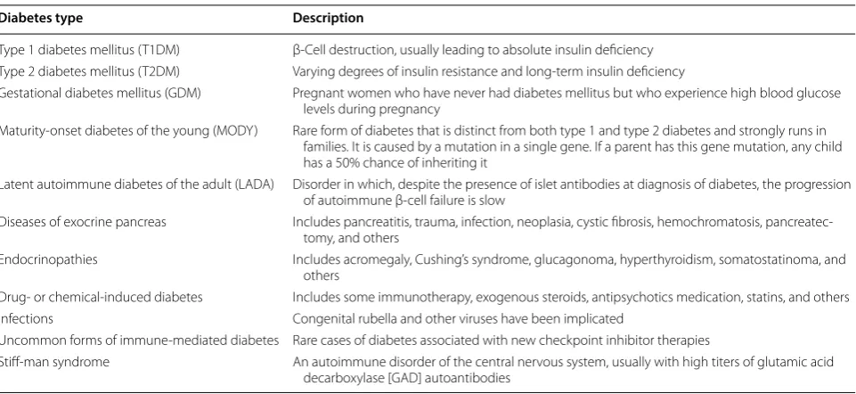
Clinical aspects and treatment of diabetes mellitus Diabetes is an endocrine disorder characterized by hyper-glycemia resulting from variable degrees of insulin resist-ance and/or deficiency [23, 24]. Several forms of diabetes have been described (Table 2). Treatment strategies for diabetes depend on, among other factors, the type of dia-betes diagnosed and the severity of the pathology.
Diabetic treatment encompasses an array of lifestyle and pharmaceutical interventions aimed at the preven-tion of disease progression, hyperglycemia control and mitigation of its micro and macrovascular complications. The treatment options in Latin America include lifestyle modification, hypoglycemic agents and insulin adminis-tration [25]. For the past 10 years, several new types of hypoglycemic agents have emerged in the market [26, 27], including metformin, alpha-glucosidase inhibitors, colesevelam, bromocriptine, sulfonylureas thiazolidinedi-ones, dipeptidyl peptidase IV (DPP-4) inhibitors, megli-tinide analogs, sodium-glucose cotransporter 2 (SGLT2) inhibitors, and a glucagon-like peptide-1 (GLP-1) recep-tor agonist. Insulin administration can comprise a sim-ple injection or a more sophisticated insulin pump and closed loop system. However, none of these strategies are able to perfectly control blood glucose, eventually lead-ing to complications. Advances in the development of new therapeutic options, through stem cells, will open
Table 1 Relevant recent estimates for diabetes mellitus in the Latin‑America and the Caribbean region
DALYS disability-adjusted life-years, CI confidence intervals
Study International Diabetes
Federation [123] Global Burden of Disease [124] NCD Risk Factor Collaboration [125]
Region Central and South America Latin-American and the Caribbean Latin-Ameri-can and the Caribbean
Year 2017 2016 2014
Number or people with undiagnosed diabetes (95% CI) 10.4 million (8.8–12.6 million) Age-adjusted comparative prevalence (95% CI) 7.6% (6.3–9.5%)
Age-standardised DALYs rate per 100 000 (95% CI) 1074.5 (1263.8–83.7) Age-standardised incidence rate per 100 000 (95% CI) 306.4 (337.9–279.0)
Age-standardised diabetes prevalence (men) (95% CI) 8% (6–12%)
the possibility of reversing hyperglycemia and alleviating the many debilitating complications of diabetes. A clear understanding of the pancreatic development and its mechanism of regeneration is critical for the discovery of appropriate treatment strategies for diabetes.
Insights into pancreatic regeneration: moving beyond conventional diabetes mellitus treatments through stem cell therapy
The pancreas is the major organ that systematically regulates glucose homeostasis. Pancreatic development involves the specific interplay of factors that, among other mechanisms, influence stem cell differentiation into pancreatic progenitor cells and the formation of the fully functional organ. Thus, most stem cell-based dif-ferentiation protocols are focused on the generation of mature, single hormone-expressing, glucose-responsive human β cells using information from studies of pancre-atic development [28, 29]. Specific signals are involved in the programming of insulin-producing β cells. The tran-scription factors SRY (sex determining region Y)-box (Sox)17 and homeobox gene HB9 (Hlxb9) are involved in the formation of the endoderm during gastrulation. Following foregut formation, fibroblast growth factor (FGF)-10, retinoic acid, SOX9, and hedgehog signaling pathways induce development of the pancreas. Pancre-atic specification and budding then occur through pan-creas-specific transcription factor-1a (Ptf-1a), pancreatic and duodenal homeobox 1 (PDX-1), NK6 homeobox 1 (Nkx6.1), neurogenin-3 (Ngn-3), and mafA [30], enabling endocrine formation and consequent stimulation of ISL
LIM homeobox 1 (Isl-1), NK2 homeobox 2 (Nkx2.2), neurogenic differentiation factor (NeuroD), paired box gene (Pax)4, and Pax6 signaling that form the islets of Langerhans. The transcription factors Sox17, hepatocyte nuclear factor (HNF)-6, and HNF-3beta (also known as forkhead box A2, Foxa2) are expressed throughout pan-creatic development. Finally, FGF-10 and notch sign-aling-induced stem cell and pancreatic progenitor cell differentiation stimulate neogenesis to create β cells [31, 32].
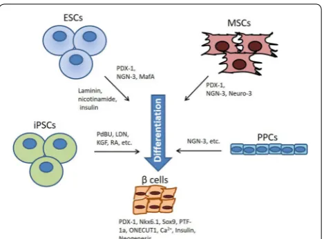
Discovery of the key factors involved in β cell devel-opment has given rise to strategies for obtaining β cells, either by inducing the expression of pancreatic-related transcription factors in distinct types of stem cells or by supplementation of soluble factors during culture. Diverse stem cell models have been used for the success-ful differentiation of β cell in vitro, including embryonic stem cells, induced pluripotent stem cells, mesenchymal stem cells, and progenitor cells.
Embryonic stem cells
The best model for pancreatic regeneration studies has been obtained from the use of embryonic stem cells (ESCs). Transgenic expression of PDX-1 and Nkx6.1 was shown to induce the differentiation of ESCs into endocrine cells that are positive for insulin, somatosta-tin, and glucagon expression [33]. Growth and extracel-lular matrix factors, including laminin, nicotinamide and insulin, lead to the formation of ESC-derived C-peptide/insulin-positive islet-like cell clusters that release insulin upon glucose stimulation and express Table 2 Diabetes classification
Diabetes type Description
Type 1 diabetes mellitus (T1DM) β-Cell destruction, usually leading to absolute insulin deficiency Type 2 diabetes mellitus (T2DM) Varying degrees of insulin resistance and long-term insulin deficiency
Gestational diabetes mellitus (GDM) Pregnant women who have never had diabetes mellitus but who experience high blood glucose levels during pregnancy
Maturity-onset diabetes of the young (MODY) Rare form of diabetes that is distinct from both type 1 and type 2 diabetes and strongly runs in families. It is caused by a mutation in a single gene. If a parent has this gene mutation, any child has a 50% chance of inheriting it
Latent autoimmune diabetes of the adult (LADA) Disorder in which, despite the presence of islet antibodies at diagnosis of diabetes, the progression of autoimmune β-cell failure is slow
Diseases of exocrine pancreas Includes pancreatitis, trauma, infection, neoplasia, cystic fibrosis, hemochromatosis, pancreatec-tomy, and others
Endocrinopathies Includes acromegaly, Cushing’s syndrome, glucagonoma, hyperthyroidism, somatostatinoma, and others
Drug- or chemical-induced diabetes Includes some immunotherapy, exogenous steroids, antipsychotics medication, statins, and others Infections Congenital rubella and other viruses have been implicated
Uncommon forms of immune-mediated diabetes Rare cases of diabetes associated with new checkpoint inhibitor therapies
Pax4 [34]. Retinoic acid (RA) has important roles in pancreatic development and is widely used to induce pancreatic differentiation of ESCs. When RA is directly added to activin A-induced human ESCs expressing CXCR4, 95% of cells become positive for the pancre-atic marker PDX-1 [35]. Animal studies have shown that human ESC-derived glucose-responsive mature β cells encapsulated in alginate and transplanted into a streptozotocin (STZ)-induced diabetic mouse model result in effective glycemic control [36]. However, the ethical implications involved with the use of ESCs have limited their further clinical application. In this respect, induced pluripotent stem cells have been proposed as a suitable alternative cell source with the same pluripo-tent characteristics as ESCs.
Induced pluripotent stem cells
Human induced pluripotent stem cells (iPSC) are obtained by reprogramming human somatic cells for the generation of stem cells with pluripotent proper-ties. Human iPSCs have been shown to be an effective cell source for deriving glucose responsive β-like cells [37–40]. Given the complex processes involved in β cell development, it has been difficult to obtain an effi-cient and replicable β cell differentiation protocol. A potential solution has been proposed to start differen-tiation from human iPSC-derived PDX-1 and SOX9-expressing pancreatic progenitor cells, which have a prolonged proliferation potential and the ability to pro-duce C-peptide positive β cells [41]. Another efficient differentiation protocol consisted of supplementation of factors involved in epidermal growth factor (EGF), transforming growth factor β (TGF-β), thyroid hor-mone, and RA signaling, as well as ɤ-secretase inhibi-tion [38], resulting in β cells with the ability to induce Ca2+ flux in response to glucose, package insulin into
secretory granules, and secrete insulin. It has recently been reported that supplementation of sodium cro-moglicate in combination with a previously described protocol causes the induction rate of insulin-positive cells to increase from a mean ± SD of 5.9 ± 1.5% (n = 3)
to 16.5 ± 2.1% (n = 3), with increased expression of
Ngn-3-positive cells at a mean ± SD of 32.6 ± 4.6%
(n = 3) compared to 14.2 ± 3.6% (n = 3) for the
non-supplemented control group [42]. Utilization of iPSCs for therapeutic applications involves other major chal-lenges, including recurrent autoimmune attacks in type 1 diabetes, the inherent risk of placing foreign tissue in the body, and potential tumor formation from cells that are not fully differentiated [2]. Fortunately, the use of mesenchymal stem cells has the potential to overcome these barriers.
Mesenchymal stem cells
Another attractive strategy for obtaining β cells is adult stem cells. Mesenchymal stem cells (MSCs) are con-sidered the most attractive cell source for regenerative medicine. MSCs have been highlighted because of their multi-potentialities, including self-renewal ability, pluri-potency, low antigenicity, reduced toxicity, and ease of culture and expansion in vitro to obtain sufficient cells for treatment. These cells are localized in diverse parts of our body, including the bone marrow, adipose tissue, amni-otic fluid, umbilical cord blood, and placenta. We have demonstrated that adipose and placenta-derived MSCs (PDMSCs) can be expanded for several passages without losing their self-renewal capacity [43, 44]. The Interna-tional Society for Cellular Therapy has provided criteria for defining MSCs. As has been previously demonstrated, MSC populations are composed of multipotent cells that are able to adhere to plastic in culture; express the cell surface markers CD105, CD73, and CD90 [45]; lack expression of CD45, CD34, CD14 or CD11b, CD79a, or CD19 and HLADR surface molecules [46]; and have the ability to differentiate into osteoblasts, adipocytes, or chondrocytes [44, 45]. MSCs have also been shown to be able to differentiate into cell types of endodermal and ectodermal lineages [47], including renal tubular cells [48], skin [49], neural cells [50], hepatocytes [51], and insulin-producing cells (IPCs) [52].
MSCs are being extensively investigated in the clini-cal setting for their immunomodulatory and tissue regenerative properties, as well as their feasibility in the context of islet transplantation [53, 54], demonstrat-ing improved engraftment of pancreatic islets through the suppression of inflammatory damage and immune-mediated rejection. MSC immunomodulatory prop-erties may be mediated through cell–cell interactions and/or secretion of soluble factors [55]. The cell-medi-ated immune response in MSCs induces T cell activa-tion and leukocyte recruitment to the inflammatory site through CD106. PDMSCs isolated from the chori-onic villi have been shown to contain a population of CD106+ cells with unique immunoregulatory
by blocking NLRP3 inflammasome activation and inflammatory agents [58]. When UC-MSCs were infused into type 2 diabetic rats, hyperglycemia was significantly ameliorated, and inflammatory activity was reduced, resulting in improved insulin sensitivity in insulin target tissues. Similarly, in adipose-derived MSCs (AD-MSCs), infusion into diabetic NOD mice reversed hyperglycemia through inducing higher serum insulin, amylin, and glucagon-like peptide 1 levels compared to untreated controls. AD-MSC treatment also reduced CD4+ T helper (Th) 1 cells, interferon-γ,
and inflammatory cell infiltration, as well as expanded Tregs in a cell contact-dependent manner in vitro and within the pancreas [59]. Administration of bone marrow-MSC-derived extracellular vesicles into mice resulted in the inhibition of antigen-presenting cell activation and suppression of Th1 and Th17 cell devel-opment, inhibiting the onset of type 1 diabetes [60]. Other studies have reported the immunomodulatory properties of bone marrow-derived MSCs (BM-MSCs) in islet xenotransplantation, as evidenced by reduced inflammatory markers and increased immune tolerance markers, demonstrating the potential of this strategy in solving transplantation issues of immune-related graft rejection, as described in more detail in the following sections [61]. Interestingly, a recent study in a UC-MSC model demonstrated that MSC-derived IPCs exhibited hypo-immunogenic characteristics in vitro but became immunogenic after transplantation to the host, possibly due to activation from the immune microenvironment [62].
In addition to their immunomodulatory effects, MSCs provide a supportive micro-environmental niche by secreting paracrine factors and depositing extracel-lular matrix [63]. Evidence has suggested a supportive role for MSCs in the regeneration of endogenous β cells. Studies have demonstrated that BM-MSCs from mice can differentiate in vitro into IPCs and that the differentiated cells express pancreas-specific marker genes [64]. Through genetic manipulation, overexpres-sion of PDX-1 in human BM-MSCs results in differen-tiation into IPCs [65]. Human BM-MSCs transfected with three genes, PDX-1, Neuro D, and Ngn-3, differ-entiate into insulin-expressing cells in vitro but lack glucose-responsive insulin expression. However, trans-plantation of these differentiated cells reduced blood glucose levels in diabetic mice. Interestingly, differen-tiated IPCs from AD-MSCs intraportally infused into patients exhibited a 30–50% decrease in their insulin requirement, with a 4- to 26-fold increase in serum C-peptide levels [66]. Umbilical cord blood derived embryonic stem cell-like cells that express stage spe-cific antigen 4 (SSEA4) and octamer 4 (Oct4) can
differentiate into insulin-producing islet-like cells that express insulin and C-peptide protein [67].
Despite the success of the differentiation proto-cols described in this review, none of these protoproto-cols are reproducible for the production of fully functional mature β cells yet. Additional research for the develop-ment of more sophisticated differentiation protocols is still required to apply these strategies clinically. None-theless, the successful generation of glucose-responsive IPCs through supplementation of crucial factors to the cell-culture medium gives hope for a diabetes treatment derived through stem cell-based cell therapy in the future (Fig. 1). Hence, due to their ease of isolation, immu-nomodulatory and tissue regenerative properties and the supportive niche they provide by secreting micro-envi-ronmental factors and depositing of extracellular matrix, MSCs are suggested to be a suitable stem cell resource for deriving in vitro β cells and for immunomodulation that may prevent graft rejection and autoimmune destruction of β cells.
Progenitor cells
Identification of progenitor cells in the adult pancreas has received increasing attention due to their pancreatic lineage characteristics that enable them to generate new functional β cells. When pancreatic progenitor cells were induced to differentiate into islets in vitro and trans-planted into STZ-induced mice, progenitor cells directly migrated into the injured pancreas, rapidly differenti-ating into IPCs that decreased glucose levels towards normoglycemia [68]. A recent study demonstrated that
progenitor cells expressing Ngn-3, which is expressed at extremely low levels in normal postnatal pancreatic tis-sues, exists in the ducts of adult mouse pancreas. Ectopic expression of Ngn-3 in pancreatic ductal cells converted them into IPCs, and treatment of human ductal and aci-nar cells with a combination of epidermal growth factor and gastrin induced neogenesis of islet β cells from the ducts, increasing the functional β cell mass [69]. In other studies, co-transplantation of purified human non-endo-crine pancreatic epithelial cells with human fetal pancre-atic tissue under the kidney capsule of immuno-deficient mice resulted in their differentiation into endocrine cells. Fetal cells seem to provide factors that support the sur-vival and differentiation of epithelial cells. Stem cell-like cells with the ability to be expanded and form clones ex vivo have also been reported. These cells have the abil-ity to proliferate and form cellular aggregates that display the capacity for endocrine and exocrine differentiation [70]. These results suggest that stem/progenitor cells exist within the pancreas and that these cells might be a source for new islets. However, identification of specific markers is urgently needed for isolation of these cell populations.
Transplantation of stem cell‑derived pancreatic cells Several types of stem cell-derived pancreatic cells have been proposed for transplantation into diabetic models, including pancreatic progenitors and insulin-secreting cells. As endocrine progenitors differentiate, they migrate cohesively and form bud-like islet precursors. Increas-ing evidence indicates that proper glucose regulation requires coordination between various islet cell types; therefore, it may be advantageous to produce whole islets in vitro rather than differentiating cells into a specific cell type. A recent study demonstrated obtaining islet precur-sors from embryonic stem cells, proposing this model to be optimal for obtaining whole islet populations [71].
When conditioned to mature in vivo, transplanted pancreatic progenitor cells produce insulin-secreting cells that prevent or reverse diabetes after transplan-tation. Transplantation of stem cell-derived pancre-atic progenitors on scaffolds that release exendin-4 has been reported to promote the engraftment of stem cell-derived pancreatic progenitors and their matu-ration toward insulin producing β cells, significantly increasing C-peptide levels and reducing blood glu-cose in STZ-induced mice [72]. Chronic hyperglycemia and an immunodeficient environment accelerate the maturation of transplanted progenitor cells under the kidney capsule in mice [73, 74]. Pancreatic progenitor cell-to-cell contact before transplantation is crucial for maturation into IPCs in vivo [75]. Nevertheless, in vivo maturation remains a critical issue to be resolved. It is
expected that mature endocrine cells generated in vitro would reverse diabetes more rapidly than pancreatic progenitor cells after transplantation. The development of novel techniques is required for in vitro differen-tiation protocols that may efficiently direct progenitor cells further down the β cell development pathway.
Stem cells treatment for diabetic complications In addition to the generation of IPCs from renewable stem cells, their immunomodulatory, self-renewal, and differentiation properties also suggests MSCs as poten-tially new therapeutic candidates in the treatment of diabetic-related complications [84]. Retinopathy, critical limb ischemia, and nephropathy are the most common and deleterious diabetic-related complications. Advances in stem cell research have shown the reversal of these complications through stem cell transplant, which will be reviewed below.
Critical limb ischemia
Peripheral arteriopathy in diabetic patients remains a serious health problem, despite enormous clinical and surgical advances over the last few decades. Stem cell therapy may be a good alternative to major amputation for restoring blood flow and attenuating ischemic dis-ease. Induction of diabetic animals with streptozotocin is a well-defined methodology for the study of periph-eral arteriopathy in diabetic models. Our laboratory has established this methodology and demonstrated that a formulated matrix gel induces regenerative neovasculo-genesis in the ischemic region (unpublished). AD-MSCs secrete angiogenic and cell survival factors and have been shown to be effective in the treatment of both coronary disease and complications of diabetes in animal and human models [85, 86]. MSC transplantation improves diabetic neuropathy via promoting direct peripheral nerve angiogenesis, neurotrophic effects, and restoration of myelination [87]. A recently reported GMP-compatible protocol described the generation of human ESC-derived endothelial cell products that improve ischemic limb per-fusion and local angiogenesis [88]. PDMSC injection into STZ-treated mice demonstrated newly formed capillar-ies, increased arterioles, and the secretion of proangio-genic factors that promoted ischemic recovery [89].
Retinopathy
Diabetic retinopathy (DR) is a microvascular complica-tion caused by hyperglycemia in which the retinal blood vessels weaken and rupture due to the chronic degenera-tion of retinal nerve tissue. Retinal glial cells and peri-cytes are the earliest-damaged cells with the highest rate of cell death during disease progression, for which cell replacement therapy will be more effective compared to conventional localized treatments. Stem cells have been studied for nerve regeneration in the retina [90]. Recent studies have demonstrated that intravitreal trans-plantation of neural stem cells originating from human UC-MSCs in DR rats resulted in the long-term preser-vation of retinal function and significantly delayed the
progression of DR for up to 8 weeks with the restoration of vision [91]. It was previously demonstrated that stem cells have protective effects against retinal vasculopathy by preventing capillary loss and retinal capillary drop-out in an STZ-induced rodent model of DR [92]. A sin-gle intravitreal dose of adipose-derived stem cells along with its secreted paracrine factors was shown to thera-peutically improve retina damage through neurovascu-lar repair that led to improved vision [93]. An efficient protocol was reported for generating highly purified human ESC-derived perivascular progenitor cells that demonstrated pericyte marker expression, neural differ-entiation potential, and improved damaged retinal vas-culature after transplantation into a STZ-induced rodent model [94]. In recent clinical studies, autologous BM-MSCs intravenously infused into patients’ eyes exhibited reduced fasting blood glucose, decreased macular thick-ness reduction, and improved the best corrected visual acuity [95]. Taken together, these studies demonstrate the potential of stem cell-based therapies in the treatment of retinovascular diseases.
Nephropathy
Diabetic nephropathy is the most common cause of end-stage renal disease and is characterized by alterations of the renal structure and function, including changes in renal tubules, stromal cells, and the incidence of glo-merular filtration. MSCs have been shown to relieve diabetic nephropathy through renal tissue repair, modu-lation of the immune response, and exertion of anti-fibrotic effects [96]. The paracrine effects of MSCs have been shown to increase the regeneration speed of renal tissue during diabetic nephropathy compared to the abil-ity of MSCs to differentiate into renal cells [97]. MSC-derived exosomes improved renal function and repaired renal tissue through autophagic mechanisms in an STZ-induced rat model [98, 99]. In addition, when MSCs are infused in combination with microRNA-124, the results demonstrate the attenuation of renal impairment as well as the inhibition of nephrocyte apoptosis during dia-betic nephropathy [100]. Injections of BM-MSCs at the early stages of diabetic nephropathy suppress renal mac-rophage and cytokine infiltration in diabetic rats, which prevented kidney dysfunction and glomerular defects [101].
Advances in stem cell research for diabetes treatment in Latin America
America for DM. Potential application of MSCs in the treatment of diabetic neuropathy was demonstrated after observing that preconditioning of human AD-MSCs with increasing concentrations of an iron chelator, deferoxam-ine, increased the abundance of hypoxia inducible factor 1 alpha, leading to upregulation of pro-angiogenic and neuroprotective factors [102]. On the other hand, stud-ies on diabetic nephropathy from Chile demonstrated that intravenously administered BM-MSCs allowed for pancreatic islet recovery, improved insulin secretion and reversed hyperglycemia in low dose STZ-induced diabetic mice [103]. Further Chilean collaborations with Argentina demonstrated that administration of MSCs to diabetic-induced mice restored pro-regenerative fac-tors, increased the renal proliferation index and anti-inflammatory cytokines levels, and reduced the renal apoptotic index, macrophage infiltration, and oxidative stress damage, resulting in preserved renal function and structure in mice with severe DM. MSC administration completely prevented retinal ganglion cell loss, improv-ing diabetic retinopathy [104]. Donor cells remained in the vitreous cavity and did not differentiate into neural or perivascular-like cells. Nevertheless, they increased the intraocular levels of several potent neurotrophic factors (nerve growth factor, basic fibroblast growth factor and glial cell line-derived neurotrophic factor) and reduced oxidative damage in the retina. MSC administration in diabetic-induced mice showed significant improvement in the functional parameters of kidneys with diabetic nephropathy, with an improved renal proliferation index, decreased renal apoptotic index and restoration of pro-regenerative factors and anti-inflammatory cytokine lev-els [105, 106]. By contrast, intravenous administration of BM-MSCs neither improved nor impaired diabetic cardiomyopathy in an obesity-induced mouse model [107]. Another research collaboration between Chile and Colombia showed that administration of allogenic BM-MSCs was insufficient for wound healing in diabetic mice, resulting in a delayed therapeutic effect, potentially explained by trophic factors secreted by MSCs being critical for skin regeneration and not the cells per se, sug-gesting that MSCs may require time to secrete these fac-tors after their administration [108]. In this respect, they showed that MSCs have the capability of restoring the balance between Th1 and Th2 immunological responses along with modification of the pancreatic microenviron-ment [109].
Pre-clinical animal studies in Brazil demonstrated that betacellulin (BTC), a ligand of the epidermal growth fac-tor recepfac-tor, promotes the growth and differentiation of pancreatic β cells and improves glucose metabolism in experimental diabetic rodent models [110]. When MSC-BTC was transplanted into STZ diabetic rats,
BTC-transfected cells ameliorated hyperglycemia from over 500 to approximately 200 mg/dL at 35 days post-cell transplantation. Administration of BM-MSCs into diabetic mice reversed hyperglycemia, improved β cell function, and modulated pancreatic cytokine levels [111]. Transplantation of stem cells obtained from murine den-tal pulp into STZ-induced type 1 diabetic mice improved pancreatic damage and renal function during diabetic neuropathy [112]. AD-MSC treatment reversed hyper-glycemia in early-onset diabetes in 78% of diabetic NOD mice, and this effect was associated with higher serum insulin, amylin, and glucagon-like peptide 1 levels com-pared to untreated controls. In addition, AD-MSC treat-ment ameliorated autoimmune diabetes pathogenesis in diabetic NOD mice by attenuating the Th1 immune response concomitant with the expansion/proliferation of Tregs, thereby contributing to the maintenance of functional β cells [59]. Co-transplantation of rat-derived BM-MSCs with pancreatic islets into mice resulted in reduced expression of inflammatory markers, such as TNFs, chemoattractant protein 1, and IL-1b, along with increased immune tolerance markers, IL-4, IL-10, and forkhead box P3, demonstrating the immunomodulatory actions of BM-MSC [61].
Brazil launched clinical trials testing the ability of autol-ogous BM-MSCs to reverse diabetes, stroke and heart conditions. Seventeen clinical trials in progress are utiliz-ing AD-MSCs, especially in cardiology, orthopedics, dia-betes and neurology. High-dose immunosuppression and hematopoietic stem cell (HSCs) transplant performed with acceptable toxicity in a small number of patients with newly diagnosed type 1 DM has shown increased β cell function in all but 1 of 18 patients with prolonged insulin independence in the majority of patients [113]. Administration of autologous HSCs into 21 type 1 dia-betic patients resulted in all patients becoming insulin independent for a period of 43 months with induced immunoregulation that consisted of lower CD3+CD4+
T cell numbers and consistent CD3+CD8+ T-cell levels,
resulting in a CD4/CD8 ratio inversion. Memory cyto-toxic T cells comprised the majority of T cells detected, and B cells returned to baseline levels 2–3 months post-transplantation. Baseline islet-specific T-cell autoreac-tivity persisted after transplantation, but regulatory T cell counts increased. Patients with lower frequencies of autoreactive islet-specific T cells remained insulin-free longer and presented greater C-peptide levels than those with lower frequencies of these cells [114].
developed by inducing expression of Nestin in MSCs, followed by a short incubation of 24 h in low glucose medium, and finally, a longer incubation of 168 h in high glucose medium [116]. Another study reported full ulcer recovery of a patient with chronic foot ulcer after MSC transplantation [117].
Findings from Argentina demonstrated that the com-bined therapy of intra-pancreatic AD-MSC infusion and hyperbaric oxygen improved metabolic control and reduced insulin requirements in patients with type 2 DM [118]. In general, Argentina’s governmental support is strong and a driving force in stem cell research. Cur-rently, 0.65% of the country’s GDP is invested in science and technology, which is a model that should be followed in additional Latin-American countries.
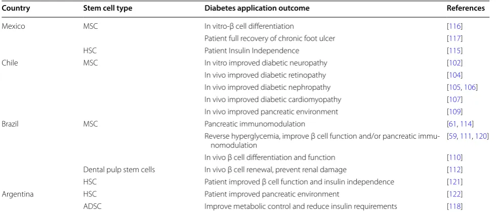
The latest strategy for the restoration of the β cell mass is through the generation and transplantation of stem cell-derived β cells [119], indicating that related research will be beneficial to Latin America (Table 3). Our shared vision is that these countries will maintain their effort in promoting innovative excellent research, establishing or improving regulations to the highest international level, increasing regional and international cooperation and identifying country- or region-specific opportunities to collaborate worldwide without diminishing identity or sovereignty.
Conclusion
Diabetes is a global health and economic burden in which the disability-adjusted life years and years of life lost rep-resent a problematic issue in Latin America. A unique
set of challenges exist for DM treatment, as the diabetic prevalence has increased over the years. Latin Amer-ica urgently needs to reorganize its health care services to optimize diabetes therapeutic goals. Recent break-throughs in deriving glucose responsive β-like cells from human stem cells has provided encouragement for β cell replacement therapy. More evidence is demonstrating the potential for embryonic stem cells, adult stem cells, and progenitor cells to produce β cells with the ability to produce insulin, reduce glucose levels in animal models, and to some extent, reverse diabetes symptoms through pancreatic regeneration. Stem cell research groups in Latin America have focused their efforts and provided important contributions to the DM field. Success in the generation of glucose-responsive IPCs and MSC-induced immunomodulation gives hope for the development of improved diabetic treatments through stem cell-based cell therapy in the near future.
Abbreviations
DM: diabetes mellitus; MODY: maturity-onset diabetes of the young; LADA: latent autoimmune diabetes of the adult; DPP-4: dipeptidyl peptidase IV; SGLT2: sodium-glucose cotransporter 2; GLP-1: glucagon-like peptide-1; NPH: neutral protomaine hagedorn; Sox: SRY (sex determining region Y)-box; Hlxb9: homeobox gene HB9; FGF-10: fibroblast growth factor 10; Ptf-1a: pancreas-specific transcription factor-1a; PDX-1: pancreatic and duodenal homeobox 1; Nkx6.1: NK6 homeobox 1; Ngn-3: neurogenin-3; Isl-1: ISL LIM homeobox 1; Nkx2.2: NK2 homeobox 2; NeuroD: neurogenic differentiation factor; Pax: paired box gene; HNF: hepatocyte nuclear factor; Foxa2: forkhead box A2; ESC: embryonic stem cells; RA: retinoic acid; STZ: streptozotocin; EGF: epidermal growth factor; TGF-β: transforming growth factor beta; iPSC: induced pluripotent stem cells; MSC: mesenchymal stem cell; PDMSCs: placenta-derived MSCs; IPCs: insulin-producing cells; TNF: tumor necrosis factor; IL: interleukin; HGF: hepatocyte growth factor; PGE2: prostaglandin E2; Table 3 Stem cell research in diabetes in some Latin‑American countries
MSC mesenchymal stem cell, HSC hematopoietic stem cell, ADSC adipose-derived stem cell
Country Stem cell type Diabetes application outcome References
Mexico MSC In vitro-β cell differentiation [116]
Patient full recovery of chronic foot ulcer [117]
HSC Patient Insulin Independence [115]
Chile MSC In vitro improved diabetic neuropathy [102]
In vivo improved diabetic retinopathy [104] In vivo improved diabetic nephropathy [105, 106] In vivo improved diabetic cardiomyopathy [107] In vivo improved pancreatic environment [109]
Brazil MSC Pancreatic immunomodulation [61, 114]
Reverse hyperglycemia, improve β cell function and/or pancreatic
immu-nomodulation [59, 111, 120]
In vivo β cell differentiation and function [110] Dental pulp stem cells In vivo β cell renewal, prevent renal damage [112] HSC Patient improved β cell function and insulin independence [121]
Argentina HSC Patient improved pancreatic environment [122]
UC-MSC: umbilical cord-derived MSCs; Th: T helper; BTC: betacellulin; BMSC: bone marrow-derived MSC; SSEA4: stage specific antigen 4; Oct4: octamer 4; DR: diabetic retinopathy; NSC: neural stem cell; HSC: hematopoietic stem cell; PPC: pancreatic progenitor cell.
Authors’ contributions
MAS designed the manuscript. MAS, IMV, and RC wrote and revised the manuscript. LLH revised the manuscript. All authors read and approved the final manuscript.
Author details
1 Gorgas Memorial Institute for Health Studies, Panama City, Republic of Panama. 2 Department of Medicine, Warren Alpert School of Medicine, Brown University, Rhode Island, USA. 3 Department of Medicine, University of Arizona College of Medicine, Phoenix, AZ, USA. 4 Department of Biotechnol-ogy and Bioindustry Sciences, National Cheng Kung University, Tainan, Taiwan. 5 Institute of Clinical Medicine, College of Medicine, National Cheng Kung University, Tainan, Taiwan. 6 Research Center of Excellence in Regenerative Medicine, National Cheng Kung University, Tainan, Taiwan.
Competing interests
The authors declare that they have no competing interests.
Availability of data and materials
All data generated or analyzed during this study are included in this manuscript.
Funding
This work was supported by Gorgas Memorial Institute for Health Studies Research Grant and Secretaria Nacional de Ciencia, Tecnología, e Innovación research grant 09-2018-ITE17-R1-001 from Panama. IMV is supported by the Sistema Nacional de Investigación (SNI, SENACYT). The funding sources had no involvement in the manuscript design, writing, or in the decision to submit the article for publication.
Publisher’s Note
Springer Nature remains neutral with regard to jurisdictional claims in pub-lished maps and institutional affiliations.
Received: 7 December 2018 Accepted: 13 February 2019
References
1. Weir GC, Bonner-Weir S, Leahy JL. Islet mass and function in diabetes and transplantation. Diabetes. 1990;39:401–5.
2. Aguayo-Mazzucato C, Bonner-Weir S. Pancreatic β cell regeneration as a possible therapy for diabetes. Cell Metab. 2018;27(1):57–67.
3. Ryan EA, Lakey JRT, Rajotte RV, Korbutt GS, Kin T, Imes S, et al. Clinical outcomes and insulin secretion after islet transplantation with the edmonton protocol. Diabetes. 2001;50:710–9.
4. Shapiro AMJ, Lakey JRT, Ryan EA, Korbutt GS, Toth E, Warnock GL, et al. Islet transplantation in seven patients with type 1 diabetes mellitus using a glucocorticoid-free immunosuppressive regimen. N Engl J Med. 2000;343:230–8.
5. Keymeulen B, Gillard P, Mathieu C, Movahedi B, Maleux G, Delvaux G, et al. Correlation between β cell mass and glycemic control in type 1 diabetic recipients of islet cell graft. Proc Natl Acad Sci. 2006;103:17444–9.
6. Rodriguez F, Cuero C, Delgado E, Camargo I, Tuñon R. Diagnóstico de la enfermedad renal crónica y factores de riesgo asociados en áreas seleccionadas de la provincia de Coclé, Panamá. Rev Med Panamá. 2014;34:31–8.
7. The Lancet. Life, death, and disability in 2016. Lancet. 2017;390:1083. 8. GBD 2016 Causes of Death Collaborators. Global, regional, and national
age-sex specific mortality for 264 causes of death, 1980–2016: a systematic analysis for the global burden of disease study 2016. Lancet. 2017;390:1151–210.
9. GBD 2016 DALYs and HALE Collaborators. Global, regional, and national disability-adjusted life-years (DALYs) for 333 diseases and injuries and healthy life expectancy (HALE) for 195 countries and territories, 1990–2016: a systematic analysis for the global burden of disease study 2016. Lancet. 2017;390:1260–344.
10. Sobers-Grannum N, Murphy MM, Nielsen A, Guell C, Samuels TA, Bishop L, et al. Female gender is a social determinant of diabetes in the Caribbean: a systematic review and meta-analysis. PLoS ONE. 2015;10:e0126799.
11. Sacerdote C, Ricceri F, Rolandsson O, Baldi I, Chirlaque MD, Feskens E, et al. Lower educational level is a predictor of incident type 2 diabetes in European countries: the EPIC-InterAct study. Int J Epidemiol. 2012;41:1162–73.
12. Schneiderman N, Llabre M, Cowie CC, Barnhart J, Carnethon M, Gallo LC, et al. Prevalence of diabetes among Hispanics/Latinos from diverse backgrounds: the Hispanic community health study/study of Latinos (HCHS/SOL). Diabetes Care. 2014;37:2233–9.
13. Mc Donald Posso AJ, Meza RAB, Morales EAM, Jaen Y, Ortega AC, Posada EJM. Diabetes in Panama: epidemiology, risk factors, and clinical management. Ann Glob Health. 2015;81:754–64.
14. Motta JA, Ortega-Paz LG, Gordón CA, Gómez B, Castillo E, Herrera Ballesteros V, et al. Diabetes mortality in Panama and related biological and socioeconomic risk factors. Revista Panamericana de Salud Pública. 2013;34:114–20.
15. Irazola V, Rubinstein A, Bazzano L, Calandrelli M, Chung-Shiuan C, Elor-riaga N, et al. Prevalence, awareness, treatment and control of diabetes and impaired fasting glucose in the Southern Cone of Latin America. PLoS ONE. 2017;12:e0183953.
16. Fowler MJ. Microvascular and macrovascular complications of diabetes. Clin Diabetes. 2008;26:77–82.
17. Barcelo A, Arredondo A, Gordillo-Tobar A, Segovia J, Qiang A. The cost of diabetes in Latin America and the Caribbean in 2015: evidence for decision and policy makers. J Glob Health. 2017;7:020410.
18. Bertoldi AD, Kanavos P, França GVA, Carraro A, Tejada CAO, Hallal PC, et al. Epidemiology, management, complications and costs associated with type 2 diabetes in Brazil: a comprehensive literature review. Glob Health. 2013;9:62.
19. Chatterjee S, Davies MJ, Heller S, Speight J, Snoek FJ, Khunti K. Diabetes structured self-management education programmes: a narrative review and current innovations. Lancet Diabetes Endocrinol. 2018;6:130–42. 20. Pauly MV, Zweifel P, Scheffler RM, Preker AS, Bassett M. Private health
insurance in developing countries. Health Aff. 2006;25:369–79. 21. Smith-Spangler CM, Bhattacharya J, Goldhaber-Fiebert JD. Diabetes,
its treatment, and catastrophic medical spending in 35 developing countries. Diabetes Care. 2012;35:319–26.
22. Chow CK, Ramasundarahettige C, Hu W, AlHabib KF, Avezum A Jr, Cheng X, et al. Availability and affordability of essential medicines for diabetes across high-income, middle-income, and low-income coun-tries: a prospective epidemiological study. Lancet Diabetes Endocrinol. 2018;6:798–808.
23. Association AD. Standards of medical care in diabetes-2014. Diabetes Care. 2014;37:S14–80.
24. Association AD. Diagnosis and classification of diabetes mellitus. Diabe-tes Care. 2014;37:S81–90.
25. Hippisley-Cox J, Pringle M. Prevalence, care, and outcomes for patients with diet-controlled diabetes in general practice: cross sectional survey. Lancet. 2004;364:423–8.
26. Bennett WL, Maruthur NM, Singh S, Segal JB, Wilson LM, Chatterjee R, et al. Comparative effectiveness and safety of medications for type 2 diabetes: an update including new drugs and 2-drug combinations. Ann Intern Med. 2011;154:602–13.
27. Fradkin JE, Rodgers GP. Glycemic therapy for type 2 diabetes: choices expand, data lag behind. Ann Intern Med. 2017;166:309–10. 28. Jorgensen MC, Ahnfelt-Ronne J, Hald J, Madsen OD, Serup P,
Hecksher-Sorensen J. An illustrated review of early pancreas development in the mouse. Endocr Rev. 2007;28:685–705.
29. Murtaugh LC. Pancreas and beta-cell development: from the actual to the possible. Development. 2007;134:427–38.
31. Bhushan A, Itoh N, Kato S, Thiery JP, Czernichow P, Bellusci S, et al. Fgf10 is essential for maintaining the proliferative capacity of epithelial progenitor cells during early pancreatic organogenesis. Development. 2001;128:5109–17.
32. Hald J, Hjorth JP, German MS, Madsen OD, Serup P, Jensen J. Activated Notch1 prevents differentiation of pancreatic acinar cells and attenuate endocrine development. Dev Biol. 2003;260:426–37.
33. Ida H, Akiyama T, Ishiguro K, Goparaju SK, Nakatake Y, Chikazawa-Nohtomi N, et al. Establishment of a rapid and footprint-free protocol for differentiation of human embryonic stem cells into pancreatic endocrine cells with synthetic mRNAs encoding transcription factors. Stem Cell Res Ther. 2018;9:277.
34. Schroeder IS, Rolletschek A, Blyszczuk P, Kania G, Wobus AM. Differentia-tion of mouse embryonic stem cells to insulin-producing cells. Nat Protoc. 2006;1:495–507.
35. Cai J, Yu C, Liu Y, Chen S, Guo Y, Yong J, et al. Generation of homoge-neous PDX1+ pancreatic progenitors from human ES cell-derived endoderm cells. J Mol Cell Biol. 2010;2:50–60.
36. Vegas AJ, Veiseh O, Gürtler M, Millman JR, Pagliuca FW, Bader AR, et al. Long-term glycemic control using polymer-encapsulated human stem cell-derived beta cells in immune-competent mice. Nat Med. 2016;22:306.
37. Korytnikov R, Nostro MC. Generation of polyhormonal and multipotent pancreatic progenitor lineages from human pluripotent stem cells. Methods. 2016;101:56–64.
38. Pagliuca Felicia W, Millman Jeffrey R, Gürtler M, Segel M, Van Dervort A, Ryu JH, et al. Generation of functional human pancreatic β cells in vitro. Cell. 2014;159:428–39.
39. Rezania A, Bruin JE, Arora P, Rubin A, Batushansky I, Asadi A, et al. Rever-sal of diabetes with insulin-producing cells derived in vitro from human pluripotent stem cells. Nat Biotechnol. 2014;32:1121–33.
40. Russ HA, Parent AV, Ringler JJ, Hennings TG, Nair GG, Shveygert M, et al. Controlled induction of human pancreatic progenitors produces functional beta-like cells in vitro. EMBO J. 2015;34:1759–72.
41. Trott J, Tan EK, Ong S, Titmarsh DM, Denil SLIJ, Giam M, et al. Long-term culture of self-renewing pancreatic progenitors derived from human pluripotent stem cells. Stem Cell Rep. 2017;8:1675–88.
42. Kondo Y, Toyoda T, Ito R, Funato M, Hosokawa Y, Matsui S, et al. Identifi-cation of a small molecule that facilitates the differentiation of human iPSCs/ESCs and mouse embryonic pancreatic explants into pancreatic endocrine cells. Diabetologia. 2017;60:1454–66.
43. Chen PY, Huang LLH, Hsieh HJ. Hyaluronan preserves the proliferation and differentiation potentials of long-term cultured murine adipose-derived stromal cells. Biochem Biophys Res Commun. 2007;360:1–6. 44. Wong TY, Chang CH, Yu CH, Huang LLH. Hyaluronan keeps
mesenchy-mal stem cells quiescent and maintains the differentiation potential over time. Aging Cell. 2017;16:451–60.
45. Solis MA, Wei Y-H, Chang C-H, Yu C-H, Kuo P-L, Huang LLH. Hyaluronan upregulates mitochondrial biogenesis and reduces adenoside triphos-phate production for efficient mitochondrial function in slow-proliferat-ing human mesenchymal stem cells. Stem Cells. 2016;34:2512–24. 46. Liu CM, Chang CH, Yu CH, Hsu CC, Huang L. Hyaluronan substratum
induces multidrug resistance in human mesenchymal stem cells via CD44 signaling. Cell Tissue Res. 2009;336:465–75.
47. Figliuzzi M, Bonandrini B, Silvani S, Remuzzi A. Mesenchymal stem cells help pancreatic islet transplantation to control type 1 diabetes. World J Stem Cells. 2014;6:163–72.
48. Morigi M, Imberti B, Zoja C, Corna D, Tomasoni S, Abbate M, et al. Mesenchymal stem cells are renotropic, helping to repair the kidney and improve function in acute renal failure. J Am Soc Nephrol. 2004;15:1794–804.
49. Nakagawa H, Akita S, Fukui M, Fujii T, Akino K. Human mesenchymal stem cells successfully improve skin-substitute wound healing. Br J Dermatol. 2005;153:29–36.
50. Munoz-Elias G, Marcus AJ, Coyne TM, Woodbury D, Black IB. Adult bone marrow stromal cells in the embryonic brain: engraftment, migration, differentiation, and long-term survival. J Neurosci. 2004;24:4585–95. 51. Schwartz RE, Reyes M, Koodie L, Jiang Y, Blackstad M, Lund T, et al.
Multipotent adult progenitor cells from bone marrow differentiate into functional hepatocyte-like cells. J Clin Invest. 2002;109:1291–302.
52. Jun H-S, Park E-Y. Adult stem cells as a renewable source of insulin-producing cells. Int J Stem Cells. 2009;2:115–21.
53. Sakata N, Chan NK, Chrisler J, Obenaus A, Hathout E. Bone marrow cell cotransplantation with islets improves their vascularization and func-tion. Transplantafunc-tion. 2010;89:686–93.
54. Zang L, Hao H, Liu J, Li Y, Han W, Mu Y. Mesenchymal stem cell therapy in type 2 diabetes mellitus. Diabetol Metab Syndr. 2017;9:36. 55. Lu LL, Liu YJ, Yang SG, Zhao QJ, Wang X, Gong W, et al. Isolation and
characterization of human umbilical cord mesenchymal stem cells with hematopoiesis-supportive function and other potentials. Haemato-logica. 2006;91:1017–26.
56. Yang ZX, Han ZB, Ji YR, Wang YW, Liang L, Chi Y, et al. CD106 identifies a subpopulation of mesenchymal stem cells with unique immunomodu-latory properties. PLoS ONE. 2013;8:e59354.
57. Abumaree MH, Abomaray FM, Alshabibi MA, AlAskar AS, Kalionis B. Immunomodulatory properties of human placental mesenchymal stem/stromal cells. Placenta. 2017;59:87–95.
58. Sun X, Hao H, Han Q, Song X, Liu J, Dong L, et al. Human umbilical cord-derived mesenchymal stem cells ameliorate insulin resistance by suppressing NLRP3 inflammasome-mediated inflammation in type 2 diabetes rats. Stem Cell Res Ther. 2017;8:241.
59. Bassi EJ, Moraes-Vieira PM, Moreira-Sa CS, Almeida DC, Vieira LM, Cunha CS, et al. Immune regulatory properties of allogeneic adipose-derived mesenchymal stem cells in the treatment of experimental autoimmune diabetes. Diabetes. 2012;61:2534–45.
60. Shigemoto-Kuroda T, Oh JY, Kim D-K, Jeong HJ, Park SY, Lee HJ, et al. MSC-derived extracellular vesicles attenuate immune responses in two autoimmune murine models: type 1 diabetes and uveoretinitis. Stem Cell Rep. 2017;8:1214–25.
61. Corradi-Perini C, Santos TM, Camara NOS, Riella MC, Aita CAM. Co-transplantation of xenogeneic bone marrow-derived mesenchymal stem cells alleviates rejection of pancreatic islets in non-obese diabetic mice. Transplant Proc. 2017;49:902–5.
62. Yang XF, Chen T, Ren LW, Yang L, Qi H, Li FR. Immunogenicity of insulin-producing cells derived from human umbilical cord mesenchymal stem cells. Exp Ther Med. 2017;13:1456–64.
63. Hematti P, Kim J, Stein AP, Kaufman D. Potential role of mesenchy-mal stromesenchy-mal cells in pancreatic islet transplantation. Transplant Rev. 2013;27:21–9.
64. Tang D-Q, Cao L-Z, Burkhardt BR, Xia C-Q, Litherland SA, Atkinson MA, et al. In vivo and in vitro characterization of insulin-producing cells obtained from murine bone marrow. Diabetes. 2004;53:1721–32. 65. Karnieli O, Izhar-Prato Y, Bulvik S, Efrat S. Generation of
insulin-pro-ducing cells from human bone marrow mesenchymal stem cells by genetic manipulation. Stem Cells. 2007;25:2837–44.
66. Trivedi HL, Vanikar AV, Thakker U, Firoze A, Dave SD, Patel CN, et al. Human adipose tissue-derived mesenchymal stem cells combined with hematopoietic stem cell transplantation synthesize insulin. Trans-plant Proc. 2008;40:1135–9.
67. Sun B, Roh KH, Lee SR, Lee YS, Kang KS. Induction of human umbilical cord blood-derived stem cells with embryonic stem cell phenotypes into insulin producing islet-like structure. Biochem Biophys Res Com-mun. 2007;354:919–23.
68. Srivastava A, Dadheech N, Vakani M, Gupta S. Pancreatic resident endo-crine progenitors demonstrate high islet neogenic fidelity and commit-ted homing towards diabetic mice pancreas. J Cell Physiol. 2018. https ://doi.org/10.1002/jcp.27568 .
69. Rooman I, Bouwens L. Combined gastrin and epidermal growth factor treatment induces islet regeneration and restores normoglycaemia in C57Bl6/J mice treated with alloxan. Diabetologia. 2004;47:259–65. 70. Rovira M, Scott SG, Liss AS, Jensen J, Thayer SP, Leach SD. Isolation and
characterization of centroacinar/terminal ductal progenitor cells in adult mouse pancreas. Proc Natl Acad Sci USA. 2010;107:75–80. 71. Sharon N, Chawla R, Mueller J, Vanderhooft J, Whitehorn LJ, Rosenthal
B, et al. A Peninsular structure coordinates asynchronous differ-entiation with morphogenesis to generate pancreatic islets. Cell. 2019;176:790–804.
73. Bruin JE, Saber N, Braun N, Fox JK, Mojibian M, Asadi A, et al. Treating diet-induced diabetes and obesity with human embryonic stem cell-derived pancreatic progenitor cells and antidiabetic drugs. Stem Cell Rep. 2015;4:605–20.
74. Bruin JE, Asadi A, Fox JK, Erener S, Rezania A, Kieffer TJ. Accelerated maturation of human stem cell-derived pancreatic progenitor cells into insulin-secreting cells in immunodeficient rats relative to mice. Stem Cell Rep. 2015;5:1081–96.
75. Beattie GM, Rubin JS, Mally MI, Otonkoski T, Hayek A. Regulation of proliferation and differentiation of human fetal pancreatic islet cells by extracellular matrix, hepatocyte growth factor, and cell-cell contact. Diabetes. 1996;45:1223–8.
76. Belame Shivakumar S, Bharti D, Baregundi Subbarao R, Park J-M, Son Y-B, Ullah I, et al. Pancreatic endocrine-like cells differentiated from human umbilical cords Wharton’s jelly mesenchymal stem cells using small molecules. J Cell Physiol. 2019;234:3933–47.
77. Southard SM, Kotipatruni RP, Rust WL. Generation and selection of pluripotent stem cells for robust differentiation to insulin-secreting cells capable of reversing diabetes in rodents. PLoS ONE. 2018;13:e0203126. 78. Skurikhin EG, Ermakova NN, Khmelevskaya ES, Pershina OV, Krupin VA,
Ermolaeva LA, et al. Differentiation of pancreatic stem and progenitor beta-cells into insulin secreting cells in mice with diabetes mellitus. Bull Exp Biol Med. 2014;156:726–30.
79. Skurikhin EG, Pakhomova AV, Epanchintsev AA, Stronin OV, Ermakova NN, Pershina OV, et al. Role of β cell precursors in the regeneration of insulin-producing pancreatic β cells under the influence of glucagon-like peptide 1. Bull Exp Biol Med. 2018;165:644–8.
80. Rhee M, Lee S-H, Kim J-W, Ham D-S, Park H-S, Yang HK, et al. Preadipo-cyte factor 1 induces pancreatic ductal cell differentiation into insulin-producing cells. Sci Rep. 2016;6:23960.
81. Gopurappilly R, Bhat V, Bhonde R. Pancreatic tissue resident mesenchy-mal stromesenchy-mal cell (MSC)-like cells as a source of in vitro islet neogenesis. J Cell Biochem. 2013;114:2240–7.
82. Chen W, Begum S, Opare-Addo L, Garyu J, Gibson TF, Bothwell AL, et al. Promotion of beta-cell differentiation in pancreatic precursor cells by adult islet cells. Endocrinology. 2009;150:570–9.
83. Sharma A, Rani R. Do we really need to differentiate mesenchymal stem cells into insulin-producing cells for attenuation of the autoimmune responses in type 1 diabetes: immunoprophylactic effects of precursors to insulin-producing cells. Stem Cell Res Ther. 2017;8:167.
84. Katuchova J, Harvanova D, Spakova T, Kalanin R, Farkas D, Durny P, et al. Mesenchymal stem cells in the treatment of type 1 diabetes mellitus. Endocr Pathol. 2015;26:95–103.
85. Kim SW, Han H, Chae GT, Lee SH, Bo S, Yoon JH, et al. Successful stem cell therapy using umbilical cord blood-derived multipotent stem cells for Buerger’s disease and ischemic limb disease animal model. Stem Cells. 2006;24:1620–6.
86. Williams AR, Trachtenberg B, Velazquez DL, McNiece I, Altman P, Rouy D, et al. Intramyocardial stem cell injection in patients with ischemic cardiomyopathy: functional recovery and reverse remodeling. Circ Res. 2011;108:792–6.
87. Xia N, Xu JM, Zhao N, Zhao QS, Li M, Cheng ZF. Human mesenchymal stem cells improve the neurodegeneration of femoral nerve in a dia-betic foot ulceration rats. Neurosci Lett. 2015;597:84–9.
88. MacAskill MG, Saif J, Condie A, Jansen MA, MacGillivray TJ, Tavares AAS, et al. Robust revascularization in models of limb ischemia using a clinically translatable human stem cell-derived endothelial cell product. Mol Ther. 2018;26:1669–84.
89. Liang L, Li Z, Ma T, Han Z, Du W, Geng J, et al. Transplantation of human placenta-derived mesenchymal stem cells alleviates critical limb ischemia in diabetic nude rats. Cell Transplant. 2017;26:45–61. 90. Ezquer F, Ezquer M, Arango-Rodriguez M, Conget P. Could donor
multipotent mesenchymal stromal cells prevent or delay the onset of diabetic retinopathy? Acta Ophthalmol. 2014;92:e86–95.
91. Chen S, Zhang W, Wang JM, Duan HT, Kong JH, Wang YX, et al. Differen-tiation of isolated human umbilical cord mesenchymal stem cells into neural stem cells. Int J Ophthalmol. 2016;9:41–7.
92. Zhang W, Wang Y, Kong J, Dong M, Duan H, Chen S. Therapeutic efficacy of neural stem cells originating from umbilical cord-derived mesenchymal stem cells in diabetic retinopathy. Sci Rep. 2017;7:408.
93. Elshaer SL, Evans W, Pentecost M, Lenin R, Periasamy R, Jha KA, et al. Adipose stem cells and their paracrine factors are therapeutic for early retinal complications of diabetes in the Ins2Akita mouse. Stem Cell Res Ther. 2018;9:322.
94. Kim JM, Hong KS, Song WK, Bae D, Hwang IK, Kim JS, et al. Perivascular progenitor cells derived from human embryonic stem cells exhibit functional characteristics of pericytes and improve the retinal vascula-ture in a rodent model of diabetic retinopathy. Stem Cells Transl Med. 2016;5:1268–76.
95. Gu X, Yu X, Zhao C, Duan P, Zhao T, Liu Y, et al. Efficacy and safety of autologous bone marrow mesenchymal stem cell transplantation in patients with diabetic retinopathy. Cell Physiol Biochem. 2018;49:40–52. 96. Li Y, Liu J, Liao G, Zhang J, Chen Y, Li L, et al. Early intervention with
mesenchymal stem cells prevents nephropathy in diabetic rats by ameliorating the inflammatory microenvironment. Int J Mol Med. 2018;41:2629–39.
97. Nagaishi K, Mizue Y, Chikenji T, Otani M, Nakano M, Saijo Y, et al. Umbili-cal cord extracts improve diabetic abnormalities in bone marrow-derived mesenchymal stem cells and increase their therapeutic effects on diabetic nephropathy. Sci Rep. 2017;7:8484.
98. Ebrahim N, Ahmed I, Hussien N, Dessouky A, Farid A, Elshazly A, et al. Mesenchymal stem cell-derived exosomes ameliorated diabetic nephropathy by autophagy induction through the mTOR signaling pathway. Cells. 2018;7:226.
99. Rashed LA, Elattar S, Eltablawy N, Ashour H, Mahmoud LM, El-Esawy Y. Mesenchymal stem cells pretreated with melatonin ameliorate kidney functions in a rat model of diabetic nephropathy. Biochem Cell Biol. 2018;96:564–71.
100. Sun J, Zhao F, Zhang W, Lv J, Lv J, Yin A. BMSCs and miR-124a amelio-rated diabetic nephropathy via inhibiting notch signalling pathway. J Cell Mol Med. 2018;22:4840–55.
101. Li H, Rong P, Ma X, Nie W, Chen C, Yang C, et al. Paracrine effect of mesenchymal stem cell as a novel therapeutic strategy for diabetic nephropathy. Life Sci. 2018;215:113–8.
102. Oses C, Olivares B, Ezquer M, Acosta C, Bosch P, Donoso M, et al. Precon-ditioning of adipose tissue-derived mesenchymal stem cells with defer-oxamine increases the production of pro-angiogenic, neuroprotective and anti-inflammatory factors: potential application in the treatment of diabetic neuropathy. PLoS ONE. 2017;12:e0178011.
103. Ezquer FE, Ezquer ME, Parrau DB, Carpio D, Yañez AJ, Conget PA. Sys-temic administration of multipotent mesenchymal stromal cells reverts hyperglycemia and prevents nephropathy in type 1 diabetic mice. Biol Blood Marrow Transplant. 2008;14:631–40.
104. Ezquer M, Urzua CA, Montecino S, Leal K, Conget P, Ezquer F. Intravitreal administration of multipotent mesenchymal stromal cells triggers a cytoprotective microenvironment in the retina of diabetic mice. Stem Cell Res Ther. 2016;7:42.
105. Ezquer F, Giraud-Billoud M, Carpio D, Cabezas F, Conget P, Ezquer M. Proregenerative microenvironment triggered by donor mesenchymal stem cells preserves renal function and structure in mice with severe diabetes mellitus. Biomed Res Int. 2015;2015:164703.
106. Ezquer F, Ezquer M, Simon V, Pardo F, Yanez A, Carpio D, et al. End-ovenous administration of bone-marrow-derived multipotent mesen-chymal stromal cells prevents renal failure in diabetic mice. Biol Blood Marrow Transplant. 2009;15:1354–65.
107. Calligaris SD, Conget P. Intravenous administration of bone marrow-derived multipotent mesenchymal stromal cells has a neutral effect on obesity-induced diabetic cardiomyopathy. Biol Res. 2013;46:251–5. 108. de Mayo T, Conget P, Becerra-Bayona S, Sossa CL, Galvis V,
Arango-Rodriguez ML. The role of bone marrow mesenchymal stromal cell derivatives in skin wound healing in diabetic mice. 2017;12:e0177533. 109. Ezquer F, Ezquer M, Contador D, Ricca M, Simon V, Conget P. The
antidiabetic effect of mesenchymal stem cells is unrelated to their transdifferentiation potential but to their capability to restore Th1/Th2 balance and to modify the pancreatic microenvironment. Stem Cells. 2012;30:1664–74.
•fast, convenient online submission
•
thorough peer review by experienced researchers in your field
• rapid publication on acceptance
• support for research data, including large and complex data types
•
gold Open Access which fosters wider collaboration and increased citations maximum visibility for your research: over 100M website views per year
•
At BMC, research is always in progress.
Learn more biomedcentral.com/submissions
Ready to submit your research? Choose BMC and benefit from:
111. Yaochite JNU, de Lima KWA, Caliari-Oliveira C, Palma PVB, Couri CEB, Simões BP, et al. Multipotent mesenchymal stromal cells from patients with newly diagnosed type 1 diabetes mellitus exhibit preserved in vitro and in vivo immunomodulatory properties. Stem Cell Res Ther. 2016;7:14.
112. Guimaraes ET, Cruz Gda S, Almeida TF, Souza BS, Kaneto CM, Vascon-celos JF, et al. Transplantation of stem cells obtained from murine dental pulp improves pancreatic damage, renal function, and painful diabetic neuropathy in diabetic type 1 mouse model. Cell Transplant. 2013;22:2345–54.
113. Couri CB, Oliveira MB, Stracieri AL, et al. C-peptide levels and insulin independence following autologous nonmyeloablative hematopoietic stem cell transplantation in newly diagnosed type 1 diabetes mellitus. JAMA. 2009;301:1573–9.
114. Malmegrim KC, de Azevedo JT, Arruda LC, Abreu JR, Couri CE, de Oliveira GL, et al. Immunological balance is associated with clinical outcome after autologous hematopoietic stem cell transplantation in type 1 diabetes. Front Immunol. 2017;8:167.
115. Cantu-Rodriguez OG, Lavalle-Gonzalez F, Herrera-Rojas MA, Jaime-Perez JC, Hawing-Zarate JA, Gutierrez-Aguirre CH, et al. Long-term insulin independence in type 1 diabetes mellitus using a simplified autologous stem cell transplant. J Clin Endocrinol Metab. 2016;101:2141–8. 116. Martinez-Gamboa M, Cruz-Vega DE, Moreno-Cuevas J, Gonzalez-Garza
MT. Induction of nestin early expression as a hallmark for mesenchy-mal stem cells expression of PDX-1 as a pre-disposing factor for their conversion into insulin producing cells. Int J Stem Cells. 2017;10:76–82. 117. Benitez-Arvizu G, Palma-Lara I, Vazquez-Campos R, Sesma-Villalpando
RA, Parra-Barrera A, Gutierrez-Iglesias G. Autologous mesenchymal stem cells and cutaneus autograft as a treatment for chronic ulcer secondary to diabetes mellitus 2. Cir Cir. 2015;83:532–6.
118. Estrada EJ, Valacchi F, Nicora E, Brieva S, Esteve C, Echevarria L, et al. Combined treatment of intrapancreatic autologous bone marrow stem cells and hyperbaric oxygen in type 2 diabetes mellitus. Cell Transplant. 2008;17:1295–304.
119. Suarez-Rodriguez R, Belkind-Gerson J. Cultured nestin-positive cells from postnatal mouse small bowel differentiate ex vivo into neurons, glia, and smooth muscle. Stem Cells. 2004;22:1373–85.
120. Yaochite JN, Caliari-Oliveira C, de Souza LE, Neto LS, Palma PV, Covas DT, et al. Therapeutic efficacy and biodistribution of allogeneic mesenchy-mal stem cells delivered by intrasplenic and intrapancreatic routes in streptozotocin-induced diabetic mice. Stem Cell Res Ther. 2015;6:31. 121. Voltarelli JC, Couri CE, Stracieri AB, Oliveira MC, Moraes DA, Pieroni
F, et al. Autologous nonmyeloablative hematopoietic stem cell transplantation in newly diagnosed type 1 diabetes mellitus. JAMA. 2007;297:1568–76.
122. Mesples A, Majeed N, Zhang Y, Hu X. Early immunotherapy using autologous adult stem cells reversed the effect of anti-pancreatic islets in recently diagnosed type 1 diabetes mellitus: preliminary results. Med Sci Monit. 2013;19:852–7.
123. International Diabetes Federation. IDF diabetes atlas, 8th edition. http:// www.diabe tesat las.org/resou rces/2017-atlas .html. Accessed 4 Jan 2018. 124. Global Burden of Disease Collaborative Network. Global burden of
disease study 2016 (GBD 2016) results. Seattle, United States: Institute for Health Metrics and Evaluation (IHME), 2017. http://ghdx.healt hdata .org/gbd-resul ts-tool. Accessed 4 Jan 2018.