R E S E A R C H A R T I C L E
Open Access
Partial axillary lymph node dissection
inferior to the intercostobrachial nerves
complements sentinel node biopsy in
patients with clinically node-negative
breast cancer
Jianyi Li, Shi Jia, Wenhai Zhang
*, Fang Qiu, Yang Zhang, Xi Gu and Jinqi Xue
Abstract
Background:The practice of breast cancer diagnosis and treatment in China varies to that in western developed countries. With the unavailability of radioactive tracer technique for sentinel lymph nodes biopsy (SLNB), using blue dye alone has been the only option in China. Also, the diagnosis of breast malignant tumor in most Chinese centres heavily relies on intraoperative instant frozen histology which is normally followed by sentinel lymph nodes mapping, SLNB and the potential breast and axillary operations in one consecutive session. This practice appears to cause a high false negative rate (FNR) for SLNB. The present study aimed to investigate the impact of the current practice in China on the accuracy of SLNB, and whether partial axillary lymph node dissection (PALND), dissection of lymph nodes inferior to the intercostobrachial nerve (ICBN), was a good complementary procedure following SLNB using blue dye.
Methods:289 patients with clinically node-negative breast cancer were identified and recruited. Tumorectomy,
intraoperative instant frozen histological diagnosis, SLNB using methylene blue dye, and PALND or complete axillary node dissection (ALND) were performed in one consecutive operative session. The choice of SLNB only, SLNB followed by PALND or by ALND was based on the pre-determined protocol and preoperative choice by the patient. Clinical parameters were analyzed and survival analysis was performed.
Results:37 % patients with clinically negative nodes were found nodes positive. 59 patients with positive SLN underwent ALND, including 47 patients with up to two positive nodes which were all located inferior to the ICBN. 9 patients had failed SLNB and underwent PALND. Among them, 3 (33.3 %) patients were found to have one metastatic node. 149 patients showed negative SLNB but chose PALND. Among them, 30 (20.1 %), 14 (9.4) and 1 (0.7 %) patients were found to have one, two and three metastatic node(s), respectively. PALND detected 48 (30.4 %) patients who had either failed SLNB or negative SLNB to have additional positive nodes. All the patients with up to two positive nodes had their nodes located inferior to the ICBN. The FNR of SLNB was 43 %. The accuracy rate was 58 %. The follow-up ranged 12–33 months. The incidence of lymphedema for SLNB, PALND, and ALND was 0 %, 0 %, and 25.4 %, respectively (P< 0.005). The disease-free survivals for SLNB, PALND, and ALND groups were 95.8 %, 96.8 %, and 94.9 %, respectively (p> 0.05).
Conclusions:Under the circumstances of current practice in China, PALND is a good complementary procedure
following SLNB in clinically node-negative breast cancer.
* Correspondence:[email protected]
Department of Breast Surgery, Shengjing Hospital of China Medical University, 36 Sanhao Street, Shenyang, Liaoning Province, China
Background
Since the description of the radical mastectomy for patients with breast cancer 100 years ago, axillary lymph node dissection (ALND) has been a necessary part of the operative procedure [1]. Despite the high incidence of arm lymphedema after ALND, before the 1970s most surgeons mainly focused on ensuring patients’ survival rather than minimising this severe complication [2]. In 1980s, advances in chemotherapy, radiotherapy and endocrine therapy significantly improved long term sur-vival [3, 4]. Improving the quality of life for patients with early-stage breast cancer became an important issue. In 1983, Rosen et al. subdivided the axillary lymph nodes (ALNs) into three levels according to their location in relation to the pectoralis minor muscle, and recom-mended that ALND be carried out from Level I to Level III in a stepwise manner [5]. In the mid-1990s, sentinel lymph node biopsy (SLNB) was introduced [6] and proven to be the best diagnostic modality to accurately stage axilla and select patients with early-stage breast cancer who can be spare of unnecessary ALND and its possible risk of arm lymphedema [7]. The key to SLNB is to accurately identify sentinel lymph nodes (SLNs). Al-though different methodologies can be used for SLNB, the combined technique of blue dye plus radioactive tracer is significantly superior to blue dye alone tech-nique in terms of negative predictive value and overall accuracy for identification of SLNs [7]. Thus, the com-bined technique of blue dye with radioactive tracer is generally considered as a gold standard for SLNB.
China is a developing country with considerably un-even economic development and rural–urban inequities in healthcare resources across regions. Compared with western developed countries, the practice of breast can-cer diagnosis and treatment in China varies remarkably. With the radioactive tracer technique being applied mainly in research and not being permitted in clinical surgeries, using blue dye alone has been the major tech-nique in large urban medical centres in China where the majority of rural hospitals are unable to provide SLNB service [8]. Meanwhile, with an aim to shorten waiting time and hospital stays to tackle the increasing shortage in healthcare resources, tumorectomy, intraoperative in-stant frozen histology diagnosis, the potential SLNs mapping and SLNB, and axillary lymph node interven-tion are normally performed in one consecutive opera-tive session in most medical centres. Low preoperaopera-tive biopsy rate means that the primary diagnosis of breast cancer relies heavily on intraoperative instant frozen histology. However, the practice of the performing tumorectomy and instant frozen histology prior to SLNs mapping leads to a potentially high false negative rate (FNR) for SLNB because of the effect of open incision on lymphatic drainage. As a result, it has been one of
the major goals for the Chinese breast cancer experts to find an easier and more effective method addressing SLN intervention under such circumstances specific to China.
Since 2007, our surgical team has been investigating the metastasis patterns of lymph nodes and distribution of ALNs in relation to the intercostobrachial nerves (ICBNs), which resulted in two publications. We found that ICBN can be an anatomic marker dividing axillary spaces into superior and inferior parts [9]. Short-term follow-up showed that partial axillary lymph node dis-section (PALND), disdis-section of lymph nodes inferior to the ICBNs traced by dye, was a good supplementary pro-cedure to follow SLNB. PALND effectively assessed the status of ALNs and prevented lymphedema after surgery [10]. The long term effects of PALND, however, still re-main unknown. The present study aimed to investigate the impact of the current practice in China on the accur-acy of SLNB, and whether PALND is a good comple-mentary procedure following SLNB using blue dye. Data from patients with clinically node-negative breast cancer were analyzed to determine the FNR and accuracy rate for SLNB and nodes dissection in relation to ICBN. Sur-vival analysis was also conducted in patients who under-went SLNB only, SLNB followed by PALND or by ALND.
Methods
Study population
Between July 2009 and April 2012, a total of 475 patients with suspected malignant breast tumor by preoperative imaging were investigated and approached for consent to participate in the study at the Shengjing Hospital of China Medical University. None of the patients under-went preoperative biopsy. Among them, 289 patients with clinically node-negative breast cancer were identi-fied by intraoperative frozen instant histology and subse-quently recruited in this study. Inclusive criteria were: (1) clinically node-negative breast cancer, defined as negative on preoperative axillary palpation, ultra sound examination and CT scan with contrast; (2) no previous history of breast cancer or other malignancies; (3) no neoadjuvant therapy; (4) no pregnancy. Those who had a benign tumor and those who did not meet the above cri-teria were excluded from the study.
Study protocols
technique and our ongoing study on PALND. The pa-tient was informed that although blue dye alone is con-sidered acceptable for staging and prognostication based on the current guidelines, it may cause false negative re-sult which potentially misses out positive nodes; PALND following SLNB may offset the disadvantage but its long-term complications are unclear. Patients were given enough time to make voluntary decision. An informed consent form and the preference of choice on surgical procedures if the tumor was malignant were obtained. All surgical procedures were performed by the same surgical team. The study was approved by the ethics committee of Shengjing Hospital of China Medical University.
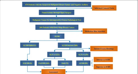
All the procedures were done in one consecutive op-erative session. Firstly, tumorectomy was performed through open surgery and the tumor was examined by intraoperative instant frozen histology analysis. SLN mapping and SLNB were followed if the tumor was proved to be malignant. 2–3 sections were examined for each node investigated by instant frozen histology. If SLNB failed (no blue staining nodes identified), PALND was subsequently performed. If SLNB succeeded and SLN was negative for cancer cells by instant frozen hist-ology, either PALND or no axillary intervention (SLNB
only) was followed based on the patient’s preoperative choice. If SLNB succeeded and SLNs showed positive, the patients were offered ALND (Fig. 1).
Operative techniques
Once the malignant tumor was diagnosed by instant fro-zen histology, SLNB mapping using methylene blue dye (3–4 ml, 25 mg/ml) was immediately followed as de-scribed previously [11]. Briefly, 2/3 of the dye dose was injected into the tumor bed and 1/3 into areola of the breast if the tumor was located in the outer quadrant; if the tumor was located in the inner quadrant, 1/3 of the dose was injected into the tumor bed and 2/3 into areola of the breast. The breast and axillary operations were then performed following 10–15 min massage. We understood that the protocol of performing SLN map-ping after tumorectomy would significantly affect the ac-curacy of SLNB, depending on the location and size of the open incision. However, because this was the com-mon practice in most centres (including ours) in China, one of the aims of the study was to investigate its impact on the accuracy of SLNB.
For the axillary operation, the ICBN was exposed com-pletely in the axillary space. All the anatomic locations of soft tissues and lymph nodes removed in relation to
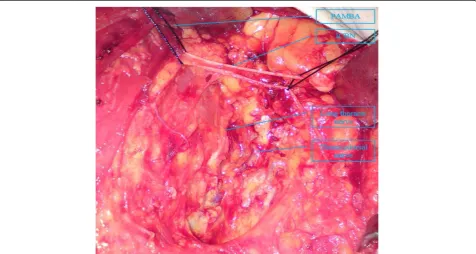
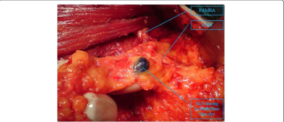
ICBN were recorded. The lymph nodes adjoining the ICBN belong to the lower lymph nodes. The long thor-acic and thoracodorsal nerves were identified and pre-served during the operation [Fig. 2]. The anatomical variations of ICBN were explored during the procedure, and recorded if any was identified. PALND technique in-volved removal of all the axillary fat pad and lymph nodes inferior to the ICBN. Drainage tube with the same calibre was placed in the axillary space at the end of the operation. All the nodes harvested were postoperatively examined for metastasis by histology.
Clinical assessment
When the drainage volume was less than 10 ml per 24 h, the tube was removed, and the extubation time and the total volume of drainage were recorded. Body mass index (BMI) and arm circumference at the point of 10 cm proximal to the medial epicondyle were mea-sured before surgery and again one year after surgery. The changes in ipsilateral upper extremity circumfer-ence, corrected for any change in the contralateral upper extremity, were calculated using the increase in arm circumference based on the formula introduced by McLaughlin et al. [12]. Severe lymphedema was diag-nosed if the increase in arm circumference was greater than or equal to 2 cm during the follow-up period, and mild lymphedema was defined as increase less than 2 cm. We observed substantial weigh gains in many
patients after surgery which unexpectedly altered the arm circumference, making this objective tool less ac-curate. Thus, a subjective method assessing long-term arm lymphedema was proposed and applied: if the pa-tient felt their ipsilateral arms were as good as the contralateral arm, they were defined as no lymphedema; if the patient did not feel that their ipsilateral arms were as good as the contralateral arms but did not affect their daily life, they were defined as mild lymph-edema; if their arm lymphedema affect their daily life, they were defined as severe lymphedema.
The collected clinical parameters included: number of nodes, postoperative extubation time, axillary drainage volume, changes in BMI and arm circumference, and other conventional indicators.
Statistical analysis
All statistical analyses were carried out using SPSS soft-ware (version 17.0 for Windows, Chicago, Illinois, USA). The correlation analyses among groups and the various biological factors were examined using the Chi-Square test or analysis of variance (ANOVA). For survival analysis, disease-free survival (DFS) were deter-mined using the Kaplan–Meier curves. The log-rank test was used to compare survival differences among the groups. P value < 0.05 was considered statistically significant.
Results
Patient distribution in groups with different intervention
Of the total of 289 recruited patients, no blue-staining SLN was identified in 9 patients who subsequently under-went PALND; therefore the success rate of dye-tracing was 96.9 % (280/289). In 280 patients with successful SLNB, 221 (78.9 %) patients had negative SLN, among whom 149 patients chose PALND and 72 patients opted for no axillary intervention. 59 patients were found SLN positive and underwent ALND as a result. Altogether, 107 out of 289 (37 %) patients with clinically negative nodes were found nodes positive by histology [Fig. 1].
Characteristics of patients
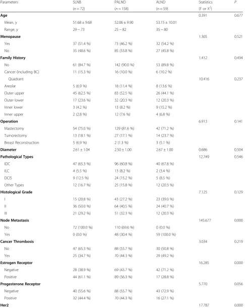
As shown in Table 1, no differences among groups were found in age, menopause, family history, quadrant distri-bution, operation type, tumour diameter, pathological types, histological grades, cancer thrombosis, biomarkers (PgR, Ki67 and P53) and targeted therapy (p> 0.05). PALND and ALND had higher node metastasis rates (30.4 % and 100 % respectively) compared with the SLNB group (0 %) (p< 0.005). The positive Estrogen re-ceptor rates were higher in the SLNB (61.1 %) and PALND (56.3 %) groups than in the ALND group (28.8 %) (p< 0.005). The ALND group showed higher positive Her2 rate, higher IIB stage rate, more chemo-therapy cycles and higher radiochemo-therapy rate than its two counterparts (p< 0.005).
Clinical outcomes
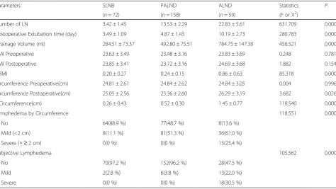
The number of removed axillary lymph nodes in the ALND group (22.83 ± 5.61) was greater than in the SLNB (3.42 ± 1.45) and PALND (13.53 ± 2.29) groups (p< 0.005). Also, the ALND group presented longer postoperative extubation time, greater total drainage vol-ume, and greater increase in BMI and arm circumfer-ence compared with its two counterparts (p< 0.005) (Table 2). Severe lymphedema evaluated using the abso-lute changes in arm circumference was not found in the SLNB and PALND groups, 25.4 % severe lymphedema was noted in the ALND group (p< 0.005). When using the subjective criteria, severe lymphedema was not re-ported by any patients in the SLNB and PALND groups, but reported by 30.5 % of patients in the ALND group (p< 0.005) (Table 2).
Anatomic location of blue staining SLNs
All the methylene blue staining SLNs were found around or inferior to the ICBN in 286/289 (99 %) patients [Fig. 3]. For the anatomical variations of ICBN, we only found one case of ICBN defect. According to a previous study, the pectoralis minor branch of thoracoacromial artery (PMBTA) can substitute ICBN as anatomic landmark [8].
Distribution of metastatic nodes in patients underwent ALND
As mentioned, 59 patients with positive SLN underwent ALND. Among them, there were 56 patients whose metastatic axillary nodes were all located inferior to the ICBN, including all the patients with one or two meta-static nodes (n= 47) and 9 patients with three metastatic nodes. Three patients were found to have metastatic nodes both inferior and superior the ICBN (2 patients with three metastatic nodes and 1 patient with four metastatic nodes) (Table 3).
Distribution of metastatic nodes in patients underwent PALND
As stated, 9 patients had failed SLNB and underwent PALND. Among them, 6 (66.7 %) patients were found no metastatic nodes and 3 (33.3 %) patients were found to have one metastatic node postoperatively. Meanwhile, 149 patients showed negative SLNB but chose PALND. Among them, 104 (69.8 %) patients were proved node metastasis free. However, 30 (20.1 %), 14 (9.4) and 1 (0.7 %) patients were found to have one, two and three metastatic node(s), respectively (Table 4). Putting to-gether, PALND detected 48 (30.4 %) out of 158 patients who had either failed SLNB or negative SLNB to have additional positive axillary nodes.
FNR of SLNB
If the nine patients with failed SLNB were not included in the analysis, the FNR of SLNB using blue dye in the present study was 43 % (45/104). The accuracy rate was 58 % (163/280) (Table 4).
Outcomes of follow-up
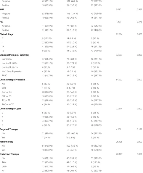
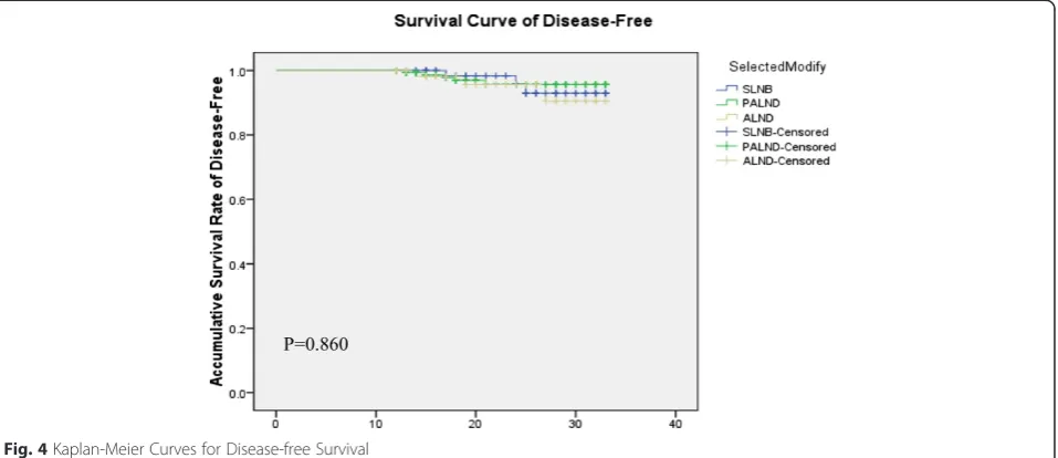
The follow-up time ranged from 12 to 33 months in each group with median 24 months, 20.5 months and 22 months for SLNB, PALND and ALND, respectively (p> 0.05). The actual DFS of SLNB, PALND, and ALND groups were 95.8 %, 96.8 %, and 94.9 %, respectively (p> 0.05), and the median DFS time was 30 months in each group [Table 5]. No differences were observed among groups in the curves for DFS [Fig. 4]. There was no difference in local recurrence among groups.
Discussion
Table 1Characteristics of patients and treatments in all groups
Parameters SLNB PALND ALND Statistics P
(n= 72) (n= 158) (n= 59) (F or X2)
Age 0.391 0.677
Mean, y 51.68 ± 9.68 52.06 ± 9.90 53.15 ± 10.01
Range, y 29 ~ 73 25 ~ 82 35 ~ 80
Menopause 1.305 0.521
Yes 37 (51.4 %) 73 (46.2 %) 32 (54.2 %)
No 35 (48.6 %) 85 (53.8 %) 27 (45.8 %)
Family History 1.412 0.494
No 61 (84.7 %) 142 (90.0 %) 53 (89.8 %)
Cancer (Including BC) 11 (15.3 %) 16 (10.0 %) 6 (10.2 %)
Quadrant 10.416 0.237
Areolar 5 (6.9 %) 18 (11.4 %) 8 (13.6 %)
Outer upper 45 (62.5 %) 83 (52.5 %) 26 (44.1 %)
Outer lower 17 (23.6 %) 32 (20.3 %) 12 (20.3 %)
Inner lower 3 (4.2 %) 13 (8.2 %) 9 (15.2 %)
Inner upper 2 (2.8 %) 12 (7.6 %) 4 (6.8 %)
Operation 6.913 0.141
Mastectomy 54 (75.0 %) 129 (81.6 %) 42 (71.2 %)
Tumorectomy 13 (18.1 %) 27 (17.1 %) 14 (23.7 %)
Breast Reconstruction 5 (6.9 %) 2 (1.3 %) 3 (5.1 %)
Diameter 2.61 ± 1.04 2.50 ± 1.00 2.67 ± 1.00 0.686 0.504
Pathological Types 12.749 0.546
IDC 47 (65.3 %) 96 (60.8 %) 40 (67.8 %)
ILC 4 (5.5 %) 13 (8.2 %) 2 (3.4 %)
DCIS 9 (12.5 %) 24 (15.2 %) 5 (8.5 %)
Other Types 12 (16.7 %) 25 (15.8 %) 12 (20.3 %)
Histological Grade 7.125 0.129
I 15 (20.8 %) 43 (27.2 %) 23 (39.0 %)
II 36 (50.0 %) 64 (40.5 %) 24 (40.7 %)
III 21 (29.2 %) 51 (32.3 %) 12 (20.3 %)
Node Metastasis 145.677 0.000
No 72 (100.0 %) 110 (69.6 %) 0 (0.0 %)
Yes 0 (0.0 %) 48 (30.4 %) 59 (100.0 %)
Cancer Thrombosis 3.034 0.219
No 47 (65.3 %) 88 (55.7 %) 30 (50.8 %)
Yes 25 (34.7 %) 70 (44.3 %) 29 (49.2 %)
Estrogen Receptor 16.285 0.000
Negative 28 (38.9 %) 69 (43.7 %) 42 (71.2 %)
Positive 44 (61.1 %) 89 (56.3 %) 17 (28.8 %)
Progesterone Receptor 5.770 0.056
Negative 40 (55.6 %) 88 (55.7 %) 43 (72.9 %)
Positive 32 (44.4 %) 70 (44.3 %) 16 (27.1 %)
Table 1Characteristics of patients and treatments in all groups(Continued)
Negative 62 (86.1 %) 137 (86.7 %) 37 (62.7 %)
Positive 10 (13.9 %) 21 (13.3 %) 22 (37.3 %)
Ki67 0.010 0.995
Negative 53 (73.6 %) 116 (73.4 %) 43 (72.9 %)
Positive 19 (26.4 %) 42 (26.6 %) 16 (27.1 %)
P53 1.497 0.473
Negative 41 (56.9 %) 77 (48.7 %) 32 (54.2 %)
Positive 31 (43.1 %) 81 (51.3 %) 27 (45.8 %)
Clinical Stage 92.884 0.000
0 9 (12.5 %) 14 (8.9 %) 0 (0.0 %)
I 22 (30.6 %) 49 (31.0 %) 0 (0.0 %)
IIA 41 (56.9 %) 51 (32.3 %) 16 (27.1 %)
IIB 0 (0.0 %) 44 (27.8 %) 43 (72.9 %)
Clinicopathological Subtypes 32.593 0.000
Luminal A 37 (51.4 %) 76 (48.1 %) 16 (27.1 %)
Luminal B Ki67+ 13 (18.1 %) 27 (17.1 %) 7 (11.9 %)
Luminal B Her2+ 6 (8.3 %) 9 (5.7 %) 3 (5.1 %)
Her2 Over-Expression 4 (5.5 %) 12 (7.6 %) 19 (32.2 %)
TNBC 12 (16.7 %) 34 (21.5 %) 14 (23.7 %)
Chemotherapy Protocols 84.222 0.000
No 6 (8.3 %) 15 (9.5 %) 5 (8.5 %)
CMF 1 (1.4 %) 8 (5.1 %) 0 (0.0 %)
CAF or AC 20 (27.8 %) 26 (16.5 %) 0 (0.0 %)
CEF or EC 18 (25.0 %) 36 (22.8 %) 0 (0.0 %)
TC or TP 23 (31.9 %) 37 (23.3 %) 14 (23.7 %)
TAC or AC-T 4 (5.6 %) 36 (22.8 %) 40 (67.8 %)
Chemotherapy Cycle 72.874 0.000
0 6 (8.3 %) 15 (9.5 %) 5 (8.5 %)
4 19 (26.4 %) 26 (16.5 %) 0 (0.0 %)
6 43 (59.7 %) 81 (51.2 %) 14 (23.7 %)
8 4 (5.6 %) 36 (22.8 %) 40 (67.8 %)
Targeted Therapy 4.201 0.122
No 71 (98.6 %) 152 (96.2 %) 54 (91.5 %)
Yes 1 (1.4 %) 6 (3.8 %) 5 (8.5 %)
Radiotherapy 26.425 0.000
No 54 (75.0 %) 100 (63.3 %) 19 (32.2 %)
Yes 18 (25.0 %) 58 (36.7 %) 40 (67.8 %)
Endocrine Therapy 20.478 0.002
No 16 (22.1 %) 46 (29.1 %) 33 (55.9 %)
TAM 22 (30.6 %) 49 (31.0 %) 9 (15.3 %)
LHRH 12 (16.7 %) 17 (10.8 %) 5 (8.5 %)
AI 22 (30.6 %) 46 (29.1 %) 12 (20.3 %)
inferior to the superior axillary as divided by ICBN [9]. This is similar to the findings by Clough et al. who discov-ered that nearly 89 % SLNs were inferior to the ICBN and few were located superior to ICBN and at the outside of the subscapular artery when traced by the combined method [14]. A recent study demonstrated that the node draining the arm lymph vessels was localized in the lateral pillar of the axillary, and was always found superior to or at the level of the second ICBN [15]. These findings indi-cate that the localization of nodes draining the breast and the arm lymphatic vessels is not overlapping and is largely divided by ICBN. Theoretically, postoperative arm lymph-edema is preventable if the tissue and nodes superior to ICBN are preserved. PALND, which only involves dissec-tion of nodes inferior to ICBN, should have similar lymph-edema occurrence rate with SLNB but lower rate than ALND. In the present study, no postoperative severe arm lymphedema was found after one year follow-up in the SLNB and PALND groups with over 15 % of severe lymphedema in the ALND group, suggesting PALND has the same protective effect as SLNB on arm lymphedema and is superior to ALND because the lymphatic drainage of the arm was preserved. The high level of chemotherapy, later clinical stage, and the poorer clinicopathological sub-types in the ALND group may have also contributed to the lymphedema observed in this group.
We carried out survival analysis according to the types of axillary intervention. There was no difference among the SLNB, PALND and ALND groups in DFS with a zero recurrence rate in all groups. Patients in the ALND group might have benefited from chemotherapy and radiotherapy. Our findings are consistent with a study by Kodama et al. who compared overall 5-year survival rates in patients undergoing PALND and his-torical controls undergoing conventional dissection [16]. Thus, there is sufficient evidence to suggest that PALND can significantly prevent arm lymphedema with a satisfying DFS.
Blue dye is so far the first choice for SLNB in China because it is cheap, safe without the need for nuclear medicine department and gamma probes. However, al-though blue dye alone technique is generally considered as a valid and acceptable tool to assess axillary nodes status and staging [17], there is growing evidence sug-gesting the combined technique of blue dye plus radio-active tracer is more superior. In a randomized study by Radovanovic et al., the false rate of blue dye was 17.6 % and accuracy was 68 % [7]. Other studies in the litera-ture showed the similar false-negative rates for the blue dye alone method which were generally higher than the recommended 5 % rate [18, 19]. The combined blue dye and radioactive tracer had a significantly lower FNR
doxorubicin + cyclophosphamide follow docetaxel,TAMtamoxifen,LHRHluteinising-hormone-releasing hormone,AIaromatase inhibitors. All nodes would be histopathological diagnosed to detect macrometastasis
Table 2Clinical outcomes in groups with different axillary intervention
Parameters SLNB PALND ALND Statistics P
(n= 72) (n= 158) (n= 59) (F or X2)
Number of LN 3.42 ± 1.45 13.53 ± 2.29 22.83 ± 5.61 631.709 0.000
Postoperative Extubation time (day) 3.49 ± 1.09 4.87 ± 1.43 10.19 ± 2.73 280.783 0.000
Drainage Volume (ml) 284.51 ± 73.37 492.80 ± 75.51 784.75 ± 147.38 458.521 0.000
BMI Preoperative 23.63 ± 3.49 23.48 ± 3.16 23.83 ± 3.69 0.248 0.781
BMI Postoperative 23.85 ± 3.41 23.72 ± 3.16 24.69 ± 3.68 1.882 0.154
△BMI 0.20 ± 0.27 0.24 ± 0.15 0.86 ± 0.63 85.318 0.000
Circumference Preoperative(cm) 24.81 ± 2.61 24.84 ± 2.62 24.84 ± 3.05 0.004 0.996
Circumference Postoperative(cm) 25.05 ± 2.56 25.36 ± 2.60 26.29 ± 3.19 3.682 0.026
△Circumference(cm) 0.26 ± 0.43 0.52 ± 0.30 1.45 ± 0.77 118.540 0.000
Lymphedema by Circumference 118.551 0.000
No 64(88.9 %) 77(48.7 %) 8(13.6 %)
Mild (<2 cm) 8(11.1 %) 81(51.3 %) 36(61.0 %)
Severe (△≥2 cm) 0(0 %) 0(0 %) 15(25.4 %)
Subjective Lymphedema 105.562 0.000
No 70(97.2 %) 152(96.2 %) 28(47.5 %)
Mild 2(2.8 %) 6(3.8 %) 13(22.0 %)
Severe 0(0 %) 0(0 %) 18(30.5 %)
(4.5 %) with higher accuracy [7]. In the present study, the FNR of blue dye technique was as high as 43 % and the accuracy rate was only 58 %, which appeared to be unexpected and unacceptable. We believe that the prac-tice of performing SLN mapping/SLNB after tumorectoy is largely responsible for the high FNR and low accuracy rate as open incision and the tumor removal procedure are potentially destroy breast lymphatic network, thus reduce breast lymphatic drainage towards axilla. The lack of other intensive node exploration technique (e.g. continuous multiple sections and lymph node imprint) in the present study may have also been a contributor. Different from western countries where malignance of a tumor is determined by preoperative biopsy and surgical plan is normally determined preoperatively, such prac-tice is common in China where malignance of a tumor is largely identified by intraoperative instant frozen hist-ology and the therapeutic plan is determined intraopera-tively. With unknown biological behaviour of a tumor and uncertain time frame before the result of intraopera-tive frozen histology was available, it appeared inappro-priate to perform SLNB prior to tumorectomy. Our
study showed an unacceptably higher FNR using blue dye alone under such circumstances.
We found that 48 (30.4 %) patients who showed either failed SLNB (33.3 %) or negative SLNB (30.2 %) using blue dye were found additional positive nodes by PALND. These data demonstrated that there may be an over 30 % chance of leaving behind positive nodes for those with failed or negative SLNB using blue dye alone. In a country where blue dye is the only option for SLNB and intraoperative instant frozen histology is the major method for primary diagnosis of breast cancer, PALND can be considered as a good complementary procedure to SLNB in patients with clinically-negative node breast cancer. Our findings are in keeping with an Indian study Fig. 3Sentinel lymph nodes located inferior to the intercostobrachial nerve. Illustration: SLN mostly located under the intercostalbrachial
nerve (ICBN)
Table 3Anatomic location of metastatic nodes in patients underwent ALND
Number of LNM Under the ICBN Above the ICBN Patients
1 + - 16
2 + - 31
3 + - 9
+ + 2
4 + + 1
LNMlymph node metastases,ICBNintercostobrachial nerve
Table 4Distribution of metastatic nodes in patients of all groups
Number of LNM
SLNB failed
SLNB negative SLNB positive
X2 P
PALND after SLNB(9)
PALND after SLNB(149)
End after SLNB(72)
ALND after SLNB (59)
0 6
(66.7 %) 104 (69.8 %)
- 0 181.568 0.000
1 3
(33.3 %) 30 (20.1 %)
- 16
(27.1 %)
2 0 14
(9.4 %)
- 31
(52.5 %)
3 0 1 (0.7 %) - 11
(18.6 %)
4 0 0 - 1
(1.7 %)
which showed low axillary sampling is accurate in pre-dicting axillary nodes status in women with clinically node negative operable breast cancer [20]. In the light of the high FNR of SLNB, we are now trialling SLNB prior to tumorectomy to minimise the effect of open incision on breast lymphatic drainage in all patients with sus-pected breast cancer. The FNR and accuracy rate are yet to be evaluated.
Recently, a presentation and related publications of the American College of Surgeons Oncology Group (ACO-SOG) Z0011 have provoked controversy around the world regarding the need for ALND in patients with SLN-positive breast cancer [21–23]. It seems that less than or
equal to two positive nodes does not warrant dissection. In our study, all the patients with less than or equal to two positive SLNs had all their metastatic nodes located infer-ior to the ICBN. Only three patients with more than two metastatic nodes had nodes found superior to the ICBNs. Therefore, PALND is considered as a way of biopsy for clinically node-negative breast cancer: if there are two or fewer positive nodes, then ALND is not necessary and the PALND is sufficient; if there are more than two positive nodes, then ALND is necessary. A recent study reported that for patients with positive nodes who do not undergo ALND, adjuvant therapy can have the same effect on over-all survival as the ALND procedure performed on similar Table 5Survival analysis according to axillary intervention
Parameters SLNB PALND ALND Statistics P
(n= 72) (n= 158) (n= 59) (F or X2)
Overall Survival 97.2 % 98.1 % 96.6 % 0.445 0.796
Deaths (n) 2 3 2
Median Survival Time (mo) 30.0 30.0 30.0 0.251 0.882
Disease-Free Survival 95.8 % 96.8 % 94.9 % 9.033 0.340
Event (n) 3 5 3
Local Recurrence 0 0 0
Contralateral breast cancer 0 2 0
Bone Metastasis 1 0 0
Lung Metastasis 0 0 1
Multi-Organ 2 3 2
Disease-Free Survival (mo) 30.0 30.0 30.0 0.303 0.860
Follow-Up Time 1.633 0.197
Median (mo) 24.0 20.5 22.0
Range (mo) 12-33 12-33 12-33
SLNBsentinel lymph node biopsy,PALNDpartial axillary lymph node dissection,ALNDaxillary lymph node dissection,momonths
node-positive patients [24]. However, the presence of posi-tive nodes is one of the important factors to be taken into account when considering radiotherapy and chemother-apy. Perhaps in the near future we can develop a novel im-aging technique instead of SLNB for the assessment of ALNs, we will enter a new era when the axillary surgery is not necessary. However, at the present time, PALND could be considered as a procedure complementary to SLNB in clinically node-negative breast cancer, especially in China where the combined technique is not available for SALN detection. This technique is so simple that a surgeon could easily perform without the need of compli-cated equipment. In fact, we prefer the name“functional axillary lymph node dissection (FALND) to PALND, a term which is coined when SLNs are not located. Our further investigation is ongoing on the clinical difference between FALND and ALND when there are two or fewer positive nodes and the results will be available in the near future.
In conclusion, there are considerable differences in the practice of breast cancer diagnosis and treatment between China and western developed countries. In a country where blue dye is the only technique for SLNB and intra-operative SLNB is normally performed after tumorectomy, the FNR of SLNB is unacceptably high. Under such cir-cumstances specific to China, axillary node dissection in-ferior to the ICBN is a good complementary procedure following SLNB in clinically node-negative breast cancer because it provides better assessment of nodes status, re-duces the occurrence rate for postoperative arm lymph-edema with satisfying long-term disease free survival.
Conclusions
The current practice in China where SLNB is generally performed after tumorectomy and intraoperative instant frozen histology reduces the accuracy of SLNB using blue dye. Under such specific circumstances, PALND is a good complementary procedure following SLNB in clinically node-negative breast cancer.
Competing interests
The authors declare that they have no competing interests.
Authors’contributions
LJ and ZW performed the operation, conducted data analysis and drafted the manuscript. JS, QF, ZY, GX and XJ participated in the follow-up. LJ and ZW participated in the study design and performed the statistical analysis. JS contributed to the ideas of the study, participated in the design and coordination. All authors read and approved the final manuscript.
Acknowledgements
The funding for this study was provided by the Nature and Science Foundation of Liaoning Province, No. 2013021009. The design, execution and analysis of the study are the sole responsibility of the authors.
Received: 23 November 2013 Accepted: 18 June 2015
References
1. Halsted WS. The Results of Radical Operations for the Cure of Carcinoma of the Breast. Ann Surg. 1907;46(1):1–19.
2. Mamaril AP. Preventing complications after radical mastectomy. Am J Nurs. 1974;74(11):2000–3.
3. Bonadonna G, Rossi A, Valagussa P. The CMF program for operable breast cancer with positive axillary nodes. Updated analysis on the disease-free interval, site of relapse and drug tolerance. Cancer. 1977;39(6 Suppl):2904–15. 4. Corkery J, Leonard RC, Henderson IC. Tamoxifen and aminoglutethimide in
advanced breast cancer. Cancer Res. 1982;42(8 Suppl):3409s–14. 5. Rosen PP, Lesser ML, Kinne DW. Discontinuous or "skip" metastases in
breast carcinoma. Analysis of 1228 axillary dissections. Ann Surg. 1983;197(3):276–83.
6. Giuliano AE, Kirgan DM, Guenther JM. Lymphatic mapping and sentinel lymphadenectomy for breast cancer. Ann Surg. 1994;220(3):391–8. discussion 398–401.
7. Radovanovic Z, Golubovic A, Plzak A. Blue dye versus combined blue dye-radioactive tracer technique in detection of sentinel lymph node in breast cancer. Eur J Surg Oncol. 2004;30(9):913–7.
8. Chinese Anti - Cancer Association & Professional Committee of Breast. Chinese guidelines for the diagnosis and treatment of breast cancer (2013 version). Chin Oncol. 2013;23(8):637–97.
9. Li J, Zhang Y, Zhang W. Intercostobrachial nerves as a novel anatomic landmark for dividing the axillary space in lymph node dissection. ISRN Oncol. 2013;2013:279013. doi:10.1155/2013/279013. Epub 2013 Jan 20. 10. Li J, Jia S, Zhang W. A New Technique That Complements Sentinel Lymph
Node Biopsy: Lymph Node Dissection Under the Intercostobrachial Nerves in Early-Stage Breast Cancer. Clin Breast Cancer.
2013;13(3):212–8.
11. Mathelin C, Croce S, Brasse D. Methylene blue dye, an accurate dye for sentinel lymph node identification in early breast cancer. Anticancer Res. 2009;29(10):4119–25.
12. McLaughlin SA, Wright MJ, Morris KT. Prevalence of lymphedema in women with breast cancer 5 years after sentinel lymph node biopsy or axillary dissection: objective measurements. J Clin Oncol. 2008;26(32):5213–9. Epub 2008 Oct 6. Erratum in:J ClinOncol.2010 Apr 1;28(10):1808.
13. Freeman SR, Washington SJ, Pritchard T. Long term results of a randomised prospective study of preservation of the intercostobrachial nerve. Eur J Surg Oncol. 2003;29(3):213–5.
14. Clough KB, Nasr R, Nos C. New anatomical classification of the axilla with implications for sentinel node biopsy. Br J Surg. 2010;97(11):1659–65. 15. Han JW, Seo YJ, Choi JE. The efficacy of arm node preserving surgery using
axillary reverse mapping for preventing lymphedema in patients with breast cancer. J Breast Cancer. 2012;15(1):91–7.
16. Kodama H, Mise K, Kan N. Partial Lower Axillary Dissection for Patients with Clinically Node-negative Breast Cancer. J Int Med Res. 2012;40(6):2336–45. 17. Morrow M, Rademaker AW, Bethke KP. Learning sentinel node biopsy:
Results of a prospective randomized trial of two techniques. Surgery. 1999;126(4):714–20.
18. Rufino AC, Baracat FF, Madeiro AP. Identification of the sentinel lymph node stained with blue dye in breast cancer patients. Eur J Gynaecol Oncol. 2003;24(5):387–90.
19. Meyer-Rochow GY, Martin RC, Harman CR. Sentinel node biopsy in breast cancer: validation study and comparison of blue dye alone with triple modality localization. ANZ J Surg. 2003;73(10):815–8.
20. Parmar V, Hawaldar R, Nair NS. Sentinel node biopsy versus low axillary sampling in women with clinically node negative operable breast cancer. The Breast. 2013;22:1081–6.
21. Gatzemeier W, Bruce Mann G. Which sentinel lymph-node (SLN) positive breast cancer patient needs an axillary lymphnode dissection (ALND) -ACOSOG Z0011 results and beyond. Breast. 2013;22(3):211–6. 22. Delpech Y, Bricou A, Lousquy R. The Exportability of the ACOSOG Z0011
criteria for omitting axillary lymph node dissection after positive sentinel lymph node biopsy findings: A multicenter study. Ann Surg Oncol. 2013;20(8):2556–61.
23. Nakamura S. Axillary lymph node dissection in sentinel node positive breast cancer: is it necessary? Curr Opin Obstet Gynecol.2013. [Epub ahead of print].