R E S E A R C H A R T I C L E
Open Access
Clinicopathological evaluation of anoxic mucosal
injury in strangulation ileus
Ryuji Takahashi
1*, Yoshito Akagi
1, Takaho Tanaka
2, Atsushi Kaibara
2, Sugako Kajiwara
3, Ichirou Shima
4, Jun Taguchi
5,
Tomoaki Mizobe
1, Tatsuyuki Kakuma
6and Kazuo Shirouzu
1Abstract
Background:In patients with strangulation ileus, the severity of bowel ischemia is unpredictable before surgery. To consider a grading scale of anoxic damage, we evaluated the pathological findings and investigated predictive factors for bowel gangrene.
Methods:We assessed 49 patients with strangulation ileus who underwent a laparotomy between January 2004 and November 2012. Laboratory tests and the contrast computed tomography (CT) were evaluated before surgery. According to the degree of mucosal degeneration, we classified anoxic damages into the following 3 grades. Ggrade 1 shows mild mucosal degeneration with extended subepithelial space. Grade 2 shows moderate degeneration and mucosal deciduation with residual mucosa on the muscularis mucosae. Grade 3 shows severe degeneration and mucosal digestion with disintegration of lamina propria.
Results:Resected bowel specimens were obtained from the 36 patients with severe ischemia, while the remaining 13 patients avoided bowel resection. The mucosal injury showed grade 1 in 11 cases, grade 2 in 10 cases, and grade 3 in 15 cases. The patients were divided into two groups. One group included grade 1 and non-resected patients (n = 24) while the other included grades 2 and 3 (n = 25). When comparing the clinical findings for these groups, elevated creatine kinase (P= 0.017), a low base excess (P= 0.021), and decreased bowel enhancement on the contrast CT (P= 0.001) were associated with severe mucosal injury.
Conclusion:In strangulation ileus, anoxic mucosal injury progresses gradually after rapid spreading of bowel congestion. Before surgical intervention, creatine kinase, base excess, and bowel enhancement on the contrast CT could indicate the severity of anoxic damage. These biomarkers could be the predictor for bowel resection before surgery.
Keywords:Strangulation ileus, Anoxic injury, Mucosal injury, Creatine kinase, Base excess
Background
Strangulation ileus is a critical condition leading to emer-gency surgeries, in which patients require early diagnosis and surgical decision making. Even when an immediate laparotomy is performed, many patients must undergo a bowel resection due to severe anoxic injury. To avoid un-desirable outcomes for the patients, the clinicians must detect bowel strangulation as soon as possible using diag-nostic equipment. Although previous studies have investi-gated several diagnostic parameters for strangulation ileus
[1-6], the severity of bowel ischemia remains to be unpre-dictable before surgery. To consider a grading scale of anoxic injury, we evaluated the pathological findings in resected bowel specimens and investigated predictive factors for bowel gangrene.
Methods
Study design and obtained approval
This study was retrospective observational study based on the medical records. We concentrated consecutive patients with strangulation ileus who underwent a laparotomy at two hospitals in Japan affiliated with Kurume University School of Medicine (Social Insurance Tagawa Hospital, Tagawa, and Asakura Medical Association Hospital, Asakura) * Correspondence:[email protected]
1
Department of Surgery, Kurume University School of Medicine, Kurume, Japan
Full list of author information is available at the end of the article
between January 2004 and November 2012. Resected bowel specimens were obtained from a total of 36 pa-tients. Blood samples were obtained from a total of 49 patients. Subsequently, we evaluated laboratory data and contrast computed tomography (CT) findings. This observational study was approved by the local eth-ics committees (The Etheth-ics Committee of Social Insurance Tagawa Hospital, Tagawa, and The Ethics Committee of Asakura Medical Association Hospital, Asakura) and pa-tients had given written informed consent for both the treatment and publication of the data.
Patient eligibility and data acquisition
We assessed 49 patients with strangulation ileus who underwent a laparotomy at the two hospitals. In all cases, the strangulated small bowel was discovered during sur-gery. Each patient received a physical examination, labora-tory tests, and a contrast computed tomography (CT) before surgery. Laboratory tests, including a white blood cell (WBC), absolute neutrophil count, C-reactive protein (CRP), lactate dehydrogenase (LDH), creatine kinase (CK), pH and base excess in blood gas analysis, were conducted within 24 hours prior to the operation. The absolute neu-trophil count was calculated by multiplying the WBC by the cell frequency in the visual observation method using May-Grünwald-Giemsa stain. In addition, we assessed the contrast CT finding, time to surgery from onset time, dur-ation of hospitalizdur-ation, and postoperative complicdur-ation. Onset time is the time at which the patient began to feel acute abdominal pain.
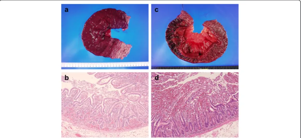
Pathological evaluation of resected bowel specimens To evaluate the degree of anoxic injury, we assessed resected bowel specimens (Hematoxylin and eosin stain; H & E stain). Transversally resected specimens were ob-tained from the strangulated bowel. Pathological findings were evaluated by the first author of this manuscript and a pathological specialist in hepatology and digestive disease. First, we observed the anoxic changes in bowel specimens pathologically and noticed the degree of mucosal degener-ation in the mucosal layer. Then we classified the severity of ischemic injury into 3 grades according to the degree of mucosal degeneration. Our grading scale is compatible with a previous report [7] which categorizes ischemic changes of the mucosal lesion into 6 grades. Our grade 1 shows mild mucosal degeneration with extended sube-pithelial space. Grade 2 shows moderate degeneration and mucosal deciduation with residual mucosa on the muscu-laris mucosae. Grade 3 shows severe degeneration and mucosal digestion with disintegration of lamina propria. Macro- and microscopic findings of these mucosal degen-erations are shown in Figures 1 and 2 [Figure 1a and b show mild degeneration (grade 1); Figure 1c and d show moderate degeneration (grade 2); Figure 2a and b show severe degeneration (grade 3)]. When the inhomogeneity of these mucosal damages was observed, the most severe damage was administrated.
Statistical analysis
According to the degree of mucosal injury and surgical procedure, the patients were divided into two groups.
Figure 1Macro-and microscopic findings of grades 1 and 2. aThe mucosa of grade 1 shows a segmental color change to deep red.
Patients’characteristics, laboratory data, and radiographic findings for the two groups were compared using the Mann–Whitney U test for continuous variables and the Fisher-Freeman-Halton exact test for categorical variables, respectively. All tests were two-sided andP values less than 0.05 were considered to be significant. Statistical tests were performed using JMP version 10 (SAS Institute Inc., Cary, NC, USA) and StatXact version 8 (Cytel Inc. Cambridge, MA, USA).
Results
Patients’overall characteristics
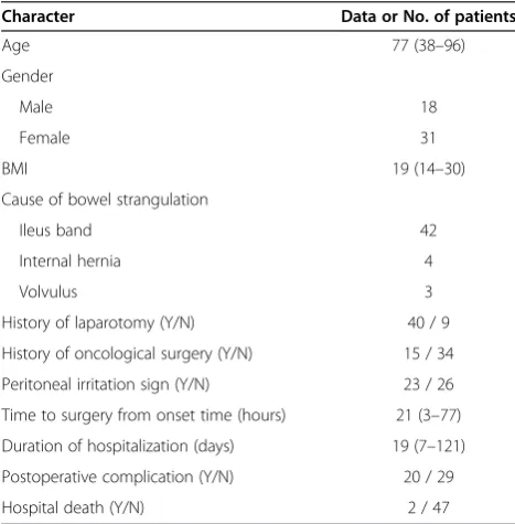
The patients’overall characteristics are shown in Table 1. Eighteen male and 31 female patients were enrolled in this study. Their median age was 77 year-old and median body mass index (BMI) was 19. The causes of bowel strangulation were ileus bands (42), internal hernias (4), and volvuluses (3). Forty patients had at least a history of laparotomy and details of the operations were as follows, gynecologic surgeries (14), appendectomies (9), gastrec-tomies (7), colecgastrec-tomies (5), cholecystecgastrec-tomies (3), surgical repairs for obturator hernias (3), low anterior resections (2), enterolyses (2), urological surgeries (2), partial bowel resection for traumatic perforation (1), ventriculoperitoneal shunting (1), and a Y-graft replacement (1). Although these operations included 15 oncological surgeries, neither can-cerous ileus nor peritoneal carcinomatosa was discovered during surgery. On the physical examination, a sign of peri-toneal irritation was shown in 23 patients before surgery.
Pathological findings of anoxic injuries
A total of 36 bowel specimens (H & E stain) were evalu-ated pathologically. The anoxic mucosal injury showed mild degeneration in 11 cases, moderate degeneration in 10 cases, and in 15 cases, the degeneration was severe. Four of the 10 cases with moderate degeneration showed Figure 2Macro- and microscopic findings of grade 3,and fibromuscular intimal thickening of the vasa recta. aThe mucosa of grade 3 shows a segmental color change to dark red.bThe mucosal injury of grade 3 shows severe degeneration and mucosal digestion with disintegration of lamina propria (H & E stain, ×10).cThe microscopic finding shows fibromuscular intimal thickening of the vasa recta without occlusive thrombus (H & E stain, ×10).
Table 1 Patients’overall characteristics (n = 49)
Character Data or No. of patients
Age 77 (38–96)
Gender
Male 18
Female 31
BMI 19 (14–30)
Cause of bowel strangulation
Ileus band 42
Internal hernia 4
Volvulus 3
History of laparotomy (Y/N) 40 / 9
History of oncological surgery (Y/N) 15 / 34
Peritoneal irritation sign (Y/N) 23 / 26
Time to surgery from onset time (hours) 21 (3–77)
Duration of hospitalization (days) 19 (7–121)
Postoperative complication (Y/N) 20 / 29
Hospital death (Y/N) 2 / 47
inhomogeneity of mucosal damages. For these 4 cases, we categorized the mucosal injury according to the se-verest finding. The other common pathological findings showed congestion and/or hemorrhage (3 mild, 9 mod-erate, and 24 severe), and neutrophil and/or lymphocyte infiltrations (10 mild, 14 moderate, and 12 severe). Four of the 36 cases showed fibromuscular intimal thickening of the vasa recta without occlusive thrombus (shown in Figure 2c).
Operative methods and postoperative complications All patients underwent a laparotomy under general anesthesia. At the time of laparotomy, the site of strangu-lation marks was successfully detected in all patients. In the cases requiring a bowel resection, we performed a par-tial or massive resection of the small bowel with a con-current end-to-end anastomosis. The median length of resected bowel was 70 cm (range, 20–250). The median length of resected bowel was longer in grades 2 and 3 pa-tients than grade 1 papa-tients (120 cm, 100 cm, and 50 cm, respectively; P= 0.006) In the cases not requiring bowel resection, we only released the bowel strangulation. The median period of hospitalization was 19 days. During hospitalization, 20 patients experienced postoperative complications. The complications included surgical site infections (8), pneumonitis (7), postoperative ileus (3), liver failure (2), disseminated intravascular coagulation [DIC] (2), short bowel syndrome (1), respiratory failure (1), and renal failure (1). There was no patient who devel-oped anastomotic leakage or bowel perforation after sur-gery. We lost 2 patients during hospitalization from acute renal failure and aspiration pneumonitis, respectively.
Comparisons of patients’characteristics and laboratory data
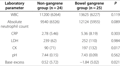
As we previously mentioned, the patients were divided into two groups. One group was defined as non-gangrene group (NG group) which included grade 1 and non-resected patients (n = 24). The other group was defined as bowel gangrene group (BG group) which included grades 2 and 3 patients (n = 25). Comparisons of patients’ char-acteristics for these groups are shown in Table 2. The NG group included a higher proportion of male patients than those in the BG group (P= 0.019). There were no significant statistical differences associated with age, BMI, history of laparotomy, history of oncological surgery, peritoneal irritation sign, time to surgery from onset time, postoperative complication, duration of hospitalization, or hospital death event. Furthermore, comparisons of labora-tory data for these groups are shown in Table 3. There were no significant statistical differences in the WBC, ab-solute neutrophil count, CRP, LDH, or pH between the two groups. However, higher levels of CK and lower levels of base excess were observed in the BG group (Wilcoxon
test,P= 0.017 and 0.021, respectively). The mean levels of CK showed 90 IU/l in the NG group and 197 IU/l in the BG group, respectively. The mean levels of base excess showed 0.52 mEq/l in the NG group and −1.84 mEq/l in the BG group, respectively. Gender was not a confounder because levels of CK and base excess were not associated with gender (Wilcoxon test,P= 0.987 and 0.717, respectively). Table 2 Comparisons of patients’characteristics for the NG group and BG group
Character Non-gangrene group (n = 24)
Bowel gangrene group (n = 25) P
Age 77 (51–94) 74 (50–96) 0.756*
Gender 0.019
Male 13 5
Female 11 20
Cause of strangulation 0.598†
Ileus band 22 20
Internal hernia 1 3
Volvulus 1 2
History of laparotomy (Y/N)
20 / 4 20 / 5 0.567
History of oncological surgery (Y/N)
10 / 14 5 / 20 0.096
Peritoneal irritation sign (Y/N)
10 / 14 13 / 12 0.571
Time to surgery from onset time (hours)
16 (3–74) 19 (5–77) 0.851
Postoperative complication (Y/N)
8 / 16 11 / 14 0.561
Duration of hospital stay (days)
18 (9–118) 18 (20–121) 0.233
Hospital death (Y/N) 1 / 23 1 / 24 0.235
Values are median (range) or No. of patients; values in parentheses are ranges. (Y/N), (Yes/No). *A Wilcoxon test.†A Fisher-Freeman-Halton exact test. The other analyses are performed by Fisher’s exact tests.
Table 3 Comparisons of laboratory data for the NG group and BG group
Laboratory parameter
Non-gangrene group (n = 24)
Bowel gangrene group (n = 25) P
WBC 11200 (6264) 13625 (6227) 0.119
Absolute neutrophil count
9540 (6326) 12124 (5955) 0.089
CRP 2.78 (5.46) 5.36 (8.19) 0.303
LDH 239 (62) 252 (110) 0.984
CK 90 (71) 197 (132) 0.017
pH 7.44 (0.15) 7.43 (0.09) 0.562
Base excess 0.52 (5.72) −1.84 (5.02) 0.021
Contrast CT findings and diagnostic accuracy
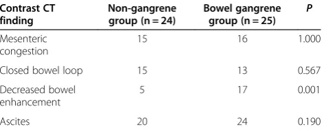
The common contrast CT findings for the NG group and BG group are shown in Table 4. The most common CT finding was ascites which was shown in 44 cases, while we detected peritoneal free air in 4 cases and hepatic portal gas in 1 case. The contrast CT finding showed mes-enteric congestion in 31 cases, closed bowel loop in 28 cases, and decreased bowel enhancement in 22 cases, re-spectively. Consequently, 36 patients were diagnosed with strangulation ileus by CT scans, while the remaining 13 patients had initially been diagnosed with adhesive ileus. These 13 patients had an ileus tube inserted upon admis-sion and subsequently underwent a laboratory, because their fluoroscopy findings showed irremediable bowel stenosis. Statistical analyses of these radiographic findings showed that decreased bowel enhancement on the con-trast CT (Fisher’s exact test,P= 0.001) was associated with severe mucosal injury. Gender was not a confounder be-cause decreased bowel enhancement on the contrast CT was not associated with gender (Fisher’s exact test, P= 1.000).
Cases presentation
Here we introduce the two patients who underwent a massive resection of the small bowel. The first case is a 61-year-old man who visited our hospital after 4 hours from onset time. Levels of CK and base excess showed normal range before surgery, and the contrast CT showed closed bowel loop, mesenteric congestion, and mild ascites (shown in Figure 3a). We resected 60 cm of his small bowel and pathological findings showed grade 1 mucosal injury, moderate congestion and hemorrhage (shown in Figure 3b). The second case is a 72-year-old woman who visited our hospital after 18 hours from on-set time. In this case, a low base excess (−4.5 mEq/l) and elevated CK (378 IU/l) were observed before surgery, and the contrast CT showed decreased bowel enhance-ment, closed bowel loop, mesenteric congestion, and mild ascites (shown in Figure 4a). We resected 195 cm of her small bowel and pathological findings showed grade 3 mucosal injury, severe congestion and hemorrhage (shown in Figure 4b).
Discussion
Can we evaluate the severity of anoxic injury in patients with strangulation ileus? We propose a grading scale for ischemic mucosal injury, which Chiu et al initially discov-ered in the dogs [7]. They categorized ischemic changes of the mucosal lesion into 6 grades (graded from 0 to 5); grade 0−normal mucosal villi; grade 1−development of subepithelial Gruenhagen’s space; Grade 2−extension of the subepithelial space with moderate lifting of epithelial layer from the lamina propria; Grade 3−massive epithelial lifting down the sides of villi; Grade 4−denuded villi with lamina propria and dilated capillaries exposed; Grade 5−digestion and disintegration of lamina propria. They confirmed that the speed of mucosal injury depended on arterial blood flow of the small bowel. Our grading scale of mucosal injury corresponded to each grade of their cri-teria, such as our grade 1 to their grade 0–2, our grade 2 to their grade 3 – 4, and our grade 3 to their grade 5, respectively.
To predict bowel strangulation and its severity, which radiographic finding is helpful? Our evaluation of con-trast CT scans found that mesenteric congestion, closed bowel loop, and decreased bowel enhancement are helpful in discovering bowel strangulation. A meta-analysis of 15 separate studies found CT scans to be 83% sensitive and 92% specific in detecting an ischemic bowel in patients with obstructive ileus [8]. Sheedy et al found that de-creased segmental enhancement was the most specific sign for small bowel ischemia [9]. Hayakawa et al men-tioned that the direct CT findings (without contrast) suggestive bowel strangulation include high-density bowel wall, mesenteric congestion, and localized pneu-matosis, while the indirect CT findings include C- or U-shaped loops with mesenteric vessels converging to-ward the obstruction site, ascites, target sign, two adja-cent collapsed round loops, and whirl sign [10]. Our results suggested that a decrease in bowel enhance-ment is associated with the degree of mucosal injury. The other diagnostic approaches include ascites ana-lysis by abdominocentesis and laparoscopic surgery/ adhesiolysis for selected patients with small bowel strangulation [11,12].
After making a decision to do a laparotomy, the surgeon must release the strangulated bowel and decide whether to resect or not. The decision regarding bowel resection or conservation is made based on the surgeon’s im-pression of the ischemic damage and viability. There-fore, it is very important to understand the progression of anoxic injuries after the strangulation. Sullins et al compared pathological changes for the arteriovenous and venous occlusions in horses with small bowel strangu-lation [13]. After 1 to 3 hour intervals, segments of ar-teriovenous occlusion became dark without edema and hemorrhage, while segments of venous occlusion were Table 4 Comparisons of contrast CT findings for the NG
group and BG group
Contrast CT finding
Non-gangrene group (n = 24)
Bowel gangrene group (n = 25) P
Mesenteric congestion
15 16 1.000
Closed bowel loop 15 13 0.567
Decreased bowel enhancement
5 17 0.001
Ascites 20 24 0.190
characterized by progressive congestion, edema, and hemorrhage especially in the mucosal layer. Park et al confirmed that ischemic periods of more than 20 mi-nutes caused villous injuries and extensive tissue injury in rats, but there was no exacerbation of tissue injury after the reperfusion [14]. Otherwise, an isolated arter-ial occlusion of 40 to 60 minutes induced significant exacerbation of tissue injuries at reperfusion [14]. Al-though these animal experiments indicate that the mesenteric arterial occlusion rapidly induces exacerba-tion of tissue injury and necrosis, only few studies have evaluated pathological findings of strangulated bowel and non-occlusive mesenteric ischemia (NOMI) in humans [15-19]. Pathologically, intimal thickening in the vasa recta and arteritis without organic vascular occlusion are associated with the development of NOMI [19]. In our study, 4 of the 36 cases (11%) showed fibromuscular intimal thickening of the vasa
recta. Generally, NOMI accounts for 20−30% of all cases of acute mesenteric ischemia with a mortality rate of nearly 50% [20]. One German article analyzed 62 patients with NOMI and concluded that leukocytosis, elevated serum lactate, and increased CK/CK-MB levels are specific parameters to diagnose NOMI [21]. Since we did not investigate serum lactate and CK-MB levels in our patients, further investigations should be performed to evaluate these biomarkers in strangulation ileus.
Ischemic or hemorrhagic bowel wall infarction results in anoxia and necrosis, providing ideal conditions for bacteria growth and multiplication [22]. Tissue damages progress with progressive mucosal epithelial cell slough, collagen disruption, edema, neutrophil infiltration of sub-mucosa and muscular layers and serosa [23]. Bowel gan-grene, caused by these types of tissue damage, promotes anaerobic acidosis and lactate production [6,24]. Fur-thermore, smooth muscle cells degenerate and eventually Figure 3Radiographic and pathological findings of the first case. aThe contrast CT shows closed bowel loop, mesenteric congestion, and mild ascites (arrows).bPathological findings show grade 1 mucosal injury, moderate congestion and hemorrhage (H&E stain, ×4).
Figure 4Radiographic and pathological findings of the second case. aThe contrast CT shows decreased bowel enhancement, closed bowel loop, mesenteric congestion, and mild ascites (arrows).bPathological findings showed grade 3 mucosal injury, severe congestion and
disrupt leading to the release of CK and its isoenzymes. Graeber et al reported that levels of serum CK and each of its isoenzymes became significantly elevated in dogs with strangulated infarction [25]. Despite the fact that we have to perform surgery as soon as possible, there was no cor-relation between time to surgery from onset time of symp-tom and bowel gangrene. Since the progression of anoxic damage showed many variations, time to surgery from on-set time could not be a reliable predictor for bowel gan-grene. Although our study evaluated a limited number of patients and blood samples at different times from onset, elevated CK and a low base excess which indicates the de-gree of acidosis could predict severe mucosal injury. These biomarkers and decreased bowel enhancement on the contrast CT could indicate the severity of anoxic mucosal injury in patients with strangulation ileus.
Conclusion
In strangulation ileus, anoxic mucosal injury progresses gradually after rapid spreading of bowel congestion. Be-fore surgical intervention, elevated CK, a low base ex-cess, and decreased bowel enhancement on the contrast CT could indicate the severity of anoxic damage. These biomarkers could be the predictor for bowel resection before surgery.
Abbreviations
WBC:White blood cell; CRP: C-reactive protein; LDH: Lactate dehydrogenase; CK: Creatine kinase; CT: Computed tomography; H & E: Hematoxylin and eosin; BMI: Body mass index; DIC: Disseminated intravascular coagulation; NG: Non-gangrene; BG: Bowel gangrene; NOMI: Non-occlusive mesenteric ischemia..
Competing interests
Disclosure: The authors declare that they have no competing interests.
Authors’contributions
RT, TT and JT are responsible for the conception and design of the study, the acquisition, analysis and interpretation of data, and drafting the work. YA is responsible for the interpretation of data and drafting the work. AK, IS, TM and KS are responsible for the interpretation of data and revising the work critically. SK and TK are responsible for the acquisition and analysis of data and revising the work critically. All authors read and approved the final manuscript.
Author details
1Department of Surgery, Kurume University School of Medicine, Kurume,
Japan.2Department of Surgery, Social Insurance Tagawa Hospital, Tagawa, Japan.3Department of Pathology, Social Insurance Tagawa Hospital, Tagawa,
Japan.4Department of Surgery, Asakura Medical Association Hospital, Asakura, Japan.5Department of Pathology, Asakura Medical Association
Hospital, Asakura, Japan.6Department of Biostatistics Center, Kurume University, Kurume, Japan.
Received: 23 May 2013 Accepted: 7 October 2014 Published: 16 October 2014
References
1. Tsumura H, Ichikawa T, Hiyama E, Murakami Y, Sueda T:Systemic inflammatory response syndrome (SIRS) as a predictive factor of strangulated small bowel obstruction.Hepatogastroenterology2004,
51:1393–1396.
2. Ayten R, Dogru O, Camci C, Aygen E, Cetinkaya Z, Akbulut H:Predictive value of procalcitonin for the diagnosis of bowel strangulation.World J Surg2005,29:187–189.
3. Jancelewicz T, Vu LT, Shawo AE, Yeh B, Gasper WJ, Harris HW:Predicting strangulated small bowel obstruction: an old problem revisited.
J Gastrointest Surg2009,13:93–99.
4. Sumi T, Katsumata K, Tsuchida A, Sonoda I, Shimazu M, Aoki T:Evaluation of sequential organ failure assessment score for patients with strangulation ileus.Langenbecks Arch Surg2010,395:27–31. 5. Markogiannakis H, Memos N, Messaris E, Dardamanis D, Larentzakis A,
Papanikolau D, Zografos GC, Manouras A:Predictive value of procalcitonin for bowel ischemia and necrosis in bowel obstruction.Surgery2011,
149:394–403.
6. Tanaka K, Hanyu N, Iida T, Watanabe A, Kawano S, Usuba T, Iino T, Mizuno R:
Lactate levels in the detection of preoperative bowel strangulation.
Am Surg2012,78:86–88.
7. Chiu CJ, McArdle AH, Brown R, Scott HJ, Gurd FN:Intestinal mucosal lesion in low-flow states. I. A morphological, hemodynamic, and metabolic reappraisal.Arch Surg1970,101:478–483.
8. Mallo RD, Salem L, Lalani T, Flum DR:Computed tomography diagnosis of ischemia and complete obstruction in small bowel obstruction: a systemic review.J Gastrointest Surg2005,9:690–694.
9. Sheedy SP, Earnest F 4th, Fletcher JG, Fidler JL, Hoskin TL:CT of small-bowel ischemia associated with obstruction in emergency department patients: diagnostic performance evaluation.Radiology2006,241:729–736. 10. Hayakawa K, Tanikake M, Yoshida S, Yamamoto A, Yamamoto E, Morimoto T:
CT findings of small bowel strangulation: the importance of contrast enhancement.Emerg Radiol2013,20:3–9.
11. Kobayashi S, Matsuura K, Matsushima K, Okubo K, Henzan E, Maeshiro M:
Effectiveness of diagnostic paracentesis and ascites analysis for suspected strangulation obstruction.J Gastrointest Surg2007,11:240–246. 12. Di Saverio S, Coccolini F, Galati M, Smerieri N, Biffl WL, Ansaloni L, Tugnoli G,
Velmahos GC, Sartelli M, Bendinelli C, Fraga GP, Kelly MD, Moore FA, Mandalà V, Mandalà S, Masetti M, Jovine E, Pinna AD, Peitzman AB, Leppaniemi A, Sugarbaker PH, Goor HV, Moore EE, Jeekel J, Catena F:
Bologna guidelines for diagnosis and management of adhesive small bowel obstruction (ASBO): 2013 update of the evidence-based guidelines from the world society of emergency surgery ASBO working group.World J Emerg Surg2013,8:42.
13. Sullins KE, Stashak TS, Mero KN:Pathologic changes associated with induced small intestinal strangulation obstruction and nonstrangulating infarction in horses.Am J Vet Res1985,46:913–916.
14. Park RO, Haglund U, Bulkley GB, Fält K:The sequence of development of intestinal tissue injury after strangulation ischemia and reperfusion.
Surgery1990,107:574–580.
15. Haruna L, Aber A, Rashid F, Barreca M:Acute mesenteric ischemia and duodenal ulcer perforation: a unique double pathology.BMC Surg2012,
12:21.
16. Grootjans J, Thuijls G, Derikx JP, van Dam RM, Dejong CH, Buurman WA:
Rapid lamina propria retraction and zipper-like construction of the epithelium preserves the epithelial lining in human small intestine exposed to ischemia-reperfusion.J Pathol2011,224:411–419.
17. Ohmori T, Ohnishi S, Okada K, Arita N:Sclerosing encapsulating peritonitis and non-occlusive mesenteric infarction found at autopsy in a man who had undergone continuous ambulatory peritoneal dialysis: a histochemical and immunohistochemical study.Pathol Int2000,50:660–666.
18. Sarda AK, Mathur M, Kapur M:Non-occlusive ischaemic enteritis.Ann R Coll Surg Engl1990,72:18–21.
19. Sugawara G, Yamaguchi A, Isogai M, Harada T, Kaneoka Y, Suzuki M, Akutagawa A, Suzumura K, Usui T:A Clinicopathological Study on 19 Operative Cases with Non Occlusive Mesenteric Ischemia.Jpn J Gastroenterol Surg2001,34:1713–1717.
20. Trompeter M, Brazda T, Remy CT, Vestring T, Reimer P:Non occlusive mesenteric ischemia: etiology, diagnosis, and interventional therapy.
Eur Radiol2002,12:1179–1187.
21. Stöckmann H, Roblick UJ, Kluge N, Kunze U, Schimmelpenning H, Kujath P, Müller G, Bruch HP:Diagnosis and therapy of non-occlusive mesenteric ischemia (NOMI).Zentralbl Chir2000,125:144–151.
23. Nathaniel White A:Intra-abdominal conditions causing colic: How they after normal physiology and why they result in pain [vt.edu web site].
2012. September 17, 2011. Available at: http://www.vetmed.vt.edu/emc/ clinicalservices/docs/Pathophysiology_of_colic_NAW.pdf#search='Intraabdominal +conditions+causing+colic%3A+How+they. Accessed June 18.
24. Juel IS, Solligård E, Skogvoll E, Aadahl P, Grønbech JE:Lactate and glycerol released to the intestinal lumen reflect mucosal injury and permeability changes caused by strangulation obstruction.Eur Surg Res2007,39:340–349.
25. Graeber GM, O’Neill JF, Wolf RE, Wukich DK, Cafferty PJ, Harmon JW:
Elevated levels of peripheral serum creatine phosphokinase with strangulated small bowel obstruction.Arch Surg1983,118:837–840.
doi:10.1186/1471-2482-14-79
Cite this article as:Takahashiet al.:Clinicopathological evaluation of anoxic mucosal injury in strangulation ileus.BMC Surgery201414:79.
Submit your next manuscript to BioMed Central and take full advantage of:
• Convenient online submission
• Thorough peer review
• No space constraints or color figure charges
• Immediate publication on acceptance
• Inclusion in PubMed, CAS, Scopus and Google Scholar
• Research which is freely available for redistribution