A COMPREHENSIVE REVIEW OF UROLITHIASIS
Dr.S.Sivakrishnan M.Pharm.,Ph.D.,
1Assistant Professor, Department of Pharmacy, FEAT, Annamalai University,
Annamalai Nagar, Chidambaram-608002, Tamilnadu, India
ABSTRACT
The urinary system or renal system is the organ system that produces, store and eliminates urine. It includes kidneys, ureters, bladder and urethra[1]. Urolithiasis is defined as the formation of stony concretions in the kidney, bladder, and/or urethra. Kidney stones are a common cause of blood in the urine and pain in the abdomen, flank and groin. The development of the stones is related to decreased urine volume or increased excretion of stone forming components such as calcium, oxalate, urate, cysteine, xanthine and phosphate. Stones form twice as often in men as women. Although most patients have only one stone episode, 25% of patients experience recurrent stone formation. Urolithiasis therefore has a significant impact on quality of life and socioeconomic factors. This review article will be helpful to explore the knowledge about urolithiasis for the researchers.
Keywords: Urolithiasis, Researchers, Calcium oxalate, Dietary changes
INTRODUCTION
The urinary system is a group of organs that consists of two kidney and ureters with single bladder and urethra. This system mainly filters the blood stream excrete out the unwanted fluid and other substance through urine. Urine is liquid containing excess mineral or vitamins with waste product of metabolism. Urinary system along with other organ systems maintains homeostasis with acid base balance and water salt balance of blood. Kidney stone is one of the oldest recorded disorder of human and one of the major health burden. Now a days large number of peoples are affected with this disorder all over the world. Nephrolithiasis is a process of forming a kidney stone, a stone in the kidney. These stones are called as renal calculi which is very common and painful kidney disorder. They are formed when urine becomes concentrated, allowing minerals to crystallize and stick together which forms microscopic crystals in the loop of henle, distal tubules, or the collecting duct. This is usually in response to elevated levels of urinary solutes, such as calcium, uric acid, oxalate, and sodium, as well as decreased levels of stone inhibitor, such as citrate and magnesium, these can lead to urine super saturation and subsequent stone formation[4]. If left untreated it can cause complication like permanent kidney damage, urethritis, cystitis, pyelonephritis, urinary tract blockage, infection, urinary fistula[5]. Urolithiasis constitutes one of the commonest afflictions requiring surgical intervention in our country and by conservative estimates there are about 5-7 million patients suffering from urinary calculus disease in India.
EPIDEMIOLOGY
Kidney stone disease typically presents between the ages of 20 and 60 and is more prevalent in hot climates[6]. It affects about 10% of people over their lifetime, incidence increasing with age; 50% will have a recurrence within 5–10 years and 75% within 20 years[7]. Developed countries have seen rapid increases over the last 30 years, especially in women in whom incidence is now almost equal to that of men[8]. Urolithiasis is common in more than 1 million cases per year in India. The countries with alarming occurrence rate of nephrolithiasis is British island, central Europe, North Australia, Scandinavian and Mediterranean countries. The stone forming belt of the world is identified as Egypt, Sudan, Saudi Arabia, Iran, UAE, Philippines, India, Pakistan, Thailand, Myanmar and Indonesia with cases of renal calculi in all age group including child below 1 year of age and adults over 70 years with a male to female ratio[9] of 2:1. Approximate 2 million people in India is affected with nephrolithiasis every year and some parts of country has name denoted as a stone belt that is, Gujarat, Maharashtra, Punjab, Rajasthan, Delhi, Haryana and part of states on North East side[10] Without preventive treatment, the recurrence rate of calcium oxalate calculi increases with time and reaches 50 percent at 10 years.
ETIOLOGY AND PATHOPHYSIOLOGY
Stone growth starts with the formation of crystals in supersaturated urine which then adhere to the urothelium, thus creating the nidus for subsequent stone growth. The biological processes that anchor crystals to the urothelium are incompletely understood. Many, but not all, calcium oxalate stones develop on Randall's plaques which are composed of calcium phosphate crystals. These grow to erode the urothelium, forming a nucleus for calcium oxalate deposition.
More recent theories focus on the role of cell surface molecules which favour or inhibit crystal adhesion[12,13]. Urothelial injury and repair after a stone episode may increase surface expression of these molecules to favour further crystal adhesion.[14] Thus ‘stones beget stones’[15] because there may be a residual nucleus on which further stones may form and/or upregulation of molecules favouring crystal adhesion. Stone prevention focuses on identifying and ameliorating the risk factors for crystal formation
FACTORS AFFECTING NEPHROLITHIASIS OCCURRENCE
Age and sex: Most vulnerable age for nephrolithiasis occurrence is 20-70 years. It is widely occurred in men as compared to women.
Diet: Low fluid intake, High content of protein, sodium, refined sugars, fructose and high fructose corn syrup, oxalate, grapefruit juice, apple juice, and cola drinks and low level of calcium increases the risk of nephrolithiasis.
Mechanisms: Creates acidic urinary milieu, depletes available citrate; Promotes hyperoxaluria, Excess dietary sodium Promotes hypercalciuria
History of family: Family history of nephrolithiasis increased risk of the nephrolithiasis occurrence.
Mechanisms: Genetic predisposition
Dehydration: Nephrolithiasis predisposes with excretion of concentrated urine. Hypertension: Hypertension increases the risk of the nephrolithiasis.
Obesity: Increase in Body Mass Index (BMI) also has increased risk of kidney stone.
Mechanisms: May promote hypercalciuria; other results similar to excess dietary meat
Inflammatory bowel diseases and gastric bypass surgery: They affect the absorption of the calcium ion and increases the precipitation
of calcium and other stone forming substance which causes nephrolithiasis.[16]
Mechanisms: Promotes low urine volume; acidic urine depletes available citrate; hyperoxaluria.
Drugs: Loop diuretics, antacids, acetazolamide, glucocorticoids, theophylline, vitamins D and C etc., has incidental correlation with occurrence of renal calculi.
Low urine volume: Allows stone constituents to supersaturate.
Insulin resistance: Ammonia mishandling; alters pH of urine.
Gout: Promotes hyperuricosuria.
Vitamin: abnormalities like vitamin A deficiencies, excess vitamin D.
Primary hyperparathyroidism: Creates persistent hypercalciuria.
Prolonged immobilization: Bone turnover creates hypercalciuria.
Renal tubular acidosis (type 1): Alkaline urine promotes calcium phosphate supersaturation and loss of citrate.
TYPES OF KIDNEY STONES
Kidney stones are classified into 5 types are as follows:
Calcium oxalates stone are crystalline component of calcium oxalate monohydrate, calcium oxalate dihydrate and calcium oxalate trihydrate. Calcium phosphate stone have crystalline components like hydroxyapatite, calcium hydrogen phosphate, dihydrate, unusual form of calcium phosphate, tricalcium phosphate, ammonium magnesium, phosphate hexahydrate, ammonium magnesium, phosphate monohydrate, magnesium hydrogen, phosphate trihydrate, carbonate apatite and octacalcium phosphate. Calcium oxalate and calcium phosphate stones are prevalent with abnormality in urinary system like hypercalciuria, hypomagnesuria, hyperuricosuria, hyperoxolourea and hypocitraturia.
Uric acid stone are crystalline components of uric acid anhydrous and uric acid dehydrate, usually 5-10% of analyzed population with renal stone are affected with uric acid stone. Uric acid is a metabolic product and around 25% of patients with this stone also suffer from gout disorder. Main reason of this type of stone is low urine volume, hyperuricosuria and acidic urine pH.
Cystine stone are caused due to high level of essential amino acid, cystine in urine. Cystine stone usually occur in childhood and it is a rare inherited metabolic disorder affecting 1-3% analyzed population of renal stone.
Struvite stone are infectious urinary stone of ammonium magnesium phosphate hexahydrate or struvite. It is a fascinating inorganic phosphate mineral closely associated with chronic urinary tract infection due to some microorganism such as urease-producing bacteria. This bacterium split urea in to ammonium which is combined with magnesium and phosphate.
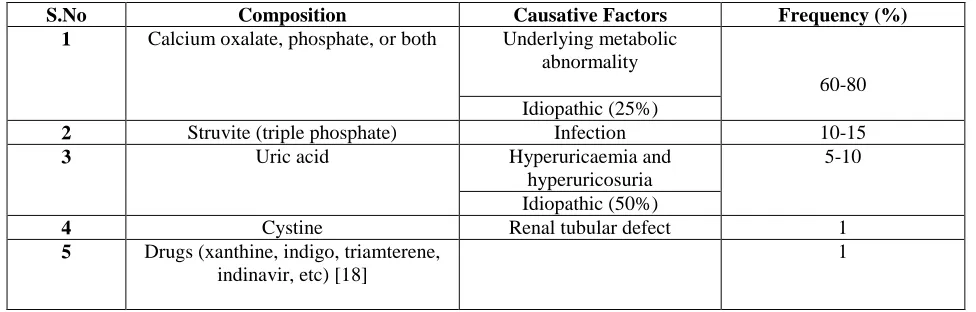
Table 1. Kidney stones composition, frequency and its causative factors
S.No Composition Causative Factors Frequency (%)
1 Calcium oxalate, phosphate, or both Underlying metabolic abnormality
60-80 Idiopathic (25%)
2 Struvite (triple phosphate) Infection 10-15
3 Uric acid Hyperuricaemia and
hyperuricosuria
5-10
Idiopathic (50%)
4 Cystine Renal tubular defect 1
5 Drugs (xanthine, indigo, triamterene, indinavir, etc) [18]
1
Table 2. High-risk stone formers [19-29]
General factors Diseases associated
with stone formation
Genetically determined stone formation
Anatomical abnormalities associated with stone formation
Early onset of urolithiasis (especially children and teenagers)
Hyper Parathyroidism
Cystinuria (type A, B and AB)
Medullary sponge kidney (tubular ectasia)
Familial stone formation Metabolic syndrome Primary hyperoxaluria (PH)
Ureteropelvic junction (UPJ) obstruction
Brushite-containing stones (CaHPO4.2H2O)
Nephrocalcinosis Renal tubular acidosis (RTA) type I
Calyceal diverticulum, calyceal cyst
Uric acid and urate-containing stones
Polycystic kidney disease (PKD)
2,8-Dihydroxyadeninuria Ureteral stricture
Infection stones Gastrointestinal diseases and bariatric
surgery
Xanthinuria Vesico-uretero-renal reflux
Solitary kidney Sarcoidosis Lesch-Nyhan syndrome Horse shoe kidney Spinal cord injury,
neurogenic bladder
Cystic fibrosis Ureterocele
Table 3. Compounds that cause drug stones
I. Calcium Stones
Calcium oxalate is considered as main constituent in the renal calculi. Approximately 70-80% of kidney stones are composed of calcium oxalate and calcium phosphate. Calcium-containing stones can occur as pure calcium oxalate (50%) or calcium phosphate (5%) but most often are mixed. Calcium oxalate stones exist in a monohydrate form, a dihydrate form or as a combination form. Many factors contribute to Calcium oxalate stones formation, i.e., hyperuricosuria, hyperoxaluria, hypocitraturia, hypomagnesuria, hypercystinuria [30]. Calcium oxalate concentration is 4 times above the normal solubility a crystal starts to form. If the Calcium oxalate concentration is 7 to 11 times higher than normal solubility the nucleation begins. In low urine volume, the presence of high calcium, high oxalate the
Active compounds crystallising in urine Substances impairing urine composition
Allopurinol/oxypurinol Acetazolamide
Amoxicillin/ampicillin Allopurinol
Ceftriaxone Aluminium magnesium hydroxide
Quinolones Ascorbic acid
Ephedrine Calcium
Indinavir Furosemide
Magnesium trisilicate Laxatives
Sulphonamides Methoxyflurane
Triamterene Vitamin D
supersaturation (SS) of Calcium oxalate is increased. Citrate in the urine forms soluble complex with urinary Ca. If urine has low citrate concentration supersaturation Calcium oxalate is promoted to form Calcium oxalate stone. If urine pH is > 6.5, proportion of divalent and trivalent ions are increased then supersaturation calcium phosphate is favorable. The levels of urinary supersaturation of the different solutes determine the specific types of stones [31-34]. Kidney stones tend to recur. Approximately 50% people who form one stone form another within 10 years.
Table 4. Common Causes of Calcium Oxalate Calculi
Abnormality Possible Mechanism
Hypercalciuria (more than 250 mg per 24 hours [6.2 mmol per day])
Absorptive hypercalciuria Increased intestinal absorption of calcium Idiopathic hypercalciuria Inherited trait
Primary hyperparathyroidism Increased bone demineralization or increased intestinal calcium absorption
Renal hypercalciuria Renal leak of calcium
Hyperoxaluria (more than 45 mg per 24 hours [500 µmol per day])
Enteric hyperoxaluria Malabsorption from any cause with increased urinary oxalate to complex with calcium
Primary hyperoxaluria Metabolic error with high level of oxalate production and urinary excretion
Hyperuricosuria (more than 800
mg per 24 hours [4.76 mmol per day])
Increased uric acid promotes calcium oxalate crystallization via the formation of nuclei
Hypocitraturia (less than 450 mg per 24 hours [2.34 mmol per day])
Idiopathic; renal tubular acidosis (types 1, 2, and 4)
II. Struvite or Magnesium Ammonium Phosphate Stones
About 10–15% of urinary calculi are composed of struvite [35] Struvite stones are also called triple phosphate stones, or infection stones. They form in the presence of upper urinary tract infections with urease-producing bacteria (most commonly Proteus and Klebsiella/the most common being Proteus mirabilis, Proteus vulgaris, and Morganella morganii and less common pathogens include Ureaplasmau realyticum, Klebsiella pneumonia, Pseudomonas aeruginosa, and Enterobacter). Normal urine is undersaturated with ammonium phosphate; struvite stone formation occurs only when ammonia production is increased and the urine pH is elevated, which decreases the solubility of phosphate. Bacterial urease is essential for the development of struvite stones because it leads to an elevation in ammonium, carbonate and urinary pH all at the same time. In this setting phosphate combines with ammonium, magnesium and carbonate to form a stone composed of magnesium ammonium phosphate (struvite) and calcium carbonate-apatite. Urease breaks down urinary urea into ammonia and carbon dioxide Resulting making urine more alkaline. Struvite stones commonly occur in patients with recurrent urinary tract infections, especially if they have abnormal urinary tract anatomy, or require frequent bladder catheterization. The stones may also occur on infected calcium, uric acid or cystine stones, especially after instrumental procedures. Struvite stones are three times more common in women than men, presumably because urinary tract infections are more common in women. They are typically very large and may be so large as to fill the renal pelvis (forming a "Staghorn calculus"). Their growth is rapid and they often grow back after surgical removal because infected fragments of stone have been left behind [36-38].
III. Uric Acid Stones
About 5–10% of all stones are formed from uric acid[39].. uric acid crystals are more common in men than in women. Pure uric acid calculi are radiolucent on plain radiographs but visible on ultrasonography or computerized tomography (CT). These stones tend to form in individuals with hyperuricosuria. Approximately 15-20% of patients with uric acid stones have a history of gout [40-43]. A diet rich in animal protein, because of its high purine content, which produces uric acid in its catabolism, may increase the risk of uric acid stone formation [44,45]. At a urinary pH of less than 5.5, uric acid is poorly soluble, but solubility increases at a pH greater than 6.5. uric acid crystals are more common in men than in women.
IV. Cystine Stones
enough to cause cystine stones). It is characterized by an inherited defect in renal cystine reabsorption expressed as b0,+ (SLC3A1 and SLC7A9). Although the defective renal tubular reabsorption affects other dibasic amino acids including arginine, lysine,
and ornithine, cystine stones are the main complication of this genetic defect due to the low solubility of cystine in the urinary environment[47]. In a recent genetic classification, cystinuria is defined as type A if mutations are found in both SLC3A1 alleles, type B if mutations are found in both SLC7A9 alleles, and type AB if one mutation is found in each gene[48]. With digenic inheritance, one would expect 50% of AB individuals to be affected by nephrolithiasis. However, cystine stones are rarely encountered in this genotype[49]. The stone are responsible for all the signs and symptoms of cystinuria, including: Hematuria , Flank , Renal colic , Obstructive uropathy and Urinary tract infections.
V. Drug-Induced Stones
Medications may lead to metabolic abnormalities that facilitate the formation of stones. Drugs that induce metabolic calculi include loop diuretics; carbonic anhydrase inhibitors; and laxatives, when abused. Urinary calculi can also be induced by medications when the drugs crystallize and become the primary component of the stones. In this case, urinary supersaturation of the agent may promote formation of the calculi. Drugs that induce calculi via this process include magnesium trisilicate; ciprofloxacin; sulfa medications; triamterene; indinavir; and ephedrine, alone or in combination with guaifenesin. Very rarely, stones can form as a result of taking certain medications or herbal products and the subsequent buildup of chemicals from those products in the urine. medications used to treat a diverse range of disease processes can induce urinary stone disease. Medication-induced calculi[50] can be composed of the drug or one of its metabolites, and their formation may be promoted by the urinary supersaturation of these substances. Alternatively, the drug may induce physiologic changes that facilitate the formation of “metabolic stones.”
SYMPTOMS
Many kidney stones are painless until they travel from the kidney, down the ureter, and into the bladder. Depending on the size of the stone, movement of the stone through the urinary tract can cause severe pain with sudden onset. Sharp pains in back, side, lower abdomen or groin, red or brown blood in urine- hematuria, a constant need to urinate, pain while urinating, inability to urinate or urinate a small amount, cloudy or bad-smelling urine, nausea, vomiting. Other symptoms include fever and chills.
MANAGEMENT
Management is appropriate should receive dietary and lifestyle advice. Drinking water is one of the easiest ways to prevent and treat kidney stones. In temperate climates, a fluid intake of at least two litres a day halves recurrence rates. Lemon juice, Basil, Apple cider vinegar, Wheatgrass juice, Celery juice or seed, Uva ursi and Kidney bean broth are the home remedies for kidney stones and avoid grapefruit juice which is linked to the development of kidney stones. A diet high in fruit and vegetables is recommended because the high potassium content promotes urinary citrate excretion. These foods are also a source of phytates which, like citrate, increase calcium salt solubility. An adequate calcium intake, with restricted animal protein, reduces urine oxalate.To limit their consumption of foods high in oxalate, such as spinach, rhubarb, Swiss chard, beets, wheat germ, and peanuts. A limited salt and sugar intake is also advised. Where possible, an underlying disorder predisposing to stone formation should be identified and treated.
DIAGNOSIS
Diagnosis of kidney stones requires a complete health history assessment and a physical exam. Urinalysis, Computed tomography (CT) scans and other tests include: blood tests for calcium, phosphorus, uric acid, and electrolytes. Blood urea nitrogen (BUN) and creatinine to assess kidney functioning. Imaging tests are usually done to confirm the diagnosis Ultrasound can detect cysts, tumors, abscesses, obstructions, fluid collection, and infection within or around the kidneys. Calculi (stones) of the kidneys and ureters may be detected by ultrasound. Ultrasound in combination with plain abdominal X-rays have been shown to be effective in diagnosing kidney stones.
Further treatment
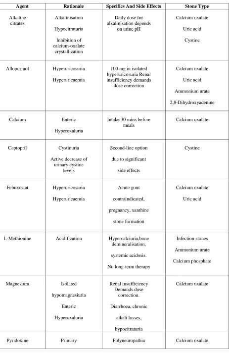
Table 5. Pharmacological substances used for stone prevention - characteristics, specifics and side-effects
Agent Rationale Specifics And Side Effects Stone Type
Alkaline citrates
Alkalinisation
Hypocitraturia
Inhibition of calcium-oxalate
crystallization
Daily dose for alkalinisation depends
on urine pH
Calcium oxalate
Uric acid
Cystine
Allopurinol Hyperuricosuria
Hyperuricaemia
100 mg in isolated hyperuricosuria Renal insufficiency demands
dose correction
Calcium oxalate
Uric acid
Ammonium urate
2,8-Dihydroxyadenine
Calcium Enteric
Hyperoxaluria
Intake 30 mins before meals
Calcium oxalate
Captopril Cystinuria
Active decrease of urinary cystine
levels
Second-line option
due to significant
side effects
Cystine
Febuxostat Hyperuricosuria
Hyperuricaemia
Acute gout
contraindicated,
pregnancy, xanthine
stone formation
Calcium oxalate
Uric acid
L-Methionine Acidification Hypercalciuria,bone demineralisation,
systemic acidosis.
No long-term therapy
Infection stones
Ammonium urate
Calcium phosphate
Magnesium Isolated
hypomagnesiuria
Enteric
Hyperoxaluria
Renal insufficiency Demands dose
correction.
Diarrhoea, chronic
alkali losses,
hypocitraturia
Calcium oxalate
Hyperoxaluria
Thiazide Hypercalciuria
Risk for agent- induced hypotonic blood pressure,
diabetes, hyperuricaemia,
hypokalaemia, followed by intracellular acidosis
and hypocitraturia
Calcium oxalate
Calcium phosphate
Tiopronin Cystinuria Active decrease of urinary
cystine levels
Risk for tachyphylaxis and proteinuria
Cystine
CONCLUSION
Kidney stone is one of the oldest recorded disorder of human and one of the major health burden. Now a days large number of peoples are affected with this disorder all over the world. The prevalence of urinary calculi is estimated to be range between 4%-20% in the general Population. In patients with all normal urinary parameters (idiopathic) the patient is advised dietary restriction and kept on periodic surveillance. In patients with multiple deranged parameters the drug approach in a permutation combination rationale is applied with periodic surveillance of the parameters at repeated intervals for dose modification or temporary discontinuation of the drug therapy. Both surgical and medical treatment is necessary for the complete management of patients of urolithiasis. In this review article, it can be concluded that the Lifestyle, dietary changes and proper use of medicines are the ideal preventive formula against kidney stones.
REFERENCES
1. Dugdale and David “Female urinary tract”. Medline plus Medical Encyclopedia. 16 september 2011, 1122.
2. Hesse A, Bra¨ndle E, Wilbert D, Ko¨hrmann K-U, Alken P. Study on the prevalence and incidence of urolithiasis in Germany comparing the years 1979 vs. 2000. Eur Urol 2003;44:709–13.
3. Lotan Y, Cadeddu JA, Roerhborn CG, Pak CY, Pearle MS. Costeffectiveness of medical management strategies for nephrolithiasis. J Urol 2004;172:2275–81.
4. Worcester EM. Inhibitors of stone formation. Semin Nephrol, 16, 1996, 474-486.
5. P Amudha , Nagalakshmi Swetha Y and Ghanapiyari Sharma.Evaluation of antinephrolithiatic activity of ethanolic extract of Rosa indica linn on ethylene glycol induced male wistar albino rats. International Journal of Phytopharmacology. 6(4), 2015, 191-195.
6. Fakheri RJ, Goldfarb DS. Ambient temperature as a contributor to kidney stone formation: implications of global warming. Kidney Int 2011;79:1178–85.
7. Moe OW. Kidney stones: pathophysiology and medical management. Review. Lancet 2006;367:333–44.
8. Lieske JC, Pena de la Vega LS, Slezak JM, et al. Renal stone epidemiology in Rochester, Minnesota: an update. Kidney Int 2006;69:760– 4
9. Lopez, M. and B. Hoppe. History, epidemiology and regional diversities of urolithiasis. Pediatr. Nephrol., 2010;25: 49-59.
10. Stamatelou, K.K., M.E. Francis, C.A. Jones, L.M. Nyberg Junior and G.C. Curhan, 2003. Time trends in reported prevalence of kidney stones in the United States: 1976-1994. KidneyInternational.,63:1817-1823.
11. Menon M, Resnick MI. Urinary lithiasis: etiology, diagnosis, and medical management. In: Campbell MF, Walsh PC, Retik AB, eds. Campbell’s Urology. 8th ed. Philadelphia, Pa.: Saunders, 2002.
12. Asselman M, Verhulst A, De Broe ME, Verkoelen CF. Calcium oxalate crystal adherence to hyaluronan–, osteopontin–, and CD44– expressing injured/regenerating tubular epithelial cells in rat kidneys. J Am Soc Nephrol 2003;14:3155–66.
13. Randall A. Recent Advances in Knowledge Relating to the Formation, Recognition and Treatment of Kidney Calculi. Bull N Y Acad Med 1944;20:473–84.
14. Asselman M, Verhulst A, Van Ballegooijen ES, et al. Hyaluronan is apically secreted and expressed by proliferating or regenerating renal tubular cells. Kidney Int 2005;68:71–83.
15. Parks JH, Coe FL. An increasing number of calcium oxalate stone events worsens treatment outcome. Kidney Int 1994;45:1722–30. 16. Stamatelou, K.K., M.E. Francis, C.A. Jones, L.M. Nyberg Junior and G.C. Curhan, 2003. Time trends in reported prevalence of kidney
stones in the United States: 1976-1994. Kidney Int., 63: 1817-1823.
19. Keoghane, S., et al. The natural history of untreated renal tract calculi. BJU Int, 2010. 105: 1627.
20. Straub, M., et al. Diagnosis and metaphylaxis of stone disease. Consensus concept of the National Working Committee on Stone Disease for the upcoming German Urolithiasis Guideline. World J Urol, 2005. 23: 309.
21. Hesse, A.T., et al. (Eds.), Urinary Stones, Diagnosis, Treatment and Prevention of Recurrence. 3rd edition. 2009, Basel.
22. Basiri, A., et al. Familial relations and recurrence pattern in nephrolithiasis: new words about old subjects. Urol J, 2010. 7: 81.
23. Goldfarb, D.S., et al. A twin study of genetic and dietary influences on nephrolithiasis: a report from the Vietnam Era Twin (VET) Registry. Kidney Int, 2005. 67: 1053.
24. Asplin, J.R., et al. Hyperoxaluria in kidney stone formers treated with modern bariatric surgery.J Urol, 2007. 177: 565. 25. Gonzalez, R.D., et al. Kidney stone risk following modern bariatric surgery. Curr Urol Rep, 2014. 15:401.
26. Rendina, D., et al. Metabolic syndrome and nephrolithiasis: a systematic review and meta-analysis of the scientific evidence. J Nephrol, 2014. 27: 371.
27. Dell’Orto, V.G., et al. Metabolic disturbances and renal stone promotion on treatment with topiramate: a systematic review. Br J Clin Pharmacol, 2014. 77: 958.
28. Mufti, U.B., et al. Nephrolithiasis in autosomal dominant polycystic kidney disease.J Endourol, 2010. 24: 1557.
29. Chen, Y., et al. Current trend and risk factors for kidney stones in persons with spinal cord injury: a longitudinal study. Spinal Cord, 2000. 38: 346.
30. Seftel A, Resnick MI: Metabolic evaluation of urolithiasis. Urol Clin North Am 1990;17: 159–169.
31. Asplin J, Parks J, Lingeman J, Kahnoski R, Mardis H, Lacey S, Goldfarb D, Grasso M, Coe F. Supersaturation and stone composition in a network of dispersed treatment sites. J Urol. 1998;159:1821–1825.
32. Asplin JR. Nephrolithiasis: introduction. Semin Nephrol. 2008;28:97–98.
33. Coe FL, Wise H, Parks JH, Asplin JR. Proportional reduction of urine supersaturation during nephrolithiasis treatment. J Urol. 2001;166:1247–1251.
34. Borghi L, Guerra A, Meschi T, Briganti A, Schianchi T, Allegri F, Novarini A. Relationship between supersaturation and calcium oxalate crystallization in normals and idiopathic calcium oxalate stone formers. Kidney Int. 1999;55:1041–1050.
35. Carl A. Osborne, D. P. (n.d.). Minnesota Urolith Center, University of Minnesota. Monitoring CaOx Urolith Prevention ,2001;1-2. 36. Flannigan R, Choy WH, Chew B, Lange D. Renal struvite stones-pathogenesis, microbiology, and management strategies. Nat Rev
Urol. 2014;11:333–341.
37. Romero-Vargas L, Barba Abad J, Rosell Costa D, Pascual Piedrola JI. Staghorn stones in renal graft. Presentation on two cases report and review the bibliography. Arch Esp Urol. 2014;67:650–653.
38. Trinchieri A. Urinary calculi and infection. Urologia. 2014;81:93–98.
39. Moe, OW. "Kidney stones: pathophysiology and medical management". The Lancet. 2006;367 (9507): 333– 44. 40. Cameron MA, Sakhaee K. Uric acid nephrolithiasis. Urol Clin North Am. 2007;34:335–346.
41. Coe FL, Parks JH, Asplin JR. The pathogenesis and treatment of kidney stones. N Engl J Med. 1992;327:1141–1152. 42. Moran ME. Uric acid stone disease. Front Biosci. 2003;8:s1339–s1355.
43. Beara-Lasic L, Pillinger MH, Goldfarb DS. Advances in the management of gout: critical appraisal of febuxostat in the control of hyperuricemia. Int J Nephrol Renovasc Dis., 2010;3:1–10.
44. Kenny JE, Goldfarb DS. Update on the pathophysiology and management of uric acid renal stones. Curr Rheumatol Rep. 2010;12:125– 129.
45. Curhan GC, Taylor EN. 24-h uric acid excretion and the risk of kidney stones. Kidney Int. 2008;73:489–496.
46. Chillarón J , Font-Llitjós M , Fort J , Zorzano A , Goldfarb DS , Nunes V , Palacín M 2010 Pathophysiology and treatment of cystinuria. Nat Rev Nephrol., 2010; 6:424–434
47. Sakhaee K 1996 Pathogenesis and medical management of cystinuria. Semin Nephrol.,1996; 16:435–447.
48. Dello Strologo L , Pras E , Pontesilli C , Beccia E , Ricci-Barbini V , de Sanctis L , Ponzone A , Gallucci M , Bisceglia L , Zelante L , Jimenez-Vidal M , Font M , Zorzano A , Rousaud F , Nunes V , Gasparini P , Palacín M , Rizzoni G 2002 Comparison between SLC3A1 and SLC7A9 cystinuria patients and carriers: a need for a new classification. J Am Soc Nephrol., 2002;13:2547–2553.
49. Font-Llitjós M , Jiménez-Vidal M , Bisceglia L , Di Perna M , de Sanctis L , Rousaud F , Zelante L , Palacín M , Nunes V 2005 New insights into cystinuria: 40 new mutations, genotype-phenotype correlation, and digenic inheritance causing partial phenotype. J Med Genet., 2005; 42:58–68.