Foreign Body Granuloma of the External Auditory Canal
Kevin C. Harris, MD*; Stephen F. Conley, MD*‡; and Joseph E. Kerschner, MD*
ABSTRACT. External auditory canal polyps are most commonly inflammatory in nature but may also manifest more severe disease. Prolonged conservative therapy may delay the correct diagnosis and appropriate inter-vention. A case is presented of a child with chronic otorrhea treated for 4 months with topical drops and antibiotics. On referral, a large external auditory canal polyp was confirmed to represent a foreign body granu-loma covering a large electrical cap, with erosion ap-proaching the facial nerve. External auditory canal pol-yps that fail to respond promptly to conservative medical therapy warrant a computed tomography scan and surgi-cal exploration with biopsy.Pediatrics2004;113:e371–e373. URL: http://www.pediatrics.org/cgi/content/full/113/4/e371; aural polyp, external auditory canal, EAC, foreign body gran-uloma.
ABBREVIATIONS. EAC, external auditory canal; CT, computed tomography; LCH, Langerhans’ cell histiocytosis.
A
ural polyps typically present in the external auditory canal (EAC) but may arise from the external ear, middle ear, or adjacent struc-tures. The polyps usually are associated with otor-rhea and present as chronic otitis media with or without cholesteatoma.1 Conservative treatmentwith topical steroids and antibiotics is recommended before surgical intervention.2 However, prolonged
topical therapy for persistent aural polyps may delay diagnosis of more serious disease.1This case report
emphasizes the importance of additional patient evaluation in patients with persistent aural polyps and explores the differential diagnoses.
CASE REPORT
A 9-year-old boy presented with a 4-month history of painless left otorrhea diagnosed as chronic otitis media by his pediatrician. There was no prior history of external otitis, otitis media, trauma, or otologic surgery. Treatment over 4 months with repeated courses of oral antibiotics and topical antimicrobial/steroid drops were without effect. On referral, the left external ear and mastoid were not tender to palpation. A large granulation tissue polyp was noted in the EAC, originating from the posterior-inferior area. The visible portion of the tympanic membrane was mobile without apparent middle ear effusion. A computed tomography (CT) scan of the temporal bones confirmed an EAC mass with partial mastoid air cell opacification but without signs of mastoid bone destruction. The patient underwent surgical exploration, with a
granulation tissue polyp found to conceal a plastic electrical cap measuring 0.6⫻1.8 cm (Fig 1). After removal of the foreign body, the inferior EAC defect was packed with topical antimicrobial/ steroid drop-soaked Gelfoam and left to heal by secondary inten-tion. Review of the preoperative CT scan verified the conical object eroding inferiorly through the bony EAC toward the styloid pro-cess adjacent to the facial nerve (Fig 2). Facial nerve function remained normal after surgery. Final histopathology revealed for-eign body giant cell reaction with chronic inflammation, consistent with a foreign body granuloma. At the 2-month follow-up exam-ination, the patient’s EAC defect had healed and completely epi-thelialized. Prolonged clinical surveillance over 2 years confirmed no persistent defect of the EAC or evidence of EAC cholesteatoma.
DISCUSSION
An EAC polyp may arise from the external ear, middle ear, or adjacent structures such as the parotid and temporomandibular joint. These polyps are as-sociated with numerous pathologies ranging from inflammatory to malignant processes. The most com-mon cause of aural polyps in children is chronic otitis media with or without cholesteatoma.1The incidence
of cholesteatoma in ears presenting with polyps var-ies from 25% to 45%, increasing up to 60% in chil-dren.3 Gliklich et al4reviewed 35 pediatric patients
presenting with primary aural polyps and found 43% with the diagnosis of chronic otitis media with effusion, 29% with cholesteatoma, and 23% with re-tained ventilation tubes. Although no malignant tu-mors were identified in that series, the overall inci-dence of either granulomatous or neoplastic disease is estimated at up to 3% of surgical specimens in some studies.4,5
Foreign bodies of the EAC follow chronic otitis media in frequency as an inflammatory etiology for an EAC polyp. Such foreign bodies may take many forms and are a relatively common problem seen and managed by a variety of physicians and other health care providers. More difficult cases are often referred to otolaryngologists for removal. In their retrospec-tive review of 698 cases of pediatric EAC foreign bodies, Schulze et al6suggest otolaryngologic
refer-ral for specific foreign body types, location near the tympanic membrane, presence for⬎24 hours, patient age of ⬍4 years, and for those foreign bodies that have failed prior removal attempts. Presenting symptoms of an aural foreign body may include pain, drainage, bleeding, hearing loss, or a sugges-tive history by the guardian. On examination, the foreign body is usually evident; however, a second-ary otitis externa may obscure the diagnosis. If the process is prolonged, an inflammatory granulation tissue polyp may be seen, as occurred in this re-ported case.
Foreign body granulomas arise from a
well-de-From the Departments of *Otolaryngology and Communication Sciences and ‡Pediatrics, Medical College of Wisconsin, Milwaukee, Wisconsin. Received for publication Jun 20, 2003; accepted Nov 20, 2003.
Address correspondence to Stephen F. Conley, MD, Medical College of Wisconsin, 9000 W Wisconsin Ave, Box 1997, Milwaukee, WI 53201. E-mail: sfconley@mcw.edu
PEDIATRICS (ISSN 0031 4005). Copyright © 2004 by the American Acad-emy of Pediatrics.
scribed foreign body reaction.7 The hallmark of all
foreign body reactions is phagocytosis or attempted phagocytosis. It begins with the recognition of a “non–self-antigen” by immunogenic cells, which at-tempt to remove the foreign body via phagocytosis. If the object is too large, the phagocytes unite to form foreign body giant cells in an attempt to surround and isolate the foreign body. If the object is macro-scopic, the giant cells fail, and the inflammatory pro-cess becomes persistent, forming foreign body gran-ulomas. As seen in this report, the foreign body reaction has the ability to be an erosive process as well. Thus, foreign body granulomas are foci of per-sistent inflammation associated with material (in this case, a plastic electrical cap) that the body is unsuc-cessfully attempting to remove by phagocytosis. However, foreign body granulomas also have been described in relation to many other materials
includ-ing hair, talc, chloromycetin powder, cotton, and Dacron.7
Other morbid diseases may present less commonly as EAC polyps. Langerhans’ cell histiocytosis (LCH) is a disease of unknown etiology that unifies the entities of eosinophilic granuloma, Hand-Schuller-Christian disease, and Lettere-Siwe disease. It is a destructive process that can present as single- or multisystem disease with involvement of the head and neck in 70% to 80% of affected children.8LCH
occurs from the proliferation of histiocytic (or Lang-erhans) cells, which have antigen-processing abilities and are characterized ultrastructurally by the pres-ence of tennis-racket–shaped Birbeck granules. The disease is rare, with the estimated incidence in the pediatric population of 3 cases per 1 million children per year.9 Quraishi et al10 reported that 50% of
pa-tients with LCH had aural symptoms, of which ap-proximately half had no other lesions at the time of presentation. LCH may be misdiagnosed frequently as otitis externa or chronic otitis media.1For
single-system disease, postauricular swelling and otorrhea are most common, whereas multisystem disease presents with skin rash, otorrhea, organomegaly, lymphadenopathy, pulmonary symptoms, oral le-sions, or cerebellar dysfunction.9,11
LCH is suggested if an aural polyp is seen without disease of the middle ear. Aural polyps in LCH tend to arise in the mastoid and erode the posterior-supe-rior canal wall, often sparing the middle ear and ossicles; the tympanic membrane is usually normal. CT scanning will reveal an osteolytic lesion, but the diagnosis is made histologically. Patients with sgle-site disease can be managed with topical or in-tralesional corticosteroids, with or without polypec-tomy or curettage, whereas multisystem disease may be treated with high-dose corticosteroids with con-sideration of chemotherapy. Low-dose radiation therapy is reserved only for symptomatic control of persistent pain or when vital structures are at risk.8
Other rare inflammatory causes of aural polyps include granulomatous diseases or fungal infection.3
Although much more common in the early 20th cen-tury, tuberculous granulation tissue polyps, typically painless, continue to occur today.12 Mycobacterium
tuberculosiswas the underlying diagnosis in 1 of 35 patients in the Gliklich et al4study of children
pre-senting with aural polyps, with the histology dem-onstrating characteristic granulomas and microab-scesses with acid-fast bacilli present. In contrast, Wegener’s granulomatosis also may rarely present in children as an aural polyp, within the classic triad of necrotizing granulomas of the upper and lower re-spiratory tracts, focal or proliferative glomerulone-phritis, and systemic vasculitis of small- and medi-um-sized vessels.13 Otologic manifestations are a
frequent presenting complaint, with secretory otitis media and chronic suppurative otitis media being the most common. Finally, acquired immunodefi-ciency syndrome patients with extrapulmonary
Pneumocystis carinii infection may present with an aural polyp, possibly as the initial presentation of acquired immunodeficiency syndrome.3,14 –16
Infec-Fig 1. Plastic electrical cap retrieved from the EAC.
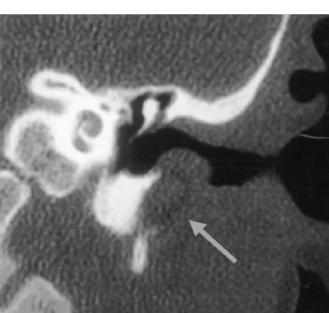
Fig 2. Coronal CT scan with the arrow indicating the foreign body eroding inferiorly through the left bony EAC toward the styloid process.
tion of the middle ear is thought to occur via retro-grade spread through the eustachian tube.
Frank neoplasms may rarely present as aural pol-yps. Rhabdomyosarcoma is a malignant tumor of mesenchymal tissue that involves the head and neck in 35% to 40% of cases.17The embryonal variation is
the most common subtype in the head and neck, whereas involvement of the temporal bone itself oc-curs in only 7% of cases.18In a study of 199 patients
with head and neck rhabdomyosarcoma, 22 origi-nated from the middle ear, and only 2 origiorigi-nated from the external ear.17 Among patients with
rhab-domyosarcoma originating in the ear, 83% presented with a mass in the external ear, 64% had ipsilateral cranial nerve palsy, and 42% had otorrhea. The di-agnosis of rhabdomyosarcoma is made histologi-cally, whereas staging is based on surgical resectabil-ity.18Treatment is radiation therapy in combination
with chemotherapy using vincristine, actinomycin, and cyclophosphamide.19Although staging depends
on completeness of surgical resection, the role of surgery may be limited to simple biopsy instead of mastoidectomy or temporal bone resection.20 When
the tumor is isolated to the external ear, the progno-sis seems favorable, thought to be secondary to prompt removal and lack of meningeal extension.21
Bearing in mind the more common infectious and inflammatory etiologies of aural polyps, initial treat-ment is appropriate culture-directed medical thera-py.4 Topical corticosteroid and antibiotic therapy
may lead to quick resolution of an aural polyp asso-ciated with chronic otitis media. In other disease processes, this therapy may reduce inflammation, polyp size, and drainage and simplify the diagnosis or surgical planning.2 For resistant disease,
knowl-edge of the differential diagnosis is important in avoiding a delay in diagnosis and potential morbid-ity. An audiogram and CT scan should be performed before any surgical intervention.4 Surgical
explora-tion, with adequate tissue specimens, is critical for a histologic diagnosis to guide therapy.
This case illustrates the potential difficulties from prolonged treatment of an aural polyp without a definitive diagnosis. Although this polyp originated from a benign process, the facial nerve was at risk from the expanding foreign body granuloma. The morbidity of long-term EAC debridement and pos-sible EAC reconstruction fortunately were avoided in this case. Delay in diagnosis of a neoplastic pro-cess has even greater potential for morbidity.
CONCLUSIONS
Aural polyps should receive initial treatment with otic drops and oral antibiotics as indicated. If the
process persists after several weeks of this therapy, strong consideration should be given to referral for additional diagnosis and treatment. CT imaging is key in assessing any associated bone destruction and may aid diagnosis. Surgical exploration and biopsy may be needed for diagnosis and should be under-taken in a timely fashion to avoid morbidity associ-ated with a delay in diagnosis.
REFERENCES
1. DeRowe A, Bernheim J, Ophir D. Eosinophilic granuloma presenting as chronic otitis media: pitfalls in the diagnosis of aural polyps in children. J Otolaryngol. 1995;24:258 –260
2. Hussain SS. Conservative treatment in the management of inflamma-tory aural polyp.J Laryngol Otol. 1992;106:313–315
3. Tay HL, Hussain SS. The management of aural polyps.J Laryngol Otol. 1997;111:212–214
4. Gliklich RE, Cunningham MJ, Eavy RD. The cause of aural polyps in children.Arch Otolaryngol Head Neck Surg. 1993;119:669 – 671 5. Williams SR, Robinson PJ, Brightwell AP. Management of the
inflam-matory aural polyp.J Laryngol Otol. 1989;103:1040 –1042
6. Schulze SL, Kerschner JE, Beste DJ. Pediatric external auditory canal foreign bodies: a review of 698 cases. Otolaryngol Head Neck Surg. 2002;127:73–78
7. Jahn AF, Hawke M. Foreign body granulomas of the ear.J Otolaryngol. 1976;5:221–226
8. Irving RM, Broadbent V, Jones NS. Langerhans’ cell histiocytosis in childhood: management of head and neck manifestations.Laryngoscope. 1994;104:64 –70
9. Angeli SI, Hoffman HT, Alcalde J, Smith RJ. Langerhans’ cell histiocy-tosis of the head and neck in children.Ann Otol Rhinol Laryngol. 1995; 104:173–180
10. Quraishi MS, Blayney AW, Breatnach FB. Aural symptoms as primary presentation of Langerhans’ cell histiocytosis.Clin Otolaryngol. 1993;18: 317–323
11. Toohill RJ, Kidder TM, Eby LG. Eosinophilic granuloma of the temporal bone.Laryngoscope. 1973;83:877– 889
12. Friedmann I. Pathological lesions of the external auditory meatus: a review.J R Soc Med. 1990;83:34 –37
13. Vartiainen E, Nuutinen J. Head and neck manifestations of Wegener’s granulomatosis.Ear Nose Throat J. 1992;71:423– 428
14. Praveen CV, Terry RM, Elmahallawy M, Horsfield C. Pneumocystis cariniiinfection in bilateral aural polyps in a human immunodeficiency virus-positive patient.J Laryngol Otol. 2002;116:288 –290
15. Patel SK, Philpott JM, McPartlin DW. An unusual case ofPneumocystis cariniipresenting as an aural mass.J Laryngol Otol. 1999;113:555–557 16. Park S, Wunderlich H, Goldenberg RA, Marshall M.Pneumocystis carinii
infection in the middle ear.Arch Otolaryngol Head Neck Surg. 1992;118: 269 –270
17. Brugler G. Tumors presenting as aural polyps: a report of four cases. Pathology. 1992;24:315–319
18. Wiatrak BJ, Pensak ML. Rhabdomyosarcoma of the ear and temporal bone.Laryngoscope. 1989;99:1188 –1192
19. Crist WM, Anderson JR, Meza JL, et al. Intergroup rhabdomyosarcoma study-IV: results for patients with nonmetastatic disease.J Clin Oncol. 2001;19:3091–3102
20. Goepfert H, Cangir A, Lindberg R, Ayala A. Rhabdomyosarcoma of the temporal bone: is surgical resection necessary?Arch Otolaryngol Head Neck Surg. 1979;105:310 –313
21. Raney RB Jr, Lawrence W Jr, Maurer HM, et al. Rhabdomyosarcoma of the ear in childhood. A report from the Intergroup Rhabdomyosarcoma Study-I.Cancer. 1983;51:2356 –2361
http://www.pediatrics.org/cgi/content/full/113/4/ at Viet Nam:AAP Sponsored on August 29, 2020 e371 e373
www.aappublications.org/news
DOI: 10.1542/peds.113.4.e371
2004;113;e371
Pediatrics
Kevin C. Harris, Stephen F. Conley and Joseph E. Kerschner
Foreign Body Granuloma of the External Auditory Canal
Services
Updated Information &
http://pediatrics.aappublications.org/content/113/4/e371 including high resolution figures, can be found at:
References
http://pediatrics.aappublications.org/content/113/4/e371#BIBL This article cites 21 articles, 1 of which you can access for free at:
Subspecialty Collections
orders_sub
http://www.aappublications.org/cgi/collection/ear_nose_-_throat_dis Ear, Nose & Throat Disorders
ub
http://www.aappublications.org/cgi/collection/dentistry:oral_health_s Dentistry/Oral Health
following collection(s):
This article, along with others on similar topics, appears in the
Permissions & Licensing
http://www.aappublications.org/site/misc/Permissions.xhtml in its entirety can be found online at:
Information about reproducing this article in parts (figures, tables) or
Reprints
http://www.aappublications.org/site/misc/reprints.xhtml Information about ordering reprints can be found online:
at Viet Nam:AAP Sponsored on August 29, 2020
www.aappublications.org/news
DOI: 10.1542/peds.113.4.e371
2004;113;e371
Pediatrics
Kevin C. Harris, Stephen F. Conley and Joseph E. Kerschner
Foreign Body Granuloma of the External Auditory Canal
http://pediatrics.aappublications.org/content/113/4/e371
located on the World Wide Web at:
The online version of this article, along with updated information and services, is
by the American Academy of Pediatrics. All rights reserved. Print ISSN: 1073-0397.
the American Academy of Pediatrics, 345 Park Avenue, Itasca, Illinois, 60143. Copyright © 2004 has been published continuously since 1948. Pediatrics is owned, published, and trademarked by Pediatrics is the official journal of the American Academy of Pediatrics. A monthly publication, it
at Viet Nam:AAP Sponsored on August 29, 2020
www.aappublications.org/news