A CRISPR Interference Platform for Efficient Genetic
Repression in
Candida albicans
Lauren Wensing,
aJehoshua Sharma,
aDeeva Uthayakumar,
aYannic Proteau,
aAlejandro Chavez,
bRebecca S. Shapiro
aaDepartment of Molecular and Cellular Biology, University of Guelph, Guelph, Ontario, Canada
bDepartment of Pathology and Cell Biology, Columbia University College of Physicians and Surgeons, New York, New York, USA
ABSTRACT
Fungal pathogens are emerging as an important cause of human
dis-ease, and
Candida albicans
is among the most common causative agents of fungal
infections. Studying this fungal pathogen is of the utmost importance and
necessi-tates the development of molecular technologies to perform comprehensive genetic
and functional genomic analysis. Here, we designed and developed a novel
clus-tered regularly interspaced short palindromic repeat interference (CRISPRi) system
for targeted genetic repression in
C. albicans
. We engineered a nuclease-dead Cas9
(dCas9) construct that, paired with a guide RNA targeted to the promoter of an
en-dogenous gene, is capable of targeting that gene for transcriptional repression. We
further optimized a favorable promoter locus to achieve repression and
demon-strated that fusion of dCas9 to an Mxi1 repressor domain was able to further
en-hance transcriptional repression. Finally, we demonstrated the application of this
CRISPRi system through genetic repression of the essential molecular chaperone
HSP90
. This is the first demonstration of a functional CRISPRi repression system in
C.
albicans
, and this valuable technology will enable many future applications in this
critical fungal pathogen.
IMPORTANCE
Fungal pathogens are an increasingly important cause of human
dis-ease and mortality, and
Candida albicans
is among the most common causes of
fun-gal disease. Studying this important funfun-gal pathogen requires a comprehensive
ge-netic toolkit to establish how different gege-netic factors play roles in the biology and
virulence of this pathogen. Here, we developed a CRISPR-based genetic regulation
platform to achieve targeted repression of
C. albicans
genes. This CRISPR
interfer-ence (CRISPRi) technology exploits a nuclease-dead Cas9 protein (dCas9) fused to
transcriptional repressors. The dCas9 fusion proteins pair with a guide RNA to target
genetic promoter regions and to repress expression from these genes. We
demon-strated the functionality of this system for repression in
C. albicans
and show that
we can apply this technology to repress essential genes. Taking the results together,
this work presents a new technology for efficient genetic repression in
C. albicans
,
with important applications for genetic analysis in this fungal pathogen.
KEYWORDS
CRISPR, CRISPRi,
Candida
,
Candida albicans
, fungal genetics, genetic
regulation, genetic technology
I
nvasive fungal infections have emerged as an important cause of human mortality,
particularly for an ever-increasing population of immunocompromised individuals
(1–3). The rise in the incidence of these opportunistic invasive infections is associated
with many factors, including the HIV/AIDS epidemic and the growing number of
patients receiving immunosuppressive therapeutics for bone marrow and organ
trans-plantations or for the treatment of autoimmune disorders (4). Invasive fungal infections
are associated with disproportionately high rates of patient mortality (
⬃
30% to 90%
mortality, depending on the pathogen and patient group [5, 6]), and with a massive
CitationWensing L, Sharma J, Uthayakumar D, Proteau Y, Chavez A, Shapiro RS. 2019. A CRISPR interference platform for efficient genetic repression inCandida albicans. mSphere 4:e00002-19.https://doi.org/10.1128/mSphere .00002-19.
EditorAaron P. Mitchell, Carnegie Mellon University
Copyright© 2019 Wensing et al. This is an open-access article distributed under the terms of theCreative Commons Attribution 4.0 International license.
Address correspondence to Rebecca S. Shapiro, [email protected].
L.W. and J.S. contributed equally to this article.
For a companion article on this topic, see
https://doi.org/10.1128/mSphere.00001-19.
Development of a new CRISPRi tool for genetic repression in Candida albicans, work by @ShapiroRebecca @UofG_MCB
Received4 January 2019 Accepted18 January 2019 Published13 February 2019
Synthetic Biology
crossm
on September 8, 2020 by guest
http://msphere.asm.org/
economic burden (
⬃
$7.2 billion in 2017) (4, 7, 8). Among these fungal pathogens,
Candida
species are among those representing the most common causes of infections,
accounting for
⬃
55% of invasive fungal infections in North America (4).
Candida
albicans
is the leading cause of invasive candidiasis and a leading cause of nosocomial
bloodstream infection (4).
C. albicans
is a polymorphic yeast species which exists as a
commensal member of the human microbiota and as an opportunistic pathogen, able
to cause disease ranging from relatively benign superficial infections to life-threatening
invasive infections.
As a critically important human fungal pathogen,
C. albicans
has been subjected to
in-depth molecular genetic analysis to uncover factors involved in its virulence,
inter-actions with the host, resistance to antifungal agents, and other important biological
processes. Previously,
C. albicans
was considered to be a highly intractable microbial
organism, due to limitations associated with genetic manipulation, including an
inabil-ity to stably maintain plasmids, an unusual form of codon usage (the CUG codon is
translated as serine instead of leucine [9]), inefficient homologous recombination, and
its diploid nature. However, in the last
⬃
10 years, new advances in functional genomic
technologies, as well as the discovery of mating-competent
C. albicans
haploid strains
(10), have enabled a growing number of large-scale functional genomic studies in this
clinically relevant pathogen. This important research has included the development of
new technologies for genetic manipulation in
C. albicans
, including genetic deletion
systems (11, 12), conditional expression systems (13), double-selection-based deletion
systems (14), and transposon mutagenesis platforms (15–17). These technologies have
been applied in a variety of innovative ways to identify genetic factors underpinning
C.
albicans
morphogenesis and biofilm formation (12, 18–21), fungus-host interactions
(12, 22), and mechanisms of antifungal drug resistance (16, 23–25) and for the
identi-fication of essential genes (13, 15, 26).
Despite this existing research repertoire of functional genomic studies in
C. albicans
,
new genetic tools continue to improve and refine our ability for targeted genetic
analysis. One example of a genetic tool that has revolutionized targeted genetic
manipulation in a diversity of fungal and other microbial species is clustered regularly
interspaced short palindromic repeat (CRISPR)-based technology (27). Recently,
CRISPR-based technologies have been applied for targeted genetic mutations and deletions in
C. albicans
(28–33), as well as in other closely related
Candida
species (34–36). Each of
these systems relies on the foundational CRISPR editing system, whereby a Cas9
endonuclease pairs with a single guide RNA (sgRNA), comprising a Cas9-binding region
(the conserved sgRNA “tail”) and a unique 20 nucleotide “N20” region complementary
to the targeted genomic locus. This sgRNA-Cas9 complex interacts with a locus based
on complementary binding of the sgRNA N20 to the target region, provided that a
necessary protospacer adjacent motif (PAM) is also present within the target locus.
After binding, the Cas9 endonuclease undergoes a conformational change, generating
a double-stranded break (DSB) within the DNA region (37). This DSB can then be
repaired via nonhomologous end joining (NHEJ) or via homology-directed repair when
repair donor DNA with homology to the region surrounding the DSB is provided. The
latter mechanism is what has been most commonly exploited for CRISPR-based genetic
manipulation in
Candida
and other yeast species (28–36, 38).
Since their development as genetic editing technologies (39), CRISPR systems have
been further modified to achieve alternative outcomes, such as base-editing (40–42),
RNA editing (43), epigenetic modifications (44, 45), and transcriptional regulation (46).
CRISPR transcriptional repression relies on a precisely mutated, nuclease-dead version
of the Cas9 endonuclease (dCas9), which is targeted to specific genomic promoter
regions by sgRNAs to achieve steric hindrance of RNA polymerase (Pol), thus blocking
transcription initiation or elongation (46–49). CRISPR interference (CRISPRi)-based
ge-netic repression was first demonstrated in mammalian cells and
Escherichia coli
(47) and
has since been applied in a diversity of other microbial species (50–53). Fusing
repressor domains to dCas9 can further enable transcriptional repression. For instance,
the Krüppel associated box (KRAB) and MeCP2 transcriptional repression domain can
on September 8, 2020 by guest
http://msphere.asm.org/
be fused to dCas9 to significantly enhance target gene repression in human cells (52),
and dCas9-Mxi1 fusions similarly enhance repression in
Saccharomyces cerevisiae
(52,
54). CRISPRi presents certain advantages in comparison to traditional CRISPR editing
systems: it facilitates the study of essential genes, enables a titratable system to
regulate the level of gene expression, is reversible, and is generally significantly easier
to engineer, as it does not rely on homology-directed repair or on the presence of
repair donor DNA templates. The CRISPRi framework can also be exploited for CRISPR
activation (CRISPRa) by fusing dCas9 to activator domains, such as VP64, to drive
transcriptional activation from a desired locus (55–57).
While CRISPR-based editing has been used to efficiently generate genetic mutations
and deletions in
Candida
species, the functionality of other CRISPR technologies, such
as CRISPRi and CRISPRa, has yet to be explored in these fungal pathogens. Here, we
present the first report detailing the design and execution of a CRISPRi platform for
genetic repression in
C. albicans
. Using
C. albicans
-optimized dCas9, we demonstrated
that CRISPRi can be used to repress gene expression in
C. albicans
and further
demonstrated that effector fusion constructs such as dCas9-Mxi1 can be used to
achieve high levels of transcriptional repression (
⬃
20-fold repression) for a target locus.
Finally, we use this optimized CRISPRi dCas9-Mxi1 system to demonstrate the ability to
repress expression of
HSP90
, the essential
C. albicans
molecular chaperone, and to
recapitulate phenotypes associated with its genetic depletion. Taken together, the
results reveal a novel genetic technology for efficient genetic repression in
C. albicans
,
with important applications for functional genomic analysis in this critical fungal
pathogen.
RESULTS
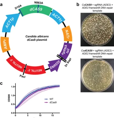
Design and preliminary validation of a CRISPRi system for
C. albicans
.
Initially,
to develop a CRISPRi system for genetic repression in
C. albicans
, we generated a
nuclease-dead version of Cas9 (dCas9), optimized for use in
C. albicans
. We exploited a
plasmid backbone that we had previously used for successful CRISPR-based genetic
deletions in
C. albicans
using Cas9 (33), modifying the
C. albicans
codon-optimized
CAS9
genes to contain two single nucleotide mutations (RuvC nuclease domain
muta-tion D10A and HNH nuclease domain mutamuta-tion N863A), previously associated with
impairment of nuclease function in Cas9 (47, 58). We incorporated this dCas9 into a
plasmid backbone to generate a single, “all-in-one” plasmid to facilitate CRISPRi
regu-lation in
C. albicans
(Fig. 1a). This plasmid contains all the required components to
achieve CRISPRi regulation in
C. albicans
and is readily modified to target any gene of
interest (Fig. 1a). The critical elements of this plasmid include the following: (i)
C.
albicans
-optimized dCas9; (ii) selection markers for bacteria (ampicillin resistance
[AMPr]) and
C. albicans
(nourseothricin resistance [NATr]); (iii) regions of homology to
the
C. albicans NEUT5L
locus to enable stable integration of the plasmid at this neutral
locus (59) upon plasmid linearization with restriction enzyme PacI; and (iv) a sgRNA
cloning locus, which contains two SapI restriction enzyme sites for efficient Golden
Gate cloning (60) of unique N20 sgRNA sequences, between the SNR52 RNA
polymer-ase III (Pol III) promoter used to drive sgRNA expression and the conserved sgRNA tail
(Fig. 1a). This permits simple, Golden Gate cloning of unique N20 sequences into the
dCas9 plasmid to target sgRNA-dCas9 to the promoter region of any gene of interest.
In order to assess whether the dCas9 construct was deficient in nuclease activity, we
compared it directly to an equivalent plasmid harboring the nonmutated
C. albicans
-optimized
CAS9
gene. We designed a sgRNA N20 targeting within the
C. albicans ADE2
open reading frame (ORF) for Cas9-mediated cleavage, as well as a repair DNA template
introducing a frameshift mutation into the
ADE2
ORF, thereby introducing a premature
stop codon, and a loss-of-function allele of
ADE2
. The
ADE2
-targeting N20 sequence
was ligated into both the Cas9 and dCas9 plasmids at the SapI sites, and the plasmids
were transformed into
C. albicans
strains along with the repair DNA. As expected, we
found that the Cas9 construct was able to mutate
ADE2
, based on the presence of red
colonies on the transformation plate (Fig. 1b). However, the dCas9 construct was
on September 8, 2020 by guest
http://msphere.asm.org/
unable to cause double-strand breaks and thus was unable mutate
ADE2
, and produced
no red colonies (Fig. 1b), indicating that dCas9 has in fact lost its nuclease activity.
Next, we assessed whether the dCas9 construct imparted any significant fitness
defect to the
C. albicans
strains. We monitored growth of a wild-type
C. albicans
strain,
compared to one harboring the dCas9 plasmid integrated at the
NEUT5L
locus. This
dCas9 strain contains an irrelevant, nontargeting sgRNA (including the
SNR52
promoter
and complete sgRNA with an sgRNA tail and an N20 that does not target the
C. albicans
genome) and the dCas9 and other components of this plasmid. Results from our
growth curve analysis indicated that strains harboring the dCas9 plasmid grew
com-parably to the wild-type strains and achieved the same maximum cell density, as
monitored by optical density (OD
600) (Fig. 1c). We subsequently used this dCas9 strain
FIG 1 Design and validation of a CRISPRi system forC. albicans. (a) dCas9 plasmid engineered for CRISPRi repression. This dCas9-based plasmid represents an all-in-one system for CRISPRi repression inC. albicans. All components have been codon optimized forC. albicans, and two nuclease mutations (D10A and N863A) have been introduced into Cas9 to render it nuclease-dead (dCas9).NEUT5Lhomology is present for integration into theC. albicansgenome upon plasmid linearization with PacI. The two SapI cloning sites allow simple sgRNA N20 cloning to generate unique sgRNAs. (b) dCas9 is deficient with respect to its nuclease function. Side-by-side comparisons ofC. albicanstransformation plates were performed using a Cas9 and dCas9 plasmid with sgRNAs targeting the
ADE2ORF for Cas9-mediated DSB. The two strains were cotransformed with a repair donor DNA template harboring a frameshift mutation to generate a premature stop codon in theADE2gene, leading to loss of function, and a red phenotype. Absence of observed red colonies upon transformation of the dCas9 construct suggests that it was deficient with respect to its nuclease activity. CaCAS9,C. albicansCas9. (c) The dCas9 plasmid integrated in theC. albicansgenome does not affect growth. Growth curves were performed using a wild-typeC. albicansstrain and one with the dCas9 plasmid integrated in its genome at theNEUT5Llocus. The dCas9-containing strain did not show a defect in growth compared to the wild-type (WT) strain.
on September 8, 2020 by guest
http://msphere.asm.org/
with nontargeting sgRNA as the wild-type control strain for future experiments. This
further validates the utility of the dCas9-based CRISPRi system for genetic repression in
C. albicans
.
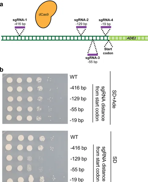
Optimization of CRISPRi for genetic repression in
C. albicans
.
Next, we aimed to
assess whether our CRISPRi system could achieve transcriptional repression of the
genes of
C. albicans
. We further aimed to assess which region of a promoter would be
optimal for targeting the sgRNA-dCas9 complex in order to achieve maximal
transcrip-tional repression. This was critical, as previous studies have found significant variability
in CRISPRi-based repression levels, depending on the region targeted by dCas9 (47, 52).
In order to determine if CRISPRi could repress transcription in
C. albicans
and the
optimal targeting locus, we designed a CRISPRi system targeting the endogenous
ADE2
gene as a reporter. We designed four unique sgRNA N20s, targeting regions 416 bp
(sgRNA-1), 129 bp (sgRNA-2), 55 bp (sgRNA-3), and 19 bp (sgRNA-4) upstream of the
ADE2
start codon, respectively (Fig. 2a). sgRNAs 1, 2, and 4 mapped to the sense DNA
strand, while sgRNA 3 mapped to the antisense strand. Each of these four N20
sequences was cloned into the dCas9 backbone (Fig. 1a) to generate four unique
plasmids targeting different regions upstream of
ADE2
for CRISPRi-based repression,
and these constructs were used to generate four CRISPRi-
ADE2 C. albicans
strains.
In order to monitor repression of
ADE2
transcription, we monitored growth of these
four
C. albicans
strains, compared to growth of a wild-type control strain, on media
containing or lacking adenine, as strains with depleted levels of
ADE2
should be
impaired in their ability to grow in the absence of supplemented adenine. We
per-formed this assay since we observed that, unlike genetic deletion or mutation of
ADE2
,
transcriptional repression was not sufficient to render cells red. Results from serial
dilution spotting assays on synthetic defined (SD) minimal media with or without
supplemented adenine indicated that while all strains grew equally well on SD plus
adenine, two of the CRISPRi
ADE2
depletion strains (those with sgRNAs targeting dCas9
129 bp and 55 bp upstream of the
ADE2
start codon) had impaired growth on medium
lacking adenine (Fig. 2b). This suggests that our CRISPRi repression system is functional
in
C. albicans
and is capable of repressing transcription from the endogenous
ADE2
locus, as indicated by the growth defect seen in the absence of supplemented adenine.
It further indicates that, for
ADE2
repression, maximal transcriptional repression is
achieved
⬃
55 to 129 bp upstream of the start codon and that CRISPRi is likely not
strand specific in
C. albicans
, as both sense and antisense sgRNAs are capable of
achieving repression (in agreement with what has been documented in
S. cerevisiae
[61]). This suggests important design principles for generation of additional CRISPRi
constructs in
C. albicans
.
Enhanced CRISPRi repression with dCas9-repressor fusion constructs.
Since we
were able to demonstrate transcriptional repression from the
ADE2
locus using a simple
dCas9 CRISPRi construct in
C. albicans
, we next wanted to assess whether we could
enhance transcriptional repression by fusing dCas9 to repressor domains. We chose
two transcriptional repressors for dCas9 fusion: (1) Mxi1, a mammalian transcriptional
repressor domain, previously reported to enhance CRISPRi-based repression in
S.
cerevisiae
(52) and suggested to interact with the yeast histone deacetylase and
transcriptional repressor Sin3 (52, 62); and (2) Mig1, a well-characterized
S. cerevisiae
transcriptional repressor protein (63) that has also been demonstrated to enhance
CRISPRi repression in
S. cerevisiae
(64). Therefore, we designed
C. albicans
codon-optimized versions of Mxi1 and Mig1 and engineered two additional dCas9 CRISPRi
plasmids with Mxi1 or Mig1 fused to C-terminal end of dCas9 (Fig. 3a; see also Fig. S1
in the supplemental material).
In order to determine if the dCas9, dCas9-Mxi1, and dCas9-Mig1 constructs would be
able to repress expression to various degrees, we cloned the sgRNA N20 targeting
129 bp upstream of the
ADE2
promoter into these plasmids. We then transformed these
constructs into
C. albicans
to generate strains with three unique CRISPRi constructs,
each targeting the same
ADE2
locus for repression (Fig. 3a; see also Fig. S1). We
on September 8, 2020 by guest
http://msphere.asm.org/
FIG 2 Optimization of CRISPRi for genetic repression inC. albicans. (a) Promoter region of theADE2gene targeted with sgRNAs. Four sgRNAs were designed at four distinct loci upstream of theADE2start codon (⫺416,⫺129,⫺55, and⫺19 bp upstream). (b) Identifying a promoter region for CRISPRi targeting.
C. albicansstrains were generated, each of which contained a dCas9 plasmid and one of the four sgRNAs described for panel a. To determine the extent ofADE2repression, growth was monitored by serial dilution spotting assays on SD media with or without supplemented adenine (Ade). Two strains (those with⫺129-bp and⫺55-bp sgRNAs) showed reduced growth on SD medium without adenine, suggesting that those strains successfully repressedADE2.
on September 8, 2020 by guest
http://msphere.asm.org/
monitored repression of
ADE2
using serial dilution spotting assays on SD minimal
media with or without adenine and confirmed that the dCas9 construct was able to
repress
ADE2
expression, based on reduced growth in the absence of supplemented
adenine (Fig. 3b). The dCas9-Mig1 strain demonstrated reduced growth in the absence
of adenine to an extent similar to that seen with the dCas9 strain, while the dCas9-Mxi1
strain showed significantly reduced growth in the absence of adenine, suggesting that
FIG 3 CRISPRi repression with dCas9-repressor fusion constructs. (a) dCas9 fusion constructs for CRISPRi repression. The diagram depicts the three dCas9 constructs (dCas9, dCas9-Mig1, and dCas9-Mxi1) engineered for CRISPRi repression inC. albicans. sgRNAs 1, 2, and 4 are on the sense strand; sgRNA 3 is antisense. RNAP, RNA polymerase. (b) dCas9-Mxi1 enhances repression from theADE2locus.C. albicansstrains were generated, with each containing a dCas9 plasmid (dCas9, dCas9-Mig1, or dCas9-Mxi1), each with the same sgRNA targetingADE2. To determine the extent ofADE2repression, growth was monitored by serial dilution spotting assays on SD media with or without supplemented adenine. While dCas9 and dCas9-Mig1 showed reduced growth on SD without adenine medium, dCas9-Mxi1 showed further growth reduction, suggesting that this construct most effectively repressedADE2expression. (c) Growth curves confirming dCas9-based repression from the ADE2locus. Wild-type, dCas9, and dCas9-Mxi1C. albicansstrains were grown in liquid SD media with or without supplemented adenine, and growth kinetics were monitored over⬃18 h. Both CRISPRi strains showed reduced growth in the absence of adenine, and the dCas9-Mxi1 strains showed further growth reduction. (d) qRT-PCR confirmed the reducedADE2transcript levels. To validate the transcriptional repression via CRISPRi, qRT-PCR was performed on wild-type, dCas9, and dCas-Mxi1 strains.ADE2transcripts were monitored and normalized to anACT1transcript as a housekeeping gene. Data were plotted as fold repression ofADE2relative to the wild-type control strain. Error bars depict standard errors of the means (SEM).
on September 8, 2020 by guest
http://msphere.asm.org/
this strain was able to repress expression of
ADE2
most effectively (Fig. 3b). Two
independently generated dCas9-Mxi1 strains were tested for
ADE2
repression via
growth on medium lacking adenine, and the two demonstrated the same phenotype
(data not shown).
To further confirm this finding, we monitored growth kinetics of wild-type, dCas9,
and dCas9-Mxi1
C. albicans
strains in liquid SD minimal media with or without adenine
over 18 h and confirmed that both the dCas9 and dCas9-Mxi1 strains were impaired in
growth in the absence of adenine, suggesting that
ADE2
was repressed in those CRISPRi
strains (Fig. 3c). And, similarly to what we observed in serial dilution spotting assays, the
dCas9-Mxi1 strain grew less well than the dCas9 strain, suggesting that this strain
achieved higher levels of
ADE2
repression (Fig. 3c). Finally, to quantify the level of
transcriptional repression achieved in the
C. albicans
dCas9 and dCas9-Mxi1 strains, we
used quantitative reverse transcription-PCR (qRT-PCR) to monitor the relative
expres-sion levels of
ADE2
in these strains. We found that the dCas9 strain was able to achieve
⬃
7-fold repression of
ADE2
, while the dCas9-Mxi1 strain showed
⬃
20-fold repression of
ADE2
(Fig. 3d). Taking the data together, this suggests that the dCas9-Mxi1 fusion
construct will be a valuable tool to efficiently repress transcription of genes in
C.
albicans
.
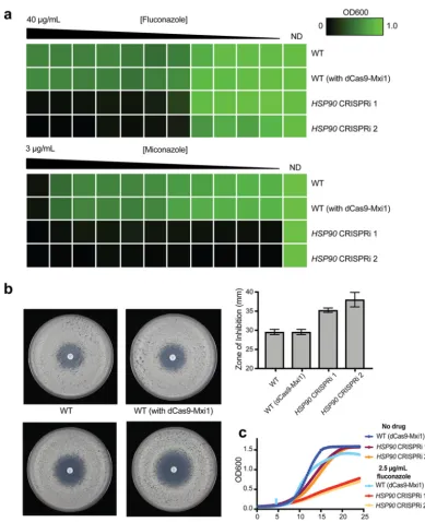
CRISPRi
-
mediated repression of an essential
C. albicans
gene.
Finally, we
vali-dated the use of this dCas9-Mxi1 CRISPRi platform for repression of an essential gene,
since the ability to target essential genes is a significant advantage of using CRISPRi
technology. For this analysis, we chose the essential molecular chaperone Hsp90, which
has been well characterized in
C. albicans
and is known to be involved in both cellular
morphogenesis and resistance to antifungal drugs (65–67). Therefore, we developed
two CRISPRi constructs, each with a distinct sgRNA, targeting
⫺
141 bp (sense strand
sgRNA) and
⫺
112 bp (antisense strand sgRNA) upstream of the
HSP90
start codon,
respectively. These regions were chosen based on our optimization with
ADE2
and were
cloned into the dCas9-Mxi1 plasmid backbone. We used these two CRISPRi constructs
to generate two
C. albicans
mutant strains, each containing one of the
HSP90
CRISPRi
repression constructs.
To validate repression of
HSP90
in these mutant
C. albicans
strains, we assessed
phenotypes known to be associated with repression of
HSP90
in CRISPRi mutant strains
compared with wild-type control strains. We monitored resistance to the azole
anti-fungals fluconazole and miconazole, as repression of
HSP90
has been shown to
abrogate resistance to these drugs (66, 68). We performed MIC assays with fluconazole
and miconazole and found that, using either of the two sgRNA constructs,
CRISPRi-based repression of
HSP90
led to increased sensitivity to both azole drugs (Fig. 4a), as
predicted based on previously observed phenotypes. We further used a fluconazole
disk diffusion assay to confirm that repression of
HSP90
led to increased susceptibility
to fluconazole, based on a larger zone of inhibition (Fig. 4b). Finally, we confirmed the
enhanced fluconazole sensitivity of these
HSP90
CRISPRi strains by monitoring growth
kinetics of these strains and of the corresponding wild-type strain, in the absence or
presence of fluconazole (Fig. 4c). Taking the results together, this demonstrates that the
HSP90
CRISPRi strains show phenotypes that correspond with depletion of
HSP90
, and
confirms the utility of this CRISPRi system for the effective genetic depletion of essential
genes.
DISCUSSION
Here, we demonstrated the first application of a CRISPRi-based genetic repression
system in the human fungal pathogen
C. albicans
. We generated nuclease-dead Cas9
(dCas9) CRISPRi plasmids, optimized for use in
C. albicans
, with a simple sgRNA cloning
site to enable efficient and rapid Golden Gate cloning of any sgRNA N20 at that locus,
to target any gene of interest for repression. We further optimized a region for
targeting sgRNA-dCas9,
⫺
55 to 129 bp upstream of the start codon, on the basis of an
ADE2
CRISPRi system. We note that while this promoter targeting region may vary for
different
C. albicans
genes, it is similar to the optimal CRISPRi targeting region for
on September 8, 2020 by guest
http://msphere.asm.org/
FIG 4 CRISPRi-based repression of the essential geneHSP90inC. albicans. (a) Reduced levels ofHSP90renderedC. albicans
more sensitive to azoles in MIC assays. MIC assays were performed with a gradient of an azole drug, namely, fluconazole (from 40g/ml to 0g/ml) or miconazole (from 3g/ml to 0g/ml), in 2-fold serial dilutions. Growth of all strains was monitored across the drug gradients and in a no-drug control (ND). The strains tested for antifungal susceptibility are indicated as follows: WT (SC5314), WT (with dCas9-Mxi1) (strain containing only dCas9-Mxi1 with nontargeting sgRNA integrated at theNEUT5Llocus [also known as fRS187]), andHSP90CRISPRi 1 (fRS221) andHSP90CRISPRi 2 (fRS222) (two independently generatedHSP90CRISPRi dCas9-Mxi1 strains, each with a unique sgRNA targetingHSP90for repression). Growth was normalized relative to the no-drug control, and data were quantitatively visualized using TreeView3. (b) Reduced levels ofHSP90renderC. albicansmore sensitive to fluconazole in disk diffusion assays. Disk diffusion assays were performed using a 25-g-fluconazole disk on Casitone agar plates. Growth of wild-type (WT) strains (includingSC5314WT and a WT strain containing only dCas9-Mxi1 with nontargeting sgRNA at theNEUT5Llocus) and of two independentHSP90
CRISPRi dCas9-Mxi1 strains (each with a unique sgRNA targetingHSP90for repression) was observed on these plates after 24 h and 48 h (48-h results are depicted here). Quantification of the zone of inhibition (measured using thediskImageR
program [87]) is depicted in the graph. (c) Growth curves confirming sensitivity ofHSP90depletion strains. The dCas9-Mxi1 wild-type strain and bothHSP90CRISPRi strains ofC. albicanswere grown in liquid YPD media with no drug or with 2.5g/ml fluconazole, and growth kinetics were monitored over⬃25 h. Both CRISPRi strains showed reduced growth in the presence of fluconazole.
on September 8, 2020 by guest
http://msphere.asm.org/
S. cerevisiae
, which is between
⬃
0 to 200 bp upstream of the transcription start site (54).
We further engineered novel CRISPRi fusion constructs for use in
C. albicans
and
demonstrated that dCas9, dCas9-Mig1, and dCas9-Mxi1 are each able to effectively
repress gene expression in
C. albicans
, with the dCas9-Mxi1 fusion being the strongest
repressor (
⬃
20-fold gene repression). Finally, we demonstrated the capability of this
system to target essential genes for genetic depletion, using
HSP90
as a candidate
essential gene target. Taking the results together, we believe this CRISPRi system will be a
powerful tool for efficient genetic repression in
C. albicans
, with many possible applications
for functional genetic studies and for the analysis of essential genes.
This work represents the first CRISPRi system for use in
C. albicans
, and, to our
knowledge, the first for use in any fungal pathogen. However, CRISPRi technologies
have been developed as powerful tools to study many other organisms, including
several important microbial species. Foundational CRISPRi studies demonstrated the
application of this technology in
E. coli
(47, 69), and CRISPRi was subsequently applied
to numerous other bacterial species. One important application of CRISPRi systems has
been the study of essential genes, as CRISPRi enables partial loss of function through
repression of these necessary genetic factors rather than a complete loss of function. In
the Gram-positive model bacterium
Bacillus subtilis
, a CRISPRi screen was used to target
all essential genes for systematic investigation of the phenotypes associated with these
factors, revealing novel genetic networks and morphological phenotypes associated
with essential genes (50). Similar CRISPRi-based gene knockdown screens were used to
identify previously uncharacterized essential genes in
Streptococcus pneumoniae
(70).
CRISPRi has also been applied in
Pseudomonas
species to study the function of essential
genes involved in cell division (53) and to identify and study the roles of essential
genes, and their interactions, in
Mycobacterium
species (51, 71). Similar CRISPRi studies
in
C. albicans
or other fungal pathogens could help unravel the function of essential
genes in these important microbial organisms.
Two valuable features of our
C. albicans
CRISPRi system are its efficiency and its
potential for scalability. We have designed the dCas9 plasmid (and dCas9-fusion
plasmids) to be a singular plasmid system, with a simple sgRNA cloning system,
requiring only the synthesis of small, 20-bp N20 sequences. The cost-effectiveness of
this system (requiring only two 23-bp oligonucleotides per gene being targeted for
repression) and the proven efficiency of the Golden Gate cloning strategy suggest that
this system could readily be scaled up to the genome level. Additionally, in
C. albicans
CRISPRi strains, the sgRNA can act as an inherent DNA barcode, containing both
conserved regions (
SNR52
promoter, sgRNA tail) and strain-specific sequences (sgRNA
N20). This could enable CRISPRi pooled screens of
C. albicans
strains similar to the
CRISPRi pooled screens employed for genome-scale functional genomic analysis in
E.
coli
(72). The use of such pooled competition assays among mutant microbial strains
has been a powerful strategy for functional-genomic profiling and chemical-genomic
analysis in
S. cerevisiae
(73–76) and
C. albicans
(12, 22), and CRISPRi could provide a
complementary approach to further enable such studies.
Finally, the development of a functional CRISPRi system in
C. albicans
facilitates the
application of other dCas9-based systems in this organism. Since we are able target
dCas9 to targeted genetic loci through deliberate sgRNA design, we can further exploit
this technology by fusing other effector domains to dCas9, as has been demonstrated
in several other systems (46). As previously described, CRISPRa enables the activation of
genes of interest (55–57, 77) and could be applied to our
C. albicans
system. Such
overexpression systems could provide a platform for antifungal drug target
identifica-tion (27), as demonstrated by the use of similar systems in
S. cerevisiae
(78). Further,
CRISPR-based epigenetic modifications can be achieved through fusion of dCas9 to
epigenetic regulators, such as histone demethylase or acetyltransferase enzymes (46).
Such CRISPR-epigenetic systems have primarily been applied in mammalian systems
(44, 45) but could similarly enable targeted epigenetic regulation in microbial
organ-isms. Additionally, improvements to this
C. albicans
CRISPRi system could be made by
fusing multiple repressor domains to dCas9 —a strategy that has been used
on September 8, 2020 by guest
http://msphere.asm.org/
fully to enhance repression in
S. cerevisiae
(64). Taking the results together, this work
has generated a new tool to enable genetic repression in
C. albicans
with potential for
adaptation for other CRISPR-based applications and for use in other related fungal
pathogens.
MATERIALS AND METHODS
Strains and culture conditions.Strains used in this study are listed in Table S1 in the supplemental material, and plasmids are listed in Table S2.C. albicansstrains were cultured on YPD (2% Bacto peptone, 1% yeast extract, 2% glucose), andE. colistrains were cultured in LB media.
Plasmid generation.The plasmid backbone used in this study was adapted from theC. albicans -optimized CRISPR-Cas9 plasmid (also known as pRS252) used in our previous study (33), containing the
NEUT5L homology site andCAS9 (79). To create a sgRNA cloning locus in this plasmid, the SNR52
promoter, SapI cloning locus, and sgRNA tail were synthesizedin vitroas gBlocks gene fragments from Integrated DNA Technologies (IDT) and were cloned into the CRISPR-Cas9 plasmid (pRS252) at the NgoMIV restriction enzyme site, using Gibson assembly, as previously described (33, 79). We have made the relevant CRISPRi (dCas9 and dCas9-Mxi1) plasmids available via Addgene (reference numbers 122377, 122378, 122379, and 122380).
Site-directed mutagenesis.Two nuclease mutations were introduced into Cas9 (D10A and N863A) to render it nuclease-dead (dCas9). These targeted mutations designed to disrupt Cas9 catalytic activity were introduced using site-directed mutagenesis as previously described (80).
dCas9 fusion construction. dCas9 fusion proteins were generated through the use of Gibson assembly. The Mxi1 effector domain was codon optimized forC. albicansexpression and synthesizedin vitroas gBlocks gene fragments from IDT, while the Mig1 coding sequence was directly amplified from
C. albicansgenomic DNA. These fragments were then cloned with Gibson assembly into the dCas9 plasmid backbone. The Mig1 gene and the Mxi1 domains were selected based on previous publications (52, 81).
sgRNA design.sgRNA N20 sequences were designed based on an efficiency score and predicted specificity using theC. albicansgenetic sequences from theCandidaGenome Database (CGD;http:// www.candidagenome.org) (82) and the sgRNA design tool Eukaryotic Pathogen CRISPR gRNA Design Tool (EuPaGDT) (83) available athttp://grna.ctegd.uga.edu.
sgRNA Golden Gate cloning.sgRNA N20 sequences were cloned into the dCas9 plasmid at the sgRNA cloning locus (containing theSNR52promoter, SapI cloning locus, and sgRNA tail) using Golden Gate cloning (60), as previously described (79). Each sgRNA N20 sequence was obtained as two oligonucleotides from IDT in forward and reverse complement orientation. Each of the two complemen-tary oligonucleotides contained a SapI cloning site, and each was reconstituted to 100M using nuclease-free duplex buffer from IDT. Equal volumes of the two complementary oligonucleotides were then combined and duplexed together by heating to 94°C for 2 min and allowed to cool to room temperature. To clone the duplexed fragment into the dCas9 plasmid, the following were combined: 10
l miniprepped dCas9 plasmid, 1l duplexed oligonucleotide, 2l 10⫻CutSmart buffer, 2l ATP, 1l SapI, 1l T4 DNA ligase, and 3l nuclease-free water. This mixture was incubated in a thermocycler under the following cycling conditions: (37°C, 2 min; 16°C, 5 min) for 99 cycles; 65°C, 15 min; 80°C, 15 min. After cycling was complete, 1l of additional SapI enzyme was added to each reaction mixture, and the mixture was incubated at 37°C for 1 h.
Bacterial transformation.Golden Gate-ligated plasmids were transformed into chemically compe-tent DH5␣Escherichia colicells. Competent cells (50l) were combined with 5l plasmid and incubated on ice for 30 min, heat shocked at 42°C for 30 s, and then incubated on ice for 5 min. This mixture was then added to 950l of Super Optimal Broth with added glucose (SOC media) and incubated at 37°C for 1 h with shaking. Transformed cells were selected on LB plates containing 100g/ml ampicillin.
Plasmid PCR validation.Ampicillin-resistant bacterial colonies were genotyped by colony PCR to confirm proper integration of the sgRNA N20 in the dCas9 plasmid. Briefly, bacterial colonies were diluted in 100l of nuclease-free water, and 5l of the mixture was added to a PCR with 2⫻Taqpolymerase mix and oligonucleotide primers. For each PCR, the primer pair was in the forward orientation N20 oligonucleotide plus TATACCATCCAAATCAATTCC and in the reverse complement orientation N20 oligonucleotide plus ACCCACTGAATTCTACATCGAAC. PCRs were run on 1% agarose gels.
C. albicanstransformation.AllC. albicansstrains were generated using a lithium acetate transfor-mation protocol, as previously described (79). Briefly, dCas9 plasmids were linearized with PacI restriction enzyme. Linearized plasmid andC. albicanscells were incubated with 800l 50% polyethylene glycol (PEG), 100l 10⫻Tris-EDTA (TE) buffer, 100l 1 M lithium acetate (pH 7.4), 40l of salmon sperm DNA, and 20l 2 M dithiothreitol (DTT). This mixture was then incubated at 30°C for 1 h and at 42°C for 45 min. Cells were grown in YPD media for 4 h at 30°C with shaking and were then selected for on YPD plates containing 200g/ml nourseothricin (NAT).
C. albicans PCR validation.NAT-resistant bacterial colonies were genotyped by colony PCR to confirm proper integration of the dCas9 plasmids at theNEUT5Llocus. Briefly,C. albicanscolonies were diluted in 100l of nuclease-free water, and 5l of this was added to a PCR with 2⫻Taqpolymerase mix and oligonucleotide primers. For each PCR, primers ACTATTAAAGAACGTGGACTCCAACGTCA (in the dCas9 plasmid) and CAAGTTTGCACTCTTTTGTCTA (in the genomicNEUT5Llocus) were used to validate integration. PCRs were run on 1% agarose gels.
Serial dilution spotting assays.C. albicansovernight cultures were diluted in 10-fold serial dilutions in sterile phosphate-buffered saline (PBS) media, and 5l of each diluted culture was spotted onto
on September 8, 2020 by guest
http://msphere.asm.org/
synthetic defined (SD; 0.67% yeast nitrogen base without amino acids, 2% glucose) agar plates with or without supplemented adenine.
Growth curve assays.C. albicanscultures were grown overnight in YPD media. Cells were diluted to an OD600of 0.05 in 96-well microtiter plates and grown at 37°C with continuous shaking, using a PerkinElmer Victor microplate reader or Bio-Rad xMark plate reader. For adenine growth curve assays, strains were grown in SD medium with or without supplemented adenine. For fluconazole growth curve assays, strains were grown in the absence of drug or with 2.5g/ml fluconazole–YPD. Each strain was grown in 3 or 4 independent wells. Optical density was measured at 600 nm every 15 min over 18 to 25 h.
Quantitative reverse transcription-PCR (qRT-PCR).To monitorADE2transcript levels, qRT-PCR was performed as previously described (84). Briefly,C. albicanscells were grown overnight in YPD at 37°C, diluted to an OD600of 0.2, and grown to an OD600of⬃1.0 at 37°C. Cultures were then pelleted and frozen overnight at – 80°C. RNA was isolated using a Geneaid yeast total RNA minikit supplemented with zymolyase. cDNA synthesis was performed using 800 ng RNA and a High Capacity cDNA reverse transcription kit (Applied Biosystems). PCR was performed using 2⫻PerfeCta SYBR green FastMix from Quanta BioScience under the following cycling conditions: 30 s at 95°C for the polymerase activation step, followed by 40 cycles of a two-step quantitative PCR (qPCR) procedure (3 s of 95°C denaturation, 30 s of 60°C combined annealing/extension). The primers used were as follows: forADE2, TTAGTGTATG CTCCTGCCAGG and GAGTTGTGAGGTCTTGGTGC; forACT1(control), GTTGGTGATGAAGCCCAATCC and CTGGATGTTCTTCTGGAGCAAC.
MIC assays.MIC assays were performed in 96-well microtiter plates, according to a standard broth microdilution protocol (85), with some modifications. Briefly, MIC tests were set up in a total volume of 200l per well with 2-fold serial dilutions of fluconazole or miconazole in YPD media. The gradients of fluconazole were 40 to 0g/ml, and the gradients of miconazole were 3 to 0g/ml, in 2-fold dilutions. The strains used for MIC analysis were grown overnight in YPD at 30°C. The cell densities of overnight cultures were determined by optical density (OD600), and dilutions were prepared such that equal numbers of cells were inoculated into all wells. MIC plates were incubated at 37°C for 24 to 48 h. After incubation, the optical density of cells in each well was determined at 600 nm using a microplate reader (PerkinElmer Victor) and growth of each strain was normalized to growth in the absence of drug. Each strain was tested in duplicate on at least three independent occasions. MIC data were quantitatively displayed with color using the program TreeView3.
Disk diffusion assays.Antifungal disk diffusion analysis was assessed using modified CLSI-M44-A2 guidelines on disk diffusion susceptibility adapted from a previous study (86). Strains were grown overnight on YPD agar at 30°C and were then resuspended in 1.5 ml of filter-sterile PBS and diluted to an OD600of 0.1. A 200-l volume of each resuspended strain was spread onto 15-ml Casitone agar plates (9 g/liter Bacto Casitone, 5 g/liter yeast extract, 11.5 g sodium citrate dehydrate, 20 g/liter glucose, 15 g/liter Bacto agar) via glass bead spreading. One 25-g fluconazole disk (Oxoid, United Kingdom) (6 mm in diameter) was placed at the center of each plate. The plates were then incubated at 30°C, and photographs were taken after 24 and 48 h. Each strain was tested on duplicate plates, on at least three independent occasions. The computational pipelinediskimageRwas used to assess the results of the antifungal diffusion assay (87).
Data availability.The relevant CRISPRi (dCas9 and dCas9-Mxi1) plasmids are available via Addgene under reference numbers 122377, 122378, 122379, and 122380.
SUPPLEMENTAL MATERIAL
Supplemental material for this article may be found at
https://doi.org/10.1128/
mSphere.00002-19
.
FIG S1
, TIF file, 1.1 MB.
TABLE S1
, XLSX file, 0.01 MB.
TABLE S2
, XLSX file, 0.01 MB.
ACKNOWLEDGMENTS
We thank members of the Shapiro laboratory and J. Pla for helpful discussions. We
thank J. Zhang at the University of Guelph Advanced Analysis Center Genomics Facility,
X. Pordel, and A. Gerstein for technical assistance.
This research was supported by a Natural Sciences and Engineering Research
Council (NSERC) Discovery Grant, NSERC Discovery Accelerator Supplement, and
Bant-ing Research Foundation Discovery Award to R.S.S.
REFERENCES
1. Denning DD. 2017. Global fungal disease burden. Eur J Clin Microbiol Infect Dis 36:923–1062.https://doi.org/10.1007/s10096-017-2909-8. 2. Bongomin F, Gago S, Oladele R, Denning D. 2017. Global and
multi-national prevalence of fungal diseases— estimate precision. J Fungi (Basel) 3:57.https://doi.org/10.3390/jof3040057.
3. Geddes-McAlister J, Shapiro RS. 2019. New pathogens, new tricks: emerging, drug-resistant fungal pathogens and future prospects for antifungal therapeutics. Ann N Y Acad Sci 1435:57–78.https://doi.org/ 10.1111/nyas.13739.
4. Webb BJ, Ferraro JP, Rea S, Kaufusi S, Goodman BE, Spalding J. 2018.
on September 8, 2020 by guest
http://msphere.asm.org/
Epidemiology and clinical features of invasive fungal infection in a US health care network. Open Forum Infect Dis 5:ofy187.https://doi.org/10 .1093/ofid/ofy187.
5. Pfaller MA, Messer SA, Boyken L, Rice C, Tendolkar S, Hollis RJ, Doern GV, Diekema DJ. 2005. Global trends in the antifungal susceptibility of
Cryptococcusneoformans (1990 to 2004). J Clin Microbiol 43:2163–2167. https://doi.org/10.1128/JCM.43.5.2163-2167.2005.
6. Pfaller MA, Diekema DJ. 2007. Epidemiology of invasive candidiasis: a persistent public health problem. Clin Microbiol Rev 20:133–163.https:// doi.org/10.1128/CMR.00029-06.
7. Drgona L, Khachatryan A, Stephens J, Charbonneau C, Kantecki M, Haider S, Barnes R. 2014. Clinical and economic burden of invasive fungal diseases in Europe: focus on pre-emptive and empirical treatment ofAspergillusandCandidaspecies. Eur J Clin Microbiol Infect Dis 33: 7–21.https://doi.org/10.1007/s10096-013-1944-3.
8. Benedict K, Jackson BR, Chiller T, Beer KD. 10 September 2018. Estima-tion of direct healthcare costs of fungal diseases in the United States. Clin Infect Dishttps://doi.org/10.1093/cid/ciy776.
9. Ohama T, Suzuki T, Mori M, Osawa S, Ueda T, Watanabe K, Nakase T. 1993. Non-universal decoding of the leucine codon CUG in several
Candida species. Nucleic Acids Res 21:4039 – 4045. https://doi.org/10 .1093/nar/21.17.4039.
10. Hickman MA, Zeng G, Forche A, Hirakawa MP, Abbey D, Harrison BD, Wang Y-M, Su C-H, Bennett RJ, Wang Y, Berman J. 2013. The “obligate diploid” Candida albicans forms mating-competent haploids. Nature 494:55–59.https://doi.org/10.1038/nature11865.
11. Homann OR, Dea J, Noble SM, Johnson AD. 2009. A phenotypic profile of theCandida albicans regulatory network. PLoS Genet 5:e1000783. https://doi.org/10.1371/journal.pgen.1000783.
12. Noble SM, French S, Kohn LA, Chen V, Johnson AD. 2010. Systematic screens of aCandida albicanshomozygous deletion library decouple morphogenetic switching and pathogenicity. Nat Genet 42:590 –598. https://doi.org/10.1038/ng.605.
13. Roemer T, Jiang B, Davison J, Ketela T, Veillette K, Breton A, Tandia F, Linteau A, Sillaots S, Marta C, Martel N, Veronneau S, Lemieux S, Kauff-man S, Becker J, Storms R, Boone C, Bussey H. 2003. Large-scale essential gene identification inCandida albicansand applications to antifungal drug discovery. Mol Microbiol 50:167–181. https://doi.org/10.1046/j .1365-2958.2003.03697.x.
14. Nobile CJ, Mitchell AP. 2009. Large-scale gene disruption using the UAU1 cassette. Methods Mol Biol 499:175–194.https://doi.org/10.1007/ 978-1-60327-151-6_17.
15. Segal ES, Gritsenko V, Levitan A, Yadav B, Dror N, Steenwyk JL, Silberberg Y, Mielich K, Rokas A, Gow NAR, Kunze R, Sharan R, Berman J. 2018. Gene essentiality analyzed by in vivo transposon mutagenesis and machine learning in a stable haploid isolate ofCandida albicans. mBio 9:e02048 -18.https://doi.org/10.1128/mBio.02048-18.
16. Gao J, Wang H, Li Z, Wong AH-H, Wang Y-Z, Guo Y, Lin X, Zeng G, Wang Y, Wang J. 2018. Candidaalbicansgains azole resistance by altering sphingolipid composition. Nat Commun 9:4495.https://doi.org/10.1038/ s41467-018-06944-1.
17. Mielich K, Shtifman-Segal E, Golz JC, Zeng G, Wang Y, Berman J, Kunze R. 2018. Maize transposable elements Ac/Ds as insertion mutagenesis tools inCandida albicans. G3 (Bethesda) 8:1139 –1145.https://doi.org/ 10.1534/g3.117.300388.
18. Ryan O, Shapiro RS, Kurat CF, Mayhew D, Baryshnikova A, Chin B, Lin Z-Y, Cox MJ, Vizeacoumar F, Cheung D, Bahr S, Tsui K, Tebbji F, Sellam A, Istel F, Schwarzmüller T, Reynolds TB, Kuchler K, Gifford DK, Whiteway M, Giaever G, Nislow C, Costanzo M, Gingras A-C, Mitra RD, Andrews B, Fink GR, Cowen LE, Boone C. 2012. Global gene deletion analysis exploring yeast filamentous growth. Science 337:1353–1356. https://doi.org/10 .1126/science.1224339.
19. Lee JA, Robbins N, Xie JL, Ketela T, Cowen LE. 2016. Functional genomic analysis ofCandida albicansadherence reveals a key role for the Arp2/3 complex in cell wall remodelling and biofilm formation. PLoS Genet 12:e1006452.https://doi.org/10.1371/journal.pgen.1006452.
20. Nobile CJ, Fox EP, Nett JE, Sorrells TR, Mitrovich QM, Hernday AD, Tuch BB, Andes DR, Johnson AD. 2012. A recently evolved transcriptional network controls biofilm development inCandida albicans. Cell 148: 126 –138.https://doi.org/10.1016/j.cell.2011.10.048.
21. Lohse MB, Ene IV, Craik VB, Hernday AD, Mancera E, Morschhäuser J, Bennett RJ, Johnson AD. 2016. Systematic genetic screen for transcrip-tional regulators of theCandida albicanswhite-opaque switch. Genetics 203:1679 –1692.https://doi.org/10.1534/genetics.116.190645.
22. O’Meara TR, Veri AO, Ketela T, Jiang B, Roemer T, Cowen LE. 2015. Global analysis of fungal morphology exposes mechanisms of host cell escape. Nat Commun 6:6741.https://doi.org/10.1038/ncomms7741.
23. O’Meara TR, Veri AO, Polvi EJ, Li X, Valaei SF, Diezmann S, Cowen LE. 2016. Mapping the Hsp90 genetic network reveals ergosterol biosyn-thesis and phosphatidylinositol-4-kinase signaling as core circuitry gov-erning cellular stress. PLoS Genet 12:e1006142.https://doi.org/10.1371/ journal.pgen.1006142.
24. Caplan T, Polvi EJ, Xie JL, Buckhalter S, Leach MD, Robbins N, Cowen LE. 2018. Functional genomic screening reveals core modulators of echino-candin stress responses inCandida albicans. Cell Rep 23:2292–2298. https://doi.org/10.1016/j.celrep.2018.04.084.
25. Mount HO, Revie NM, Todd RT, Anstett K, Collins C, Costanzo M, Boone C, Robbins N, Selmecki A, Cowen LE. 2018. Global analysis of genetic circuitry and adaptive mechanisms enabling resistance to the azole antifungal drugs. PLoS Genet 14:e1007319. https://doi.org/10.1371/ journal.pgen.1007319.
26. Enloe B, Diamond A, Mitchell AP. 2000. A single-transformation gene function test in diploidCandida albicans. J Bacteriol 182:5730 –5736. https://doi.org/10.1128/JB.182.20.5730-5736.2000.
27. Shapiro RS, Chavez A, Collins JJ. 2018. CRISPR-based genomic tools for the manipulation of genetically intractable microorganisms. Nat Rev Microbiol 16:333–339.https://doi.org/10.1038/s41579-018-0002-7. 28. Ng H, Dean N. 2017. Dramatic improvement of CRISPR/Cas9 editing in
Candida albicansby increased single guide RNA expression. mSphere 2:e00385-16.https://doi.org/10.1128/mSphere.00385-16.
29. Vyas VK, Guy Bushkin G, Bernstein DA, Getz MA, Sewastianik M, Inmacu-lada Barrasa M, Bartel DP, Fink GR. 2018. New CRISPR mutagenesis strategies reveal variation in repair mechanisms among fungi. mSphere 3:e00154-18.https://doi.org/10.1128/mSphere.00154-18.
30. Vyas VK, Barrasa MI, Fink GR. 2015. ACandida albicansCRISPR system permits genetic engineering of essential genes and gene families. Sci Adv 1:e1500248.https://doi.org/10.1126/sciadv.1500248.
31. Min K, Ichikawa Y, Woolford CA, Mitchell AP. 2016.Candida albicans
gene deletion with a transient CRISPR-Cas9 system. mSphere 1:e00130 -16.https://doi.org/10.1128/mSphere.00130-16.
32. Nguyen N, Quail MMF, Hernday AD. 2017. An efficient, rapid, and recyclable system for CRISPR-mediated genome editing in Candida al-bicans. mSphere 2:e00149-17. https://doi.org/10.1128/mSphereDirect .00149-17.
33. Shapiro RS, Chavez A, Porter CBM, Hamblin M, Kaas CS, DiCarlo JE, Zeng G, Xu X, Revtovich AV, Kirienko NV, Wang Y, Church GM, Collins JJ. 2018. A CRISPR-Cas9-based gene drive platform for genetic interaction anal-ysis inCandida albicans. Nat Microbiol 3:73– 82.https://doi.org/10.1038/ s41564-017-0043-0.
34. Enkler L, Richer D, Marchand AL, Ferrandon D, Jossinet F. 2016. Genome engineering in the yeast pathogenCandida glabratausing the CRISPR-Cas9 system. Sci Rep 6:35766.https://doi.org/10.1038/srep35766. 35. Grahl N, Demers EG, Crocker AW, Hogan DA. 2017. Use of RNA-protein
complexes for genome editing in non-albicans Candida species. mSphere 2:e00218-17.https://doi.org/10.1128/mSphere.00218-17. 36. Lombardi L, Turner SA, Zhao F, Butler G. 2017. Gene editing in clinical
isolates of Candida parapsilosis using CRISPR/Cas9. Sci Rep 7:8051. https://doi.org/10.1038/s41598-017-08500-1.
37. Palermo G, Miao Y, Walker RC, Jinek M, McCammon JA. 2017. CRISPR-Cas9 conformational activation as elucidated from enhanced molecular simulations. Proc Natl Acad Sci U S A 114:7260 –7265.https://doi.org/10 .1073/pnas.1707645114.
38. DiCarlo JE, Norville JE, Mali P, Rios X, Aach J, Church GM. 2013. Genome engineering inSaccharomyces cerevisiaeusing CRISPR-Cas systems. Nu-cleic Acids Res 41:4336 – 4343.https://doi.org/10.1093/nar/gkt135. 39. Jinek M, Chylinski K, Fonfara I, Hauer M, Doudna JA, Charpentier E. 2012.
A programmable dual-RNA-guided DNA endonuclease in adaptive bac-terial immunity. Science 337:816 – 821.https://doi.org/10.1126/science .1225829.
40. Gaudelli NM, Komor AC, Rees HA, Packer MS, Badran AH, Bryson DI, Liu DR. 2017. Programmable base editing of A●T to G●C in genomic DNA without DNA cleavage. Nature 551:464 – 471.https://doi.org/10.1038/ nature24644.
41. Komor AC, Kim YB, Packer MS, Zuris JA, Liu DR. 2016. Programmable editing of a target base in genomic DNA without double-stranded DNA cleavage. Nature 533:420 – 424.https://doi.org/10.1038/nature17946. 42. Kuscu C, Parlak M, Tufan T, Yang J, Szlachta K, Wei X, Mammadov R, Adli
M. 2017. CRISPR-STOP: gene silencing through base-editing-induced
on September 8, 2020 by guest
http://msphere.asm.org/
nonsense mutations. Nat Methods 14:710 –712.https://doi.org/10.1038/ nmeth.4327.
43. Cox DBT, Gootenberg JS, Abudayyeh OO, Franklin B, Kellner MJ, Joung J, Zhang F. 2017. RNA editing with CRISPR-Cas13. Science 358: 1019 –1027.https://doi.org/10.1126/science.aaq0180.
44. Liu XS, Wu H, Ji X, Stelzer Y, Wu X, Czauderna S, Shu J, Dadon D, Young RA, Jaenisch R. 2016. Editing DNA methylation in the mammalian ge-nome. Cell 167:233–247.e17.https://doi.org/10.1016/j.cell.2016.08.056. 45. Liao H-K, Hatanaka F, Araoka T, Reddy P, Wu M-Z, Sui Y, Yamauchi T,
Sakurai M, O’Keefe DD, Núñez-Delicado E, Guillen P, Campistol JM, Wu C-J, Lu L-F, Esteban CR, Izpisua Belmonte JC. 2017.In vivotarget gene activation via CRISPR/Cas9-mediatedtrans-epigenetic modulation. Cell 171:1495–1507.e15.https://doi.org/10.1016/j.cell.2017.10.025. 46. Dominguez AA, Lim WA, Qi LS. 2016. Beyond editing: repurposing
CRISPR-Cas9 for precision genome regulation and interrogation. Nat Rev Mol Cell Biol 17:5–15.https://doi.org/10.1038/nrm.2015.2.
47. Qi LS, Larson MH, Gilbert LA, Doudna JA, Weissman JS, Arkin AP, Lim WA. 2013. Repurposing CRISPR as an RNA-guided platform for sequence-specific control of gene expression. Cell 152:1173–1183.https://doi.org/ 10.1016/j.cell.2013.02.022.
48. La Russa MF, Qi LS. 2015. The new state of the art: Cas9 for gene activation and repression. Mol Cell Biol 35:3800 –3809.https://doi.org/ 10.1128/MCB.00512-15.
49. Larson MH, Gilbert LA, Wang X, Lim WA, Weissman JS, Qi LS. 2013. CRISPR interference (CRISPRi) for sequence-specific control of gene ex-pression. Nat Protoc 8:2180 –2196. https://doi.org/10.1038/nprot.2013 .132.
50. Peters JM, Colavin A, Shi H, Czarny TL, Larson MH, Wong S, Hawkins JS, Lu CHS, Koo B-M, Marta E, Shiver AL, Whitehead EH, Weissman JS, Brown ED, Qi LS, Huang KC, Gross CA. 2016. A comprehensive, CRISPR-based functional analysis of essential genes in bacteria. Cell 165:1493–1506. https://doi.org/10.1016/j.cell.2016.05.003.
51. Rock JM, Hopkins FF, Chavez A, Diallo M, Chase MR, Gerrick ER, Pritchard JR, Church GM, Rubin EJ, Sassetti CM, Schnappinger D, Fortune SM. 2017. Programmable transcriptional repression in mycobacteria using an or-thogonal CRISPR interference platform. Nat Microbiol 2:16274.https:// doi.org/10.1038/nmicrobiol.2016.274.
52. Gilbert LA, Larson MH, Morsut L, Liu Z, Brar GA, Torres SE, Stern-Ginossar N, Brandman O, Whitehead EH, Doudna JA, Lim WA, Weissman JS, Qi LS. 2013. CRISPR-mediated modular RNA-guided regulation of transcription in eukaryotes. Cell 154:442– 451. https://doi.org/10.1016/j.cell.2013.06 .044.
53. Tan SZ, Reisch CR, Prather KLJ. 2018. A robust CRISPR interference gene repression system inPseudomonas. J Bacteriol 200:e00575-17. 54. Smith JD, Suresh S, Schlecht U, Wu M, Wagih O, Peltz G, Davis RW,
Steinmetz LM, Parts L, St Onge RP. 2016. Quantitative CRISPR interfer-ence screens in yeast identify chemical-genetic interactions and new rules for guide RNA design. Genome Biol 17:45.https://doi.org/10.1186/ s13059-016-0900-9.
55. Konermann S, Brigham MD, Trevino AE, Joung J, Abudayyeh OO, Bar-cena C, Hsu PD, Habib N, Gootenberg JS, Nishimasu H, Nureki O, Zhang F. 2015. Genome-scale transcriptional activation by an engineered CRISPR-Cas9 complex. Nature 517:583–588. https://doi.org/10.1038/ nature14136.
56. Chavez A, Scheiman J, Vora S, Pruitt BW, Tuttle M, Iyer EPR, Lin S, Kiani S, Guzman CD, Wiegand DJ, Ter-Ovanesyan D, Braff JL, Davidsohn N, Housden BE, Perrimon N, Weiss R, Aach J, Collins JJ, Church GM. 2015. Highly efficient Cas9-mediated transcriptional programming. Nat Meth-ods 12:326 –328.https://doi.org/10.1038/nmeth.3312.
57. Gilbert LA, Horlbeck MA, Adamson B, Villalta JE, Chen Y, Whitehead EH, Guimaraes C, Panning B, Ploegh HL, Bassik MC, Qi LS, Kampmann M, Weissman JS. 2014. Genome-scale CRISPR-mediated control of gene repression and activation. Cell 159:647– 661. https://doi.org/10.1016/j .cell.2014.09.029.
58. Hess GT, Tycko J, Yao D, Bassik MC. 2017. Methods and applications of CRISPR-mediated base editing in eukaryotic genomes. Mol Cell 68: 26 – 43.https://doi.org/10.1016/j.molcel.2017.09.029.
59. Gerami-Nejad M, Zacchi LF, McClellan M, Matter K, Berman J. 2013. Shuttle vectors for facile gap repair cloning and integration into a neutral locus inCandida albicans. Microbiology 159:565–579.https://doi .org/10.1099/mic.0.064097-0.
60. Engler C, Kandzia R, Marillonnet S. 2008. A one pot, one step, precision cloning method with high throughput capability. PLoS One 3:e3647. https://doi.org/10.1371/journal.pone.0003647.
61. Howe FS, Russell A, Lamstaes AR, El-Sagheer A, Nair A, Brown T, Mellor J. 2017. CRISPRi is not strand-specific at all loci and redefines the transcriptional landscape. Elife 6:e29878.https://doi.org/10.7554/eLife .29878.
62. Schreiber-Agus N, Chin L, Chen K, Torres R, Rao G, Guida P, Skoultchi AI, DePinho RA. 1995. An amino-terminal domain of Mxi1 mediates anti-Myc oncogenic activity and interacts with a homolog of the yeast transcriptional repressorSIN3. Cell 80:777–786.https://doi.org/10.1016/ 0092-8674(95)90356-9.
63. Lutfiyya LL, Iyer VR, DeRisi J, DeVit MJ, Brown PO, Johnston M. 1998. Characterization of three related glucose repressors and genes they regulate inSaccharomyces cerevisiae. Genetics 150:1377–1391. 64. Lian J, HamediRad M, Hu S, Zhao H. 2017. Combinatorial metabolic
engineering using an orthogonal tri-functional CRISPR system. Nat Com-mun 8:1688.https://doi.org/10.1038/s41467-017-01695-x.
65. Singh SD, Robbins N, Zaas AK, Schell WA, Perfect JR, Cowen LE. 2009. Hsp90 governs echinocandin resistance in the pathogenic yeastCandida albicans via calcineurin. PLoS Pathog 5:e1000532. https://doi.org/10 .1371/journal.ppat.1000532.
66. Cowen LE, Lindquist S. 2005. Hsp90 potentiates the rapid evolution of new traits: drug resistance in diverse fungi. Science 309:2185–2189. https://doi.org/10.1126/science.1118370.
67. Shapiro RS, Uppuluri P, Zaas AK, Collins C, Senn H, Perfect JR, Heitman J, Cowen LE. 2009. Hsp90 orchestrates temperature-dependentCandida albicansmorphogenesis via Ras1-PKA signaling. Curr Biol 19:621– 629. https://doi.org/10.1016/j.cub.2009.03.017.
68. Hill JA, Ammar R, Torti D, Nislow C, Cowen LE. 2013. Genetic and genomic architecture of the evolution of resistance to antifungal drug combinations. PLoS Genet 9:e1003390.https://doi.org/10.1371/journal .pgen.1003390.
69. Hawkins JS, Wong S, Peters JM, Almeida R, Qi LS. 2015. Targeted transcriptional repression in bacteria using CRISPR interference (CRIS-PRi), p 349 –362.InLundgren M, Charpentier E, Fineran PC (ed), CRISPR: methods and protocols. Springer New York, New York, NY.
70. Liu X, Gallay C, Kjos M, Domenech A, Slager J, van Kessel SP, Knoops K, Sorg RA, Zhang J-R, Veening J-W. 2017. High-throughput CRISPRi phe-notyping identifies new essential genes inStreptococcus pneumoniae. Mol Syst Biol 13:931.https://doi.org/10.15252/msb.20167449. 71. Choudhary E, Thakur P, Pareek M, Agarwal N. 2015. Gene silencing by
CRISPR interference in mycobacteria. Nat Commun 6:6267.https://doi .org/10.1038/ncomms7267.
72. Wang T, Guan C, Guo J, Liu B, Wu Y, Xie Z, Zhang C, Xing X-H. 2018. Pooled CRISPR interference screening enables genome-scale functional genomics study in bacteria with superior performance. Nat Commun 9:2475.https://doi.org/10.1038/s41467-018-04899-x.
73. Shoemaker DD, Lashkari DA, Morris D, Mittmann M, Davis RW. 1996. Quantitative phenotypic analysis of yeast deletion mutants using a highly parallel molecular bar-coding strategy. Nat Genet 14:450 – 456. https://doi.org/10.1038/ng1296-450.
74. Winzeler EA, Shoemaker DD, Astromoff A, Liang H, Anderson K, Andre B, Bangham R, Benito R, Boeke JD, Bussey H, Chu AM, Connelly C, Davis K, Dietrich F, Dow SW, El Bakkoury M, Foury F, Friend SH, Gentalen E, Giaever G, Hegemann JH, Jones T, Laub M, Liao H, Liebundguth N, Lockhart DJ, Lucau-Danila A, Lussier M, M’Rabet N, Menard P, Mittmann M, Pai C, Rebischung C, Revuelta JL, Riles L, Roberts CJ, Ross-MacDonald P, Scherens B, Snyder M, Sookhai-Mahadeo S, Storms RK, Véronneau S, Voet M, Volckaert G, Ward TR, Wysocki R, Yen GS, Yu K, Zimmermann K, Philippsen P, et al. 1999. Functional characterization of the S.cerevisiae
genome by gene deletion and parallel analysis. Science 285:901–906. https://doi.org/10.1126/science.285.5429.901.
75. Giaever G, Shoemaker DD, Jones TW, Liang H, Winzeler EA, Astromoff A, Davis RW. 1999. Genomic profiling of drug sensitivities via induced haploinsufficiency. Nat Genet 21:278 –283.https://doi.org/10.1038/6791. 76. Giaever G, Flaherty P, Kumm J, Proctor M, Nislow C, Jaramillo DF, Chu AM, Jordan MI, Arkin AP, Davis RW. 2004. Chemogenomic profiling: identifying the functional interactions of small molecules in yeast. Proc Natl Acad Sci U S A 101:793–798.https://doi.org/10.1073/pnas .0307490100.
77. Dong C, Fontana J, Patel A, Carothers JM, Zalatan JG. 2018. Synthetic CRISPR-Cas gene activators for transcriptional reprogramming in bacte-ria. Nat Commun 9:2489.https://doi.org/10.1038/s41467-018-04901-6. 78. Luesch H, Wu TYH, Ren P, Gray NS, Schultz PG, Supek F. 2005. A
genome-wide overexpression screen in yeast for small-molecule target
on September 8, 2020 by guest
http://msphere.asm.org/
identification. Chem Biol 12:55– 63.https://doi.org/10.1016/j.chembiol .2004.10.015.
79. Halder V, Porter CBM, Chavez A, Shapiro RS. Design, execution, and analysis of CRISPR-Cas9-based deletions and genetic interaction net-works in the fungal pathogenCandida albicans. Nat Protoc, in press. https://doi.org/10.1038/s41596-018-0122-6.
80. Huang Y, Zhang L. 2017. Anin vitrosingle-primer site-directed mutagen-esis method for use in biotechnology. Methods Mol Biol 1498:375–383. https://doi.org/10.1007/978-1-4939-6472-7_26.
81. Keung AJ, Bashor CJ, Kiriakov S, Collins JJ, Khalil AS. 2014. Using targeted chromatin regulators to engineer combinatorial and spatial transcriptional regulation. Cell 158:110–120.https://doi.org/10.1016/j.cell.2014.04.047. 82. Skrzypek MS, Binkley J, Binkley G, Miyasato SR, Simison M, Sherlock G.
2017. TheCandidaGenome Database (CGD): incorporation of assembly 22, systematic identifiers and visualization of high throughput sequenc-ing data. Nucleic Acids Res 45:D592–D596.https://doi.org/10.1093/nar/ gkw924.
83. Peng D, Tarleton R. 2015. EuPaGDT: a Web tool tailored to design CRISPR
guide RNAs for eukaryotic pathogens. Microb Genom 1:e000033.https:// doi.org/10.1099/mgen.0.000033.
84. Shapiro RS, Zaas AK, Betancourt-Quiroz M, Perfect JR, Cowen LE. 2012. The Hsp90 co-chaperone Sgt1 governsCandida albicansmorphogenesis and drug resistance. PLoS One 7:e44734.https://doi.org/10.1371/journal .pone.0044734.
85. Clinical and Laboratory Standards Institute. 2008. Reference method for broth dilution antifungal susceptibility testing of yeasts: approved stan-dard. CLSI, Wayne, PA.
86. Rosenberg A, Ene IV, Bibi M, Zakin S, Segal ES, Ziv N, Dahan AM, Colombo AL, Bennett RJ, Berman J. 2018. Antifungal tolerance is a subpopulation effect distinct from resistance and is associated with persistent candidemia. Nat Commun 9:2470.https://doi.org/10.1038/ s41467-018-04926-x.
87. Gerstein AC, Rosenberg A, Hecht I, Berman J. 2016. diskImageR: quan-tification of resistance and tolerance to antimicrobial drugs using disk diffusion assays. Microbiology 162:1059 –1068.https://doi.org/10.1099/ mic.0.000295.