Comparative Analysis of Bacterial
Community Composition and Structure
in Clinically Symptomatic and
Asymptomatic Central Venous Catheters
Franziska A. Stressmann,aElodie Couve-Deacon,bDelphine Chainier,b Ashwini Chauhan,a*Aimee Wessel,aSylvaine Durand-Fontanier,c,d
Marie-Christine Escande,eIrène Kriegel,fBruno Francois,b,gMarie-Cécile Ploy,b Christophe Beloin,aJean-Marc Ghigoa
Unité de Génétique des Biofilms, Département de Microbiologie, Institut Pasteur, Paris, Francea; Université
Limoges, INSERM, CHU Limoges, UMR 1092, Limoges, Franceb; CHU de Limoges, Service de chirurgie digestive
générale et endocrinienne, Limoges, Francec; Université de Limoges, Équipe de recherche médicale
appliquée, Limoges, Franced; Institut Curie, Hôpital, Laboratoire de Microbiologie Médicale, Paris, Francee;
Institut Curie, Hôpital, Service Anesthésie-Réanimation-Douleur, Paris, Francef; INSERM, CIC-1435, Limoges,
Franceg
ABSTRACT Totally implanted venous access ports (TIVAPs) are commonly used catheters for the management of acute or chronic pathologies. Although these de-vices improve health care, repeated use of this type of device for venous access over long periods of time is also associated with risk of colonization and infection by pathogenic bacteria, often originating from skin. However, although the skin mi-crobiota is composed of both pathogenic and nonpathogenic bacteria, the extent and the consequences of TIVAP colonization by nonpathogenic bacteria have rarely been studied. Here, we used culture-dependent and 16S rRNA gene-based culture-independent approaches to identify differences in bacterial colonization of TIVAPs obtained from two French hospitals. To explore the relationships be-tween nonpathogenic organisms colonizing TIVAPs and the potential risk of in-fection, we analyzed the bacterial community parameters between TIVAPs suspected (symptomatic) or not (asymptomatic) of infection. Although we did not find a particular species assemblage or community marker to distinguish infection risk on an individual sample level, we identified differences in bacterial community composition, diversity, and structure between clinically symptomatic and asymp-tomatic TIVAPs that could be explored further. This study therefore provides a new view of bacterial communities and colonization patterns in intravascular TIVAPs and suggests that microbial ecology approaches could improve our un-derstanding of device-associated infections and could be a prognostic tool to monitor the evolution of bacterial communities in implants and their potential susceptibility to infections.
IMPORTANCE Totally implanted venous access ports (TIVAPs) are commonly used implants for the management of acute or chronic pathologies. Although their use improves the patient’s health care and quality of life, they are associated with a risk of infection and subsequent clinical complications, often leading to implant removal. While all TIVAPs appear to be colonized, only a fraction become infected, and the relationship between nonpathogenic organisms colonizing TIVAPs and the potential risk of infection is unknown. We explored bacteria present on TIVAPs implanted in patients with or without signs of TIVAP infection and identified differences in phy-lum composition and community structure. Our data suggest that the microbial ecology of intravascular devices could be predictive of TIVAP infection status and
Received29 March 2017Accepted17 May 2017 Published27 September 2017 CitationStressmann FA, Couve-Deacon E, Chainier D, Chauhan A, Wessel A, Durand-Fontanier S, Escande M-C, Kriegel I, Francois B, Ploy M-C, Beloin C, Ghigo J-M. 2017. Comparative analysis of bacterial community composition and structure in clinically symptomatic and asymptomatic central venous catheters. mSphere 2:e00146-17.
https://doi.org/10.1128/mSphere.00146-17.
EditorBrandi M. Limbago, Centers for Disease Control and Prevention
Copyright© 2017 Stressmann et al. This is an open-access article distributed under the terms of theCreative Commons Attribution 4.0
International license.
Address correspondence to Christophe Beloin, [email protected], or Jean-Marc Ghigo, [email protected]. *Present address: Ashwini Chauhan, Biotechnology and Bioinformatics, NIIT University, Neemrana, Rajasthan, India.
Clinical Science and Epidemiology
crossm
on September 8, 2020 by guest
http://msphere.asm.org/
that ultimately a microbial ecological signature could be identified as a tool to pre-dict TIVAP infection susceptibility and improve clinical management.
KEYWORDS ecology, bacterial community, biofilm, catheter colonization
T
he management of acute or chronic pathologies often requires the use of periph-eral or central venous catheters, which greatly improve patient health care (1). Nevertheless, since these devices are used as access entry ports to administer nutritive or therapeutic fluids, they are also associated with a risk of bacterial surface contami-nation, often leading to the formation of bacterial or fungal biofilms that are difficult to eradicate and cause chronic and nosocomial infections (2).Among central venous catheters used in modern medicine, totally implantable venous access ports (TIVAPs) are commonly used invasive medical devices. TIVAPs are frequently inserted for parenteral nutrition, chemotherapy, and other long-term drug treatments, for periods that can last months or years (1, 3). In addition to contamination that may occur during surgical insertion, repeated use of these long-term implants to inject or draw fluids increases the risk of TIVAP contamination and therefore infections. The high morbidity and substantial risk of complications associated with TIVAP-related infection often lead clinicians to decide on the traumatic and costly removal of devices suspected of infection (4–6).
TIVAP bacterial colonization can originate from bloodstream bacteria in bacteremic patients or from skin bacteria introduced upon repeated punctures with Huber needles to access TIVAP chambers (1, 6). Consistent with external contamination, the most frequently isolated pathogens associated with TIVAP infection correspond to bacteria commonly found on the skin, such as Staphylococcus aureus and Staphylococcus epidermidis(7–10).
The predominance of skin bacteria suggests that all TIVAPs could be contaminated and colonized by both pathogenic and nonpathogenic microorganisms present on the surface of the skin (11). It has indeed been shown by culture and molecular studies that all implanted devices become colonized at some point (12–14) and that even symptom-free implants without positive microbial culture can harbor bacteria without leading to TIVAP infection (2, 15). While this suggests that TIVAP colonization could be more complex than initially suspected, it also raises the question of whether potential relationships between microorganisms colonizing TIVAP could influence the risk of infection. Unfortunately, microbiology analysis usually targets only TIVAPs suspected of infection and not devices from symptom-free patients considered sterile or free of specific pathogens. Therefore, there is currently no global view of the microbial ecology and the dynamics of bacterial communities developing in TIVAPs. Whether colonization of TIVAPs by nonpathogenic bacteria influences the development of infection remains an unanswered question.
Here, we collaborated with two French hospitals to study the differences between bacterial populations colonizing TIVAP chambers removed from patients receiving long-term cancer therapies. We used both culture– dependent and 16S rRNA-based culture-independent approaches to analyze the bacterial content of TIVAP chambers from patients suspected of infection (here called clinically “symptomatic” TIVAPs) as well as from devices removed from patients without suspicion of infection (here called clinically “asymptomatic” TIVAPs), which are not routinely analyzed in clinical practice. We found a wide range of hospital-associated and environmental bacteria on both symptomatic and asymptomatic chambers, and we compared their compositions and community structures between hospital cohorts. This study presents new findings on the colonization patterns of intra-vascular devices and on the ecological relationships between the presence of nonpatho-genic and pathononpatho-genic microorganisms and their ability to colonize catheters.
RESULTS
Bicentric sampling of TIVAP catheters from patients suspected or not of catheter-associated infections.Totally implanted venous access ports (TIVAPs) sus-pected of being contaminated by pathogenic bacteria (here referred to as clinically
on September 8, 2020 by guest
http://msphere.asm.org/
symptomatic TIVAPs) are systematically removed and subjected to a thorough micro-biology analysis. In contrast, TIVAPs removed at the end of treatment without suspicion of infection (clinically asymptomatic TIVAPs) are routinely discarded without analysis. In order to investigate the microbial colonization of TIVAPs implanted in hospitalized patients suspected or not of catheter-associated infection, we developed a collection protocol minimizing bacterial contamination at any point during sample handling (Fig. 1A). Two groups of 10 symptomatic or asymptomatic TIVAPs were collected from two French hospitals (Institut Curie Hospital, Paris, France, and Dupuytren Hospital, Limoges, France, referred to here as Curie and Limoges Hospitals, respectively; total chambers collected, n ⫽ 40) (see Table S1 in the supplemental material). Due to potential variability and increased risk for postextraction contaminants in TIVAP distal catheter parts, we chose to analyze only the bacterial content of the catheter chamber of each TIVAP (Fig. 1B).
Culture-independent evidence for environmental bacteria in culture-negative TIVAPs.To test the potential presence of bacteria in TIVAPs removed from asymptom-atic patients, asymptomasymptom-atic TIVAPs were subjected to routine culture analysis and parallel molecular analysis of bacterial 16S rRNA gene content (clone library genera-tion). Culture analysis detected no bacteria in 9 out of 10 samples from Curie Hospital and in 8 out of 10 samples from Limoges Dupuytren Hospital (Table 1). In contrast, 16S rRNA-based molecular analysis detected bacteria in all asymptomatic TIVAP samples from Curie and Limoges Hospitals (Table 1). Reagents and materials used for molecular processing and blanks (water as the template) for all experimental procedures were also subjected to 16S rRNA sequencing. This led to the identification of 7 species detected in molecular reagents or materials or introduced during DNA extraction or PCR, two of which were also detected in patient samples (Table S2A). When any of these 7 species deemed contaminants from the molecular procedure were identified in patient samples in the study, these species were excluded from all further analysis. Two species were detected in nonimplanted and nonmanipulated TIVAPs but were not detected in any of the patient samples (Table S2B). A total of 14 species were detected as present in the flush solutions used in the hospitals or in handled nonimplanted chambers (Table S2C and D). Four of these species (includingPropionibacterium acnes andRalstonia pickettii) were also detected in patient samples.
From a total of 42 detected operational taxonomic units (OTUs), 40 different OTUs/bacterial species were detected in asymptomatic TIVAP chambers in both hos-pitals, 28 in Curie Hospital and 19 in Limoges Dupuytren Hospital (Fig. 2). Although species frequently associated with clinical infections (e.g.,Escherichia coli,Enterobacter cloacae,S. aureus,S. epidermidis,Stenotrophomonas maltophilia, andStreptococcus parasanguinis) were detected in some asymptomatic TIVAP samples, the majority of species detected are generally found in the environment. For example,Aquabacterium spp.,Bradyrhizobiumspp.,Caulobacter leidyi,Delftia acidovorans,Methylobacteriumspp., Mesorhizobiumspp., andVariovorax paradoxus(Fig. 2) are commonly isolated from soil and aquatic environments.
These results suggest that the chambers of asymptomatic TIVAPs, although without history of infection, are nevertheless commonly colonized by bacteria that are mostly of environmental origin.
Environmental bacteria are also detected in culture-positive TIVAPs. To test which bacteria were present in symptomatic TIVAPs, we carried out culture-based analysis on symptomatic TIVAP chamber flushes, which detected mostly one bacterial species per sample (Table 1). Curie Hospital samples were exclusively colonized by Gram-positive bacteria (S. aureus, S. epidermidis, and Staphylococcus xylosus), with fungal colonization detected in two chambers (Table 1). Samples from Limoges Du-puytren Hospital were colonized by a mixture of Gram-positive (S. aureus,S. epidermidis, andBacillus simplex) and Gram-negative (E. coli,E. cloacae, and Pseudomonas aerugi-nosa) bacteria. The bacteria isolated from TIVAP samples of symptomatic patients were also isolated from the blood cultures.
on September 8, 2020 by guest
http://msphere.asm.org/
FIG 1 TIVAP catheter collection from patients suspected or not of catheter-related infection from two French hospitals. (A) Study design. Sterile reagents and techniques were used throughout the study, and the protocol was designed to minimize the possibilities for bacterial contamination as much as possible. Furthermore, samples of used reagents (green asterisks), materials (blue asterisks), and blanks (water used as the template in the procedure; red asterisks) as procedure controls were taken at all steps and also submitted to 16S rRNA sequencing for species identification. (B) TIVAP parts. The TIVAP is composed of a subcutaneously implanted chamber/hub (the reservoir) connected to a catheter that is inserted into a central vein (e.g., jugular or subclavian).
on September 8, 2020 by guest
http://msphere.asm.org/
In contrast to culture-based analysis, 16S rRNA analysis enabled us to detect more than 1 species in 40% of Curie and 40% of Limoges Dupuytren Hospital symptomatic samples (Table 1). Moreover, we detected at least one bacterial species in each of the chambers from symptomatic patients that were culture negative (2 from Curie Hospital and 1 from Limoges Hospital).
The dominant organism detected by 16S rRNA PCR and sequencing also corre-sponded to the bacterial species detected by culture and identified as the infection cause in all chambers but one (CS-9) (Table 1). Besides these bacteria, a total of 20
TABLE 1 Culture results for infected and noninfected TIVAP chambers from the two hospital cohorts and species (OTU) richness as detected by 16S clone library generationa
TIVAP group and sample ID
Culture-isolated organism(s)
No. of CFU/ml
No. of OTUs detected in 16S clone library
No. of clones generated for 16S clone library Curie symptomatic
CS-1 Sterile 8 48
CS-2 Staphylococcus aureus* ⬎1,000 1 48 CS-3 Staphylococcus epidermidis* 200 1 48 CS-4 Staphylococcus epidermidis* ⬎1,000 1 48 CS-5 Staphylococcus epidermidis* ⬎1,000 1 48
CS-6 Candida parapsilosis ⬎1,000 5 48
CS-7 Sterile 1 48
CS-8 Staphylococcus aureus* 130 6 45
CS-9 Staphylococcus xylosus ⬍10 4 49
CS-10 Candida kruseiandStaphylococcus epidermidis*
⬎1,000 1 48
Curie asymptomatic
CA-1 Sterile 9 48
CA-2 Sterile 10 48
CA-3 Sterile 4 49
CA-4 Sterile 4 49
CA-5 Sterile 6 49
CA-6 Sterile 4 48
CA-7 Staphylococcus epidermidis 20 3 48
CA-8 Sterile 7 48
CA-9 Sterile 9 53
CA-10 Sterile 7 48
Limoges symptomatic
LS-1 Staphylococcus epidermidis*, Bacillus simplex
1, 1 1 50
LS-2 Sterile 6 47
LS-3 Enterobacter cloacae* ⬎500 1 50
LS-4 (Staphylococcus epidermidison distal catheter)
2 48
LS-5 Staphylococcus epidermidis* ⬎500 1 50
LS-6 Escherichia coli* ⬎500 1 50
LS-7 Staphylococcus aureus* ⬍50 1 49
LS-8 Staphylococcus aureus* 1 4 48
LS-9 Pseudomonas aeruginosa* ⬍50 1 49
LS-10 Staphylococcus epidermidis* ⬍10 2 49
Limoges asymptomatic
LA-1 Sterile 7 52
LA-2 Sterile 4 49
LA-3 Sterile 5 48
LA-4 Sterile 5 50
LA-5 Sterile 3 48
LA-6 Micrococcus luteus 1 6 50
LA-7 Micrococcus luteus 1 3 50
LA-8 Sterile 8 55
LA-9 Sterile 4 48
LA-10 Sterile 2 50
aSpecies shown in bold with an asterisk represent the cultured organism that was also the dominant organism detected in the 16S rRNA gene clone library. The bacteria isolated from the TIVAP samples of symptomatic patients were also isolated from the blood cultures.
on September 8, 2020 by guest
http://msphere.asm.org/
different bacterial species were detected in symptomatic TIVAP chambers in both hospitals, 15 different species in Curie Hospital and 11 species in Limoges Hospital (Fig. 2). Many of these species can be considered of environmental origin, including Pelomonas aquatica(both hospitals), anAquabacteriumsp., aBradyrhizobium sp., Caulo-bacter leidyi,Methylobacterium jeotgali, andVariovorax boronicumulans(Curie Hospital) and Arthrobacter spp., Leifsonia naganoensis, andMethylobacterium tardum(Limoges Du-puytren Hospital). In addition to the species associated with clinical infection detected by culture, 16S rRNA-based analysis identified four additional clinical species, Achro-mobacter xylosoxidans in Curie Hospital, Acinetobacter juniiin Limoges Hospital, and Propionibacterium acnesandRalstonia pickettiiin both hospitals (Fig. 2).
Hence, although culture-independent approaches confirmed culture analysis results in the majority of cases, they also indicated the previously unsuspected presence of bacteria from environmental origin in symptomatic TIVAPs.
Culture-positive TIVAPs reveal higher bacterial load but lower species richness than culture-negative TIVAPs. Culture-independent 16S rRNA-based analysis de-tected a higher number of OTUs overall in asymptomatic TIVAPs (40 different species or OTUs) than in symptomatic TIVAPs (20 different species or OTUs). This difference was reflected in mean species richness, which was significantly higher in asymptomatic than
FIG 2 Heat map of species presence and their relative abundance detected in clone libraries from all 40 samples. The gray scale represents the frequency at which each species was detected in the clone library. Black arrowheads designate species generally considered of clinical origin. White arrowheads designate species of environmental origin.
on September 8, 2020 by guest
http://msphere.asm.org/
in symptomatic samples in Limoges Dupuytren Hospital (asymptomatic, 3.1 ⫾ 2.6 species/sample, range of 2 to 8; symptomatic, 2⫾1.7 species/sample, range of 1 to 6; P⫽0.004), as well as in Curie Hospital (asymptomatic, 6.3⫾2.5 species/sample, range of 3 to 10; symptomatic, 4.7⫾1.9 species/sample, range of 1 to 8;P⫽0.008). Moreover, mean species richness in the overall sample set was significantly higher (P⫽0.0002) in asymptomatic (5.4⫾2.3 species/sample, range of 2 to 10) than in symptomatic (2.45 ⫾2.2 species/sample, range of 1 to 8) samples.
Based on the concentration of total DNA extracted from asymptomatic TIVAPs for 16S rRNA culture-independent analysis purposes, bacterial content appeared much lower in asymptomatic samples than in symptomatic samples. In a number of asymp-tomatic samples from Curie (CA-3 and -8) and Limoges (LA-3, -5, -7, and -10) Hospitals, DNA yield was insufficient for performing quantitative real-time PCR; therefore, addi-tional chambers were collected from Limoges Dupuytren Hospital, and the septa were assessed for the presence of bacteria using a pair of nucleic acid stains and epifluo-rescence microscopy. Elements of size and shape consistent with bacteria could be detected on the septa of all analyzed samples except for one asymptomatic chamber (Fig. 3; Table S3). However, in symptomatic samples, elements consistent with bacterial cells could be seen frequently in 79% (81 of 102) of fields of view analyzed in all samples
FIG 3 Chamber septum microscopy reveals the presence of bacteria. Cell content on TIVAP septum surfaces from Limoges Hospital (after freeze-thaw treatment) was stained with nucleic acid stains SYTO9 (green, live cells) and propidium iodide (red, membrane-compromised cells). Epifluorescence and corresponding phase-contrast images of chamber septa from four symptomatic patients (A) and of chamber septa from four asymptomatic patients (B). The stain was not exclusive to bacteria; large (⬎5-m) staining patterns illustrate the presence of host cell nuclei. Bar, 20m.
on September 8, 2020 by guest
http://msphere.asm.org/
of this group. In asymptomatic samples, elements consistent with bacterial cells were detected in only 23% (23 of 100) of analyzed fields of view (Table S3). In symptomatic samples, bacterial elements were frequently observed, mostly concentrated in biofilm structures or heterogeneously spread on the septum surface (Fig. 3). On asymptomatic septa, bacterial elements could be observed in biofilm-like structures for one sample (Fig. 3B, sample 2) but overall occurred sparsely. Generally, bacterial biomass appeared lower in asymptomatic samples. In addition to bacterial elements, the nucleic acid stains also bound to host cell nuclei (Fig. 3). Taken together, these results indicate that symptomatic TIVAPs are characterized by higher bacterial biomass with evidence for biofilm-like structures but low species richness. In contrast, although asymp-tomatic TIVAPs display higher species diversity, bacterial biomass is low with little or no evidence of biofilm structures.
Bacteria detected in culture-negative samples differ between the two tested hospital cohorts. To study potential similarities among species identified in asymp-tomatic TIVAPs collected in the two hospitals, we compared species richness and occurrence. We observed that mean species richness in Curie Hospital asymptomatic TIVAP chambers (6.3 ⫾ 2.5 species/sample, range of 3 to 10) was not significantly different (P ⫽0.06) from that in Limoges Dupuytren Hospital asymptomatic TIVAPs (3.1⫾2.6 species/sample, range of 2 to 8). However, from species detected in asymptom-atic TIVAPs, only 18% (A. junii,Arthrobactersp.,E. coli,P. aquatica,P. acnes,R. pickettii, and S. epidermidis) occurred in both hospitals, while 52% and 30%, respectively, occurred in Curie or Limoges Hospital samples only. The species occurring in both hospitals’ asymptomatic TIVAP samples were also all found in symptomatic samples. We then compared the origins of the detected species in our samples. In Limoges Hospital, the five most frequently occurring species in symptomatic chambers were associated with infection (S. epidermidis,S. aureus,A. junii, andEnterobacter hormaechei, in decreasing order of occurrence) with the exception of Arthrobacter niigatensis. In asymptomatic samples from Limoges, the five most frequently occurring species were of mixed origin (infection associated,R. pickettii,A. junii, andS. epidermidis; environ-mental, P. aquatica and A. niigatensis). In Curie Hospital, the five most frequently detected species in symptomatic samples were also of mixed origin (infection associ-ated,S. epidermidis,S. aureus, andE. coli; environmental,Aquabacterium communeand D. acidovorans), whereas in asymptomatic samples all five most frequently encountered species were of environmental origin (D. acidovorans,V. paradoxus,C. leidyi, Brevundi-monas nasdae, andMesorhizobium amorphae) (Fig. 4).
These results therefore show that while species richnesses were similar between asymptomatic samples from the hospitals, species composition varied significantly, and no bacterial species assemblage specific to asymptomatic samples could be identified for both sample cohorts.
Correlation between clinical and microbiological data.In order to test whether any clinical parameters correlated with microbiological results (culture or 16S rRNA clone library data), a total of 38 variables (17 clinical and 21 microbiological, Table S4) were regressed with the nonparametric Pearson correlation coefficient. As the number of multiple comparisons was high and to eliminate false-positive results, Bonferroni corrections were carried out. After correction, only 6 significant correlations were found. Implant duration strongly correlated with the reason for catheter removal (i.e., the earlier that a catheter was removed, the more likely that it was due to infection, but no correlation was found with any tested microbial community parameters) (P⬍0.0006, r2⫽ ⫺0.54). Moreover, the lower the species richness (P⫽0.0001,r2⫽ ⫺0.569) and the
higher the percentage of the dominant species (P⬍0.0001,r2⫽0.721), the more likely
catheters were to be removed for suspicion of infection. This was in particular the case for S. aureus(P⬍0.0001,r2⫽0.639). Species richness negatively correlated with the
abun-dance of the dominant species detected by the 16S rRNA-based approach (P⬍0.0001,r2⫽
⫺0.823), i.e., the higher the abundance of the dominant species, the lower was the species richness. Finally, the detection of four OTUs was found to be correlated. WhenR. pickettii
on September 8, 2020 by guest
http://msphere.asm.org/
was identified,P. aquaticawas likely to be found in the same sample (P⬍0.0001,r2⫽
0.763), and when aPseudomonasspecies was identified, it was also likely to be found with anAcinetobacterspecies in the same sample (P⬍0.0001,r2⫽ ⫺0.88).
These results show that symptomatic samples were more likely to have low species richness and strong dominance in the community structure.
Culture-positive and culture-negative catheters display distinct community compositions.In order to compare the relative diversities for symptomatic and asymp-tomatic samples, we calculated both Simpson and Shannon diversity indices. Both diversity index values indicated a slightly higher relative diversity for asymptomatic samples (Simpson⫽0.83, Shannon⫽2.32) than for symptomatic samples (Simpson⫽ 0.77, Shannon⫽1.92) (Table S5). However, there was no significant difference between the two groups (P⫽0.63 and 0.74 for Simpson and Shannon diversity, respectively).
FIG 4 Species occurrence in all TIVAP chambers from Curie (A) and Limoges (B) Hospitals. Number of times that clones from a particular species/OTU were detected in each hospital group.
on September 8, 2020 by guest
http://msphere.asm.org/
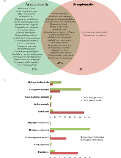
In addition to relative diversity, we also compared species and phylum composition between symptomatic and asymptomatic TIVAPs to determine whether it could reflect TIVAP infection history. 16S rRNA-based analysis revealed that only 5% of the species detected (A. xylosoxidansandP. aeruginosa) occurred in symptomatic TIVAPs only, i.e., were specific to infection, while 52% of species were detected in only asymptomatic samples and 43% of species occurred in both types of TIVAPs (Fig. 5A). Comparison of
FIG 5 Species occurrence and phylum distribution in symptomatic and asymptomatic samples from both hospitals. (A) Species occurrence. The Venn diagram shows species (OTUs) occurring in either sample group alone or in both sample groups (brown-shaded area). Percentages indicate the number of OTUs under each condition compared to the overall total. (B)Phylum distribution.
on September 8, 2020 by guest
http://msphere.asm.org/
phylum distributions of the detected species showed that symptomatic TIVAP samples contained markedly more species belonging to theFirmicutes, whereas asymptomatic samples contained many more species belonging to theBetaproteobacteria(Fig. 5B).
Hierarchical cluster analysis using the Bray-Curtis similarity index was carried out to determine how similar symptomatic and asymptomatic chambers were in terms of bacterial presence and abundance (Fig. S1). Strong clustering of symptomatic TIVAP samples from both hospitals could be observed, which was due to samples where either a single or a highly dominant species was detected. In contrast, asymptomatic samples formed much looser clusters and appeared overall more dissimilar to each other. The majority of Limoges asymptomatic samples clustered more tightly than Curie asymptomatic samples due to the relatively high abundance of P. aquatica andR. pickettii in these samples.
These results therefore show no significant difference in species diversity but reveal a distinct community composition pattern distinguishing symptomatic and asymptom-atic TIVAP samples at the phylum level. However, such a distinctive pattern could not be distinguished on a species level.
Difference in community structure revealed by culture-independent analysis is not an indicator of individual TIVAP clinical status.In order to test whether, if not community composition, community structure could differentiate asymptomatic and symptomatic TIVAPs, we plotted species rank abundance curves. The five most abun-dantly detected species in Limoges Hospital symptomatic TIVAPs were of clinical origin and corresponded to species also detected by culture (Fig. 6). In asymptomatic Limoges TIVAP samples, the five most abundant species included two species associated with infection (S. epidermidisandA. junii) and three of environmental origin. In Curie Hospital, the five most abundant species in symptomatic chambers included two species associated with clinical infection (the two main culture-detected species), as well as three species of environmental origin, whereas in Curie Hospital asymptomatic samples, all five most abundant species were of environmental origin. This demonstrates that the species detected as most abundant by 16S rRNA PCR and sequencing differed between hospitals and sample groups and did not allow the distinction between symptomatic and asymptomatic TIVAPs.
In order to compare community structures, we calculated total species evenness (E) for symptomatic and asymptomatic samples and the mean slope of the rank abun-dance curves for each sample group (Fig. 7). Asymptomatic samples showed species evenness (E⫽0.63) similar to that of symptomatic samples (E⫽0.64), but the mean slope for symptomatic samples was two times steeper (Curie,m⫽0.149; Limoges,m⫽ 0.203) than for asymptomatic samples (Curie,m⫽0.077; Limoges,m⫽0.105). When looking at individual sample rank abundance curve slopes, there was no statistical differ-ence (P⬎ 0.05) between symptomatic and asymptomatic samples in Curie Hospital or between symptomatic samples in the two hospitals. However, the slopes of individual asymptomatic and symptomatic samples in Limoges Hospital (P ⫽ 0.02), as well as asymptomatic samples in the two hospitals, differed significantly (P⫽0.03).
These results therefore indicate that symptomatic and asymptomatic sample com-munities were similarly even. The shape of the rank abundance curve, i.e., community structure, could distinguish between symptomatic and asymptomatic TIVAPs on a sample group but not an individual sample level.
DISCUSSION
We studied the bacterial colonization status of TIVAPs suspected or not of infection in two French hospitals. Whereas routine microbiology analysis yielded negative culture results for almost all asymptomatic samples, culture-independent 16S rRNA gene clone library generation identified bacteria as present in all clinically symptomatic as well as asymptomatic chambers. This is consistent with the literature showing that, irrespective of development of infection, devices become colonized after insertion (12, 15). Overall, culture and molecular data corresponded well. The latter confirmed the species iden-tified by culture as dominant OTUs in all but 5 cases. In these samples, the cultured
on September 8, 2020 by guest
http://msphere.asm.org/
FIG 6 Species abundance rank order. Species/OTUs are shown ranked according to their clone library abundance in the sample group. (A) Curie asymptomatic samples; (B) Curie symptomatic samples; (C) Limoges asymptomatic samples; (D) Limoges symptomatic samples.
on September 8, 2020 by guest
http://msphere.asm.org/
organism was detected in very low numbers. It is likely that these organisms were outcompeted or underrepresented due to PCR bias by OTUs not selected for under the culture conditions.
We identified a total of 42 different OTUs, indicating that catheter hubs were colonized by relatively complex bacterial communities. Two previous reports using molecular techniques to investigate colonization of symptomatic and asymptomatic intravascular devices also found a high number of OTUs in asymptomatic samples (13, 16). The species richness that we identified in our study was comparable to that identified by Larsen et al. (17). However, studies using high-throughput sequencing to analyze bacteria on intravascular devices found significantly higher numbers of OTUs (e.g., reference 13 identified an average of 100 OTUs per sample on peripheral venous catheters and reference 18 identified a total of 123 OTUs on needleless connectors for central venous catheters). This is not surprising, as a much greater sequencing depth and detection of low-abundance species are to be expected with these techniques.
Contamination of intravascular devices, especially long-term indwelling devices that are frequently accessed, can occur via the skin of the patient, the hospital environment, or contaminated fluids (13, 19). Consistently, the species that we identified in symp-tomatic chambers were indeed skin-associated flora, as well as human gut-associated species, hospital-associated species, and species considered of environmental origin. In asymptomatic samples, skin and clinical species were also frequently detected; how-ever, the majority of species were of environmental origin.
A number of the species identified in our study and considered of environmental origin were also detected in previous studies using molecular approaches to analyze intravascular devices. For example,Delftia acidovorans, the most frequently found species in asymptomatic patients from Curie Hospital, was detected by Zhang and colleagues (16). However, many of the species identified in our study have not previously been reported in catheter-related infections (e.g.,Blastobacter denitrificans,Herbaspirillum huttiense, andVariovorax paradoxus).
In long-term indwelling devices such as TIVAPs, contamination of the chamber or hub is the most common cause of device-related infections (1, 20). We detected bacteria in TIVAP chambers of all samples, both symptomatic and asymptomatic, in both hospitals by 16S rRNA clone library generation. However, biomass appeared to be lower in asymptomatic than in symptomatic samples. This is consistent with quantita-tive electron microscopic studies (12) that suggest that indeed most indwelling devices become colonized (to various degrees) after insertion and that only a low percentage of implants become infected (21). A number of other studies also detected bacteria by culture-independent means in samples that were culture negative (13, 16–18). How-FIG 7 Slopes of the rank abundance curves for all sample groups. For each sample group, bacterial species were ranked according to their frequency in the clone library and the slope was calculated for this rank abundance curve.
on September 8, 2020 by guest
http://msphere.asm.org/
ever, it remains difficult to determine whether these bacteria represent true coloniza-tion of the TIVAP chamber or are simply transients or reflect the presence of DNA from dead (e.g., due to use of antibiotics) bacteria.
Considering the sensitivity of molecular techniques to contamination, we took great care to identify and exclude potential contamination. When species found solely in reagents or materials used for molecular processing were also found in patient samples, these were excluded from analysis. However, Pelomonas aquatica,Ralstonia pickettii, andPropionibacterium acneswere frequently detected in patient samples in our study and were also detected as present in manipulated nonimplanted chambers and the NaCl solutions used by hospital staff to flush the chambers in order to remove the bacteria. We included these species in our analysis, as they were also detected by molecular means as dominant species in indwelling devices in references 13 and 18, respectively, andRalstonia pickettiihas indeed been shown to be a common contam-inant of hospital solutions (22, 23). As these species were not detected in nonimplanted nonmanipulated chambers but in nonimplanted chambers that were also flushed with the routinely used NaCl solution, it is uncertain whether these species could have been introduced as contaminants during sample processing or were already present in the flush solutions or whether the detection of such important levels of these species in our study represents true colonization. Furthermore, we could not analyze the solutions that were routinely introduced into the TIVAP chambers while still implanted. There-fore, we cannot draw any conclusions on potential introduction of these contaminants during routine procedures. However, it is generally possible that species potentially present in these routine working solutions could contribute significantly to the final community composition and structure as detectable by PCR-based methods, especially in the absence of a dominant pathogen such as is the case for most of the asymp-tomatic chambers. Our analysis is thus calling for increased attention to this problem. We detected significantly more species by 16S rRNA clone library generation in asymptomatic chambers than in symptomatic samples. However, the Simpson and Shannon relative diversity indices did not show this difference to be significant. Three previous molecular studies of bacterial presence on “colonized” and “uncolonized” arterial and central venous catheters (13, 16) also found no difference in relative diversity between the two groups; however, they did not detect a difference in OTU richness between the two sample types (13, 16). This could be due to truly a higher number of species being present in asymptomatic chambers in our study or could be a technical artifact. The lower bacterial biomass and absence of a highly dominant OTU in asymptomatic chambers could have led to a higher detection of different (rare) OTUs in these samples due to PCR bias (24). Due to small template amounts, we were unable to carry out quantitative confirmation such as real-time quantitative PCR or fluorescent in situhybridization (FISH) to investigate this; however, in future studies quantitative techniques should be integrated into the molecular analysis.
Our study found little overlap in bacterial presence on symptomatic and asymp-tomatic TIVAPs between the two French hospitals. We studied whether there was a potential common pool of bacteria colonizing asymptomatic TIVAPs that did not trigger infection. We found that only 18% of species occurred in asymptomatic chambers from both hospitals, all of which also occurred in symptomatic chambers. Further, the five most frequently occurring species also differed between the two asymptomatic sample cohorts. It has previously been shown that bacterial presence in different patient populations can vary dramatically between hospitals or even hospital sites and is observed in many complex polymicrobial colonization scenarios (e.g., gut [25], pros-thetic joint infection [26], and wound infections [27]). However, the hospital-specific bacterial community patterns prevented us from drawing generalizable conclusions regarding the presence of bacteria in symptomatic and asymptomatic chambers.
The diagnosis of vascular device-associated infections is notoriously difficult (28). A better understanding of the pathogenic potential of any given vascular device would therefore be crucial to improving clinical practice. Microscopy and a number of more recent molecular studies have built the case that vascular devices with and without
on September 8, 2020 by guest
http://msphere.asm.org/
symptoms are both colonized by a variety of microorganisms (12, 13, 16, 18). However, although colonization is a prerequisite for infection, it does not in itself appear to be sufficient to predict infection, as only a low percentage of catheters become infected (12). What leads to catheter infection therefore remains unknown, and different theo-ries have been proposed. For example, bacterial diversity in itself may promote increased pathogenicity or persistence in chronic infections (29, 30). Brogden et al. (31) suggested that in a polymicrobial infection, microorganisms may act synergistically and present different clinical outcomes than those in infection by a single organism. In our study, we were particularly interested in whether we could identify a bacterial com-munity parameter or signature indicative of the clinical status or fate of an implant that could potentially serve as a marker for infection.
As a predictive marker should be easily detectable and interpretable, we first investigated whether the most abundantly detected species allowed distinction be-tween symptomatic and asymptomatic samples. In neither hospital did the most abundant species allow clear distinction between either sample group; moreover, the species detected were partly hospital specific and did not allow a cross-site sample distinction either. When expanding the analysis to all species detected, we identified only 5% of species that were exclusively present in symptomatic TIVAP chambers, while other organisms, commonly associated with infection, interestingly occurred in both symptomatic and asymptomatic chambers. This is consistent with the previous findings that the simple presence of a species known to cause infection does not necessarily predict infection. We found that 52% of identified species, of mostly environmental origin, occurred in asymptomatic chambers alone. We were interested whether this could be used to predict the infection potential, e.g., whether an implant with any of these species would be more or less likely to present with clinical infection symptoms. We identified strong sample clustering, based on the presence of highly abundant hospital-associated species in symptomatic samples, and also found that the presence of highly abundantP. aquaticaorR. pickettiiled to some clustering in asymptomatic samples in Limoges which could be interesting for future studies. However, weak cross-site sample clustering and weak similarity between hospitals prevented the identification of a specific species assemblage consistently indicative of clinical status in our study. Comparison of species presences at the phylum level identified an increased presence of species belonging to theFirmicutesin symptomatic samples and a higher percentage ofBetaproteobacteriain asymptomatic samples. This is interesting, as an increase inFirmicutesmembers has been described for other infection scenarios such as in the gut (32) and should be investigated in future studies.
Finally, we investigated community structure as it was shown to be associated with clinical status in different diseases (33–36), where decreased evenness can be indicative of an imbalance, e.g., an infection, in the community. We found that, when analyzed as a group, asymptomatic samples showed similar total evenness as symptomatic samples but that in both hospitals the asymptomatic samples showed a rank abundance curve slope that was only half as steep as that for symptomatic samples. However, when rank abundance slopes were analyzed on an individual sample basis, as would be the case for a diagnostic prediction marker, they were not able to distinguish consistently between symptomatic and asymptomatic samples. This could, however, be interesting to investigate in future studies with a bigger sample group drawn from a number of different hospital sites.
Overall, our study of the bacterial colonization status of TIVAPs suspected or not of infection in two French hospitals confirms previous findings that both symptomatic and asymptomatic TIVAP chamber implants appear to be colonized by bacteria. Although we did not identify a particular species assemblage consistently predictive of infectious status of an implant, we identified a difference between symptomatic and asymptom-atic samples in species richness, phylum composition, and community structure on a sample group level, suggesting that the microbial ecology of intravascular devices could be predictive of TIVAP infection susceptibility. Hence, this improves our under-standing of the nature and evolution of intravascular device-associated infections.
on September 8, 2020 by guest
http://msphere.asm.org/
Further investigations combining culture techniques, quantitative sequencing analyses, and microscopic techniques could provide insight into the colonization dynamics of central venous catheters. Ultimately, this may lead to the definition of a microbial ecology signature that can predict infection susceptibility of individual medical im-plants.
MATERIALS AND METHODS
Patient background. Totally implantable venous access port (TIVAP) systems (Celsite [B. Braun, Melsungen, Germany] and Heliosite [Vygon, Paris, France]) were collected from the Anesthesia-Reanimation-Pain unit at the Institut Curie Hospital (Paris) between October 2008 and September 2009 and from the surgery department at the Limoges Dupuytren Hospital between August 2009 and July 2012. The medical personnel performing the TIVAP removal gave information and obtained the patient’s oral consent for sample collection.
All patients had TIVAP implants for cancer chemotherapy (see Table S1 in the supplemental material). “Asymptomatic” patients (n⫽20, 10 from each hospital) had a minimum implant duration of 6 months and were selected on the basis of absence of signs of infection from implantation to removal and absence or a minimum administration of antibiotic treatments during the last 6 months of implant indwelling. The criterion for TIVAP removal for “asymptomatic” patients (n⫽20, 10 from each hospital) was the end of treatment in the absence of suspected catheter-associated infection. We did not perform blood cultures for “asymptomatic” patients because such analysis is not included in the standard-of-care for such situations. “Symptomatic” patients were enrolled on the basis of suspicion of TIVAP implant infection, notably local and/or systemic clinical signs of infection and positive blood culture. TIVAP removal for “symptomatic” patients was based on the clinical decision of the treating physician in response to the suspected infection, with or without administration of antibiotics.
The patient group at Curie Hospital consisted of 7 males and 13 females, with a mean age of 52.3⫾ 18.9 years and a mean TIVAP implant period of 101.7 days for infected and 449.7 days for noninfected TIVAPs. The patient group at Limoges Dupuytren Hospital consisted of 8 males and 12 females, with a mean age of 62.5 ⫾10.7 years and a mean TIVAP implant period of 116.3 days for infected and 356.6 days for noninfected TIVAPs (Table S1). No significant associations were observed between TIVAP colonization and antibiotic treatment in the 4 weeks prior to TIVAP removal.
Sample collection.Inpatient TIVAP implantation, handling, and TIVAP removal were carried out under routine aseptic hospital protocols by experienced surgical staff. After removal, TIVAPs were immediately transferred to the microbiology laboratories of the respective hospitals and handled under laminar flow hoods with sterile reagents and instruments. Special arrangements were made with the hospitals to also process the noninfected TIVAPs, which are not routinely analyzed. In accordance with one of the methods for bacterial isolation from TIVAPs recommended by the European Society for Clinical Microbiology and Infectious Disease, the TIVAP chamber/hub was separated from the TIVAP catheter, disinfected on the outside with a pad soaked in povidone-iodine, and flushed with 1 ml of sterile saline solution (37, 38). This flush solution was then used for routine diagnostic culture, and 700l of the flush was immediately stored at⫺80°C for transfer to the Institut Pasteur and nucleic acid extraction. With each sample batch, an aliquot of the NaCl solution used to flush the chambers after explantation was also collected as reagent control. Control TIVAPs (nonimplanted nonmanipulated chambers and nonim-planted chambers that were manipulated like real samples, including the flush with saline solution) were collected to determine potential sources of bacterial contamination.
Routine diagnostic culture.The chamber flush was diluted and spotted on Columbia blood agar and chocolate agar for quantitative monitoring and also inoculated into liquid brain heart infusion medium and incubated for 24 to 48 h at 37°C under aerobic conditions. Identification and quantification of growing bacteria were carried out according to hospital standard operating procedures by hospital microbiologists and confirmed with Vitek mass spectrometry (MS) (matrix-assisted laser desorption ionization–time of flight [MALDI-TOF]).
DNA extraction from clinical samples. Prior to sample processing, 4 DNA extraction methods (QIAamp DNA minikit, Promega Wizard genomic extraction kit, Mo Bio PowerLyzer Ultraclean DNA isolation kit, and guanidium thiocyanate-EDTA-Sarkosyl extraction) were evaluated. The method with the best results for equal lysis of Gram-negative and Gram-positive bacteria, highest DNA yields from low starting material, and lowest intrinsic bacterial contamination was determined to be the Mo Bio PowerLyzer Ultraclean kit. Bacterial DNA was extracted from the TIVAP chamber flush liquid according to the manufacturer’s instructions. Control (DNA-free water as the template) extraction samples were run with each extraction in order to identify any potential DNA traces introduced during the extraction procedure. Extracted genomic DNA was verified by Tris-acetate-EDTA-agarose gel electrophoresis (1%), stained with GelRed, and quantified by applying 2.5l directly to a NanoDrop ND-1000 spectropho-tometer.
16S rRNA gene analysis—PCR and clone library generation.The universal primers for the domain Bacteria, 8f (5=-AGA GTT TGA TCC TGG CTC AG-3=) and 1492r (5=-GGT TAC CTT GTT ACG ACT T-3=), were used to amplify the bacterial 16S rRNA gene. Each primer was used at a final concentration of 0.2M, and 25 to 50 ng of DNA was added as the template to 50-l reaction mixtures. PCR cycling conditions were as follows: initial denaturation at 94°C for 2 min, followed by 32 cycles of denaturation at 94°C for 1 min, annealing at 56°C for 1 min, and extension at 72°C for 2 min, with a final extension step at 72°C for 10 min. 16S rRNA gene PCR products were verified on 1% agarose gels, purified with the QIAquick PCR purification kit, and cloned with the pGEM-T Easy vector system (Promega) according to the
on September 8, 2020 by guest
http://msphere.asm.org/
manufacturer’s instructions. The presence of the cloned insert was confirmed by colony PCR with vector primers gemsp6 (5=-GCT GCG ACT TCA CTA GTG AT-3=) and gemt7 (5=-GTG GCA GCG GGA ATT CGA T-3=). Clone PCR products with an insert of the correct size were purified as described above and sent for sequencing (Eurofins, Ebersberg, Germany). All reagents used for TIVAP flushing, DNA extraction, PCR, and clone library generation were subjected to 16S rRNA sequencing (Fig. 1A, green asterisks indicate all protocol steps at which reagents were tested). Materials (e.g., tubes) were flushed with DNA-free water and also sequenced (Fig. 1A, blue asterisks). Blanks using DNA-free water as the template were run for all procedures to control for handling and equipment contamination and were also subjected to 16S rRNA sequencing (Fig. 1A, red asterisks).
Clone library coverage was calculated by the formula [1⫺(n1/N2)]⫻100, wheren1is the number
of singletons detected in the clone library andN2is the total number of clones generated for this sample.
Clone libraries were generated to a minimum coverage of 95%, and 960 and 988 clones were generated for clinically symptomatic and clinically asymptomatic TIVAPs, respectively, from both hospitals (exclud-ing low-quality sequences and contaminants).
Sequence analysis. 16S rRNA sequences were manually proofread. Sequences with ambiguous bases or low quality were removed from the analysis. Primer sequences were trimmed off, and sequences were compared to GenBank (NCBI) with BLAST and to the Ribosomal Database Project with SeqMatch. A 98% similarity cutoff was used for operational taxonomic unit (OTU) determination, and a 95% cutoff was used for genus determination.
Sample staining and microscopy.Eight TIVAP chamber septa were collected separately from Limoges Hospital; four septa originated from clinically symptomatic patients, and four were from clinically asymptomatic patients. Septa were removed from their respective TIVAP chambers under sterile conditions. The septum side residing in the chamber was stained with approximately 10M SYTO9 and approximately 50M propidium iodide nucleic acid stains (LIVE/DEAD BacLight bacterial viability kits; Molecular Probes, Invitrogen) and incubated for 15 to 60 min in the dark.
Septa were imaged using a Nikon DN100 digital network camera mounted on a Nikon Eclipse E400 epifluorescence microscope equipped with a 40⫻0.75-numerical-aperture (NA) Plan Fluor objective. To excite and visualize labeled samples, a 100-W super-high-pressure mercury lamp was utilized and filtered through a green fluorescent protein (GFP) long-pass filter cube (482-/35-nm excitation filter, 505-nm dichroic mirror, 510-nm barrier filter) (Nikon France SAS). An exposure time of 17 ms was used, and a minimum of 20 fields of view was acquired for each sample. Images were manually assayed for the presence of bacteria; bacterial cells and human cells were distinguished by morphology and size.
Bacterial community and statistical analysis.We used bacterial occurrence as the determination of the presence or absence of an OTU independent of its abundance in the clone library and bacterial dominance as the abundance of an OTU either within a sample or within a sample group. Rank abundance curves were constructed by ranking the abundance of OTUs (number of clones) in a sample or sample set from most to least abundant. Linear regression was used to calculate the equation for the rank abundance curve (y⫽mx⫹p) wheremis the slope of the rank abundance curve. The Simpson diversity index was calculated as the sum of the relative species abundance (P) squared (D⫽ ⌺P2), and the Gini-Simpson index (1⫺D) was used in this paper in order to use positive correlation
values. The Shannon diversity index was calculated with the formulaHS⫽ ⫺⌺{P[ln(P)]}, wherePis the relative species abundance. Total evenness was calculated for the Shannon index asE⫽HS/Hmax.
Community similarity between samples was assessed by hierarchical cluster analysis using the Bray-Curtis similarity index [2jN/(Na⫹Nb)], whereNais the total number of OTUs in sample 1,Nbis the total number of OTUs in sample 2, and 2jNis the sum of the lower of the two abundances for species found in both sites.
Two-tailedttests for the comparison of two data sets were carried out in SPSS software, version 20 (Chicago, IL), withPbeing considered significant when it was⬍0.05. Correlation analysis was also carried out in SPSS using Pearson’s correlation coefficientRto determine linear relationships between clinical and microbiological variables and their significanceP. Bonferroni corrections were applied to eliminate false-positive results due to the high number of multiple comparisons.
Accession number(s).Sequences have been deposited in GenBank under accession numbers
MF377637toMF378601andMF399825toMF400811.
SUPPLEMENTAL MATERIAL
Supplemental material for this article may be found at https://doi.org/10.1128/ mSphere.00146-17.
FIG S1,PDF file, 0.2 MB. TABLE S1,PDF file, 0.1 MB. TABLE S2,PDF file, 0.1 MB. TABLE S3,PDF file, 0.1 MB. TABLE S4,PDF file, 0.1 MB. TABLE S5,PDF file, 0.1 MB.
ACKNOWLEDGMENTS
We thank Julie Vignaud and Roselyne Droual for their assistance during catheter collection.
on September 8, 2020 by guest
http://msphere.asm.org/
F.A.S. was a Marie Curie Fellow, and this work was supported by grants from the Institut Pasteur and Institut Pasteur-Institut Curie PTR/PIC “Nosocomial Infections pro-gram,” the Institut Pasteur-Institut Mérieux Collaborative Research program, the French Government’s Investissement d’avenir program, Laboratoire d’Excellence “Integrative Biology of Emerging Infectious Diseases” (grant ANR-10-LABX-62-IBEID), and the “Fon-dation Pour la Recherche Médicale” grant (Equipe FRM DEQ20140329508).
The funders had no role in study design, data collection and interpretation, or the decision to submit the work for publication.
REFERENCES
1. Raad I. 1998. Intravascular-catheter-related infections. Lancet 351: 893– 898.https://doi.org/10.1016/S0140-6736(97)10006-X.
2. Raad II, Hanna HA. 2002. Intravascular catheter-related infections: new horizons and recent advances. Arch Intern Med 162:871– 878.https:// doi.org/10.1001/archinte.162.8.871.
3. Mermel LA, Allon M, Bouza E, Craven DE, Flynn P, O’Grady NP, Raad II, Rijnders BJ, Sherertz RJ, Warren DK. 2009. Clinical practice guidelines for the diagnosis and management of intravascular catheter-related infection: 2009 update by the Infectious Diseases Society of America. Clin Infect Dis 49:1– 45.https://doi.org/10.1086/599376.
4. Leonidou L, Gogos CA. 2010. Catheter-related bloodstream infections: catheter management according to pathogen. Int J Antimicrob Agents 36(Suppl 2):S26 –S32.https://doi.org/10.1016/j.ijantimicag.2010.11.004. 5. Allegranzi B, Bagheri Nejad S, Combescure C, Graafmans W, Attar H,
Donaldson L, Pittet D. 2011. Burden of endemic health care-associated infection in developing countries: systematic review and meta-analysis. Lancet 377:228 –241.https://doi.org/10.1016/S0140-6736(10)61458-4. 6. Lebeaux D, Fernández-Hidalgo N, Chauhan A, Lee S, Ghigo JM, Almirante
B, Beloin C. 2014. Management of infections related to totally implant-able venous-access ports: challenges and perspectives. Lancet Infect Dis 14:146 –159.https://doi.org/10.1016/S1473-3099(13)70266-4.
7. Costerton JW, Stewart PS, Greenberg EP. 1999. Bacterial biofilms: a common cause of persistent infections. Science 284:1318 –1322.https:// doi.org/10.1126/science.284.5418.1318.
8. Donlan RM. 2001. Biofilms and device-associated infections. Emerg In-fect Dis 7:277–281.
9. O’Grady NP, Alexander M, Dellinger EP, Gerberding JL, Heard SO, Maki DG, Masur H, McCormick RD, Mermel LA, Pearson ML, Raad II, Randolph A, Weinstein RA, Healthcare Infection Control Practices Advisory Com-mittee. 2002. Guidelines for the prevention of intravascular catheter-related infections. Infect Control Hosp Epidemiol 23:759 –769.https:// doi.org/10.1086/502007.
10. Chauhan A, Lebeaux D, Decante B, Kriegel I, Escande MC, Ghigo JM, Beloin C. 2012. A rat model of central venous catheter to study estab-lishment of long-term bacterial biofilm and related acute and chronic infections. PLoS One 7:e37281. https://doi.org/10.1371/journal.pone .0037281.
11. Grice EA, Segre JA. 2011. The skin microbiome. Nat Rev Microbiol 9:244 –253.https://doi.org/10.1038/nrmicro2537.
12. Raad I, Costerton W, Sabharwal U, Sacilowski M, Anaissie E, Bodey GP. 1993. Ultrastructural analysis of indwelling vascular catheters: a quanti-tative relationship between luminal colonization and duration of place-ment. J Infect Dis 168:400 – 407.https://doi.org/10.1093/infdis/168.2.400. 13. Zhang L, Gowardman J, Morrison M, Krause L, Playford EG, Rickard CM. 2014. Molecular investigation of bacterial communities on intravascular catheters: no longer just Staphylococcus. Eur J Clin Microbiol Infect Dis 33:1189 –1198.https://doi.org/10.1007/s10096-014-2058-2.
14. Zhang L, Gowardman J, Morrison M, Runnegar N, Rickard CM. 2016. Microbial biofilms associated with intravascular catheter-related blood-stream infections in adult intensive care patients. Eur J Clin Microbiol Infect Dis 35:201–205.https://doi.org/10.1007/s10096-015-2530-7. 15. Maki DG. 1994. Infections caused by intravascular devices used for
infusion therapy: pathogenesis, prevention, and management, p 155–212.InBisno AL, Waldvogel FA (ed), Infections associated with in-dwelling medical devices, 2nd ed. American Society for Microbiology, Washington, DC.
16. Zhang L, Sriprakash KS, McMillan D, Gowardman JR, Patel B, Rickard CM. 2010. Microbiological pattern of arterial catheters in the intensive care unit. BMC Microbiol 10:266.https://doi.org/10.1186/1471-2180-10-266.
17. Larsen MK, Thomsen TR, Moser C, Høiby N, Nielsen PH. 2008. Use of cultivation-dependent and -independent techniques to assess contam-ination of central venous catheters: a pilot study. BMC Clin Pathol 8:10.
https://doi.org/10.1186/1472-6890-8-10.
18. Perez E, Williams M, Jacob JT, Reyes MD, Chernetsky Tejedor S, Steinberg JP, Rowe L, Ganakammal SR, Changayil S, Weil MR, Donlan RM. 2014. Microbial biofilms on needleless connectors for central venous catheters: comparison of standard and silver-coated devices collected from patients in an acute care hospital. J Clin Microbiol 52:823– 831.https://doi.org/10.1128/ JCM.02220-13.
19. Crnich CJ, Maki DG. 2002. The promise of novel technology for the prevention of intravascular device-related bloodstream infection. I. Pathogenesis and short-term devices. Clin Infect Dis 34:1232–1242.
https://doi.org/10.1086/339863.
20. Trautner BW, Darouiche RO. 2004. Catheter-associated infections: patho-genesis affects prevention. Arch Intern Med 164:842– 850.https://doi .org/10.1001/archinte.164.8.842.
21. Sherertz RJ, Raad II, Belani A, Koo LC, Rand KH, Pickett DL, Straub SA, Fauerbach LL. 1990. Three-year experience with sonicated vascular cath-eter cultures in a clinical microbiology laboratory. J Clin Microbiol 28: 76 – 82.
22. Maroye P, Doermann HP, Rogues AM, Gachie JP, Mégraud F. 2000. Investi-gation of an outbreak ofRalstonia pickettiiin a paediatric hospital by RAPD. J Hosp Infect 44:267–272.https://doi.org/10.1053/jhin.1999.0691. 23. Sundaram S, Lewis M, Eisenhuth J, Howard G, Jr, Larson B. 2002. Method for qualifying microbial removal performance of 0.1 micron rated filters. Part IV: retention ofHydrogenophaga pseudoflava(ATCC 700892) and Ralstonia pickettii(ATCC 700591) by 0.2 and 0.22 micron rated filters. PDA J Pharm Sci Technol 56:150 –171.
24. Polz MF, Cavanaugh CM. 1998. Bias in template-to-product ratios in multitemplate PCR. Appl Environ Microbiol 64:3724 –3730.
25. Arumugam M, Raes J, Pelletier E, Le Paslier D, Yamada T, Mende DR, Fernandes GR, Tap J, Bruls T, Batto JM, Bertalan M, Borruel N, Casellas F, Fernandez L, Gautier L, Hansen T, Hattori M, Hayashi T, Kleerebezem M, Kurokawa K, Leclerc M, Levenez F, Manichanh C, Nielsen HB, Nielsen T, Pons N, Poulain J, Qin J, Sicheritz-Ponten T, Tims S, Torrents D, Ugarte E, Zoetendal EG, Wang J, Guarner F, Pedersen O, de Vos WM, Brunak S, Doré J, MetaHIT Consortium, Antolin M, Artiguenave F, Blottiere HM, Almeida M, Brechot C, Cara C, Chervaux C, Cultrone A, Delorme C, Denariaz G, Dervyn R, Foerstner KU, Friss C, van de Guchte M, Guedon E, Haimet F, Huber W, van Hylckama-Vlieg J, Jamet A, Juste C, Kaci G, Knol J, Lakhdari O, Layec S, Le Roux K, Maguin E, Mérieux A, Melo Minardi R, M’rini C, Muller J, Oozeer R, Parkhill J, Renault P, Rescigno M, Sanchez N, Sunagawa S, Torrejon A, Turner K, Vandemeulebrouck G, Varela E, Winogradsky Y, Zeller G, Weissenbach J, Ehrlich SD, Bork P. 2011. En-terotypes of the human gut microbiome. Nature 473:174 –180.https:// doi.org/10.1038/nature09944.
26. Roger PM, Tabutin J, Blanc V, Léotard S, Brofferio P, Léculé F, Redréau B, Bernard E, Réso-Infectio-Paca-Est. 2015. Prosthetic joint infection: a plu-ridisciplinary multi-center audit bridging quality of care and outcome. Med Mal Infect 45:229 –236.https://doi.org/10.1016/j.medmal.2015.04 .003.
27. Misic AM, Gardner SE, Grice EA. 2014. The wound microbiome: modern approaches to examining the role of microorganisms in impaired chronic wound healing. Adv Wound Care 3:502–510.https://doi.org/10 .1089/wound.2012.0397.
28. Warwick S, Wilks M, Hennessy E, Powell-Tuck J, Small M, Sharp J, Millar MR. 2004. Use of quantitative 16S ribosomal DNA detection for diagnosis
on September 8, 2020 by guest
http://msphere.asm.org/
of central vascular catheter-associated bacterial infection. J Clin Micro-biol 42:1402–1408.https://doi.org/10.1128/JCM.42.4.1402-1408.2004. 29. Edwards R, Harding KG. 2004. Bacteria and wound healing. Curr Opin
Infect Dis 17:91–96.https://doi.org/10.1097/00001432-200404000-00004. 30. Ehrlich GD, Hu FZ, Shen K, Stoodley P, Post JC. 2005. Bacterial plurality as a
general mechanism driving persistence in chronic infections. Clin Orthop Relat Res (437):20 –24.https://doi.org/10.1097/00003086-200508000-00005. 31. Brogden KA, Guthmiller JM, Taylor CE. 2005. Human polymicrobial in-fections. Lancet 365:253–255. https://doi.org/10.1016/S0140-6736 (05)17745-9.
32. Kosiewicz MM, Zirnheld AL, Alard P. 2011. Gut microbiota, immunity, and disease: a complex relationship. Front Microbiol 2:180.https://doi.org/ 10.3389/fmicb.2011.00180.
33. Coburn B, Wang PW, Diaz Caballero J, Clark ST, Brahma V, Donaldson S, Zhang Y, Surendra A, Gong Y, Elizabeth Tullis D, Yau YC, Waters VJ, Hwang DM, Guttman DS. 2015. Lung microbiota across age and disease stage in cystic fibrosis. Sci Rep 5:10241.https://doi.org/10 .1038/srep10241.
34. David LA, Weil A, Ryan ET, Calderwood SB, Harris JB, Chowdhury F, Begum Y, Qadri F, LaRocque RC, Turnbaugh PJ. 2015. Gut microbial succession follows acute secretory diarrhea in humans. mBio 6:e00381-15.https://doi .org/10.1128/mBio.00381-15.
35. Kramer R, Sauer-Heilborn A, Welte T, Guzman CA, Abraham WR, Höfle MG. 2015. Cohort study of airway mycobiome in adult cystic fibrosis patients: differences in community structure between fungi and bacteria reveal predominance of transient fungal elements. J Clin Microbiol 53:2900 –2907.https://doi.org/10.1128/JCM.01094-15.
36. Sprockett DD, Ammons CG, Tuttle MS. 2015. Use of 16S rRNA sequenc-ing and quantitative PCR to correlate venous leg ulcer bacterial biobur-den dynamics with wound expansion, antibiotic therapy, and healing. Wound Repair Regen 23:765–771.https://doi.org/10.1111/wrr.12309. 37. Høiby N, Bjarnsholt T, Moser C, Bassi GL, Coenye T, Donelli G,
Hall-Stoodley L, Holá V, Imbert C, Kirketerp-Møller K, Lebeaux D, Oliver A, Ullmann AJ, Williams C, ESCMID Study Group for Biofilms and Consulting External Expert Werner Zimmerli. 2015. ESCMID guideline for the diag-nosis and treatment of biofilm infections 2014. Clin Microbiol Infect 21(Suppl 1):S1–S25.https://doi.org/10.1016/j.cmi.2014.10.024. 38. Lebeaux D, Larroque B, Gellen-Dautremer J, Leflon-Guibout V, Dreyer
C, Bialek S, Froissart A, Hentic O, Tessier C, Ruimy R, Pelletier AL, Crestani B, Fournier M, Papo T, Barry B, Zarrouk V, Fantin B. 2012. Clinical outcome after a totally implantable venous access port-related infection in cancer patients: a prospective study and review of the literature. Medicine (Baltimore) 91:309 –318. https://doi.org/10 .1097/MD.0b013e318275ffe1.