INTRODUCTION
Placenta is the communication organ that enables the exchange of various materials between mother and fetus. In addition to useful ones, transport of harmful and toxic sub-stances may also take place. These can lead to premature birth or malformations of the fetus [1]. Advancement in biomedicine is increasingly attracting more interest towards the mamma-lian development, in which one of the key roles is played by the placenta. Understanding the fundamental processes of placen-tation in different experimental animals, as well as the distinc-tion of morphologies of their placentas, is undoubtedly needed for understanding the processes of human development [2].
The blastocyst, the structure common to all the mam-mals, is composed of the embryoblast and the tropho-blast [3]. Trophotropho-blast is crucial for the maintenance of
pregnancy because it establishes the initial contact with the mucous endometrium of the uterus, assisted by expression of a series of glycoproteins on its membrane, e.g. cadherins and integrins [4]. The placental implantation in the rat occurs on the antimesometrial side of the uterus, whereas placenta is being formed on the mesometrial side of the uterus. On the 8th day of rat gestation, the trophoblast proliferates and gives
rise to the ectoplacental cone and on the 9th day maternal
lacunae without endothelium are being developed within this ectoplacental cone [5]. At the same time, an offshoot of the allantois is created. The mesenchyme of this offshoot on the 11th day of gestation establishes contact with the
ectopla-cental cone, i.e., with its basal part, which is called the cho-rion [6]. Action of allantoic mesenchyme results in angiogen-esis, and thus the fetal capillaries are being developed amongst the maternal lacunae. This process leads to the formation of the functionally most important part of the rat placenta - the labyrinth, where nutrients and gasses are being exchanged between mother and the embryo [7]. It is believed that defects in development of the labyrinth can lead to various disorders of placentation, and consequently to growth arrest of the fetus.
Structural changes in the rat placenta during the last third
of gestation discovered by stereology
Ljiljana Šerman1*, Iris Žunić1, Nina Vrsaljko1, Đurđica Grbeša2, Emil Gjurčević3, Željka Matašin3,
Tamara Nikuševa Martić1, Floriana Bulić Jakuš1, Ivana Tlak Gajger3, Alan Šerman4
1Department of Biology and, 2Department of Histology and Embryology, University of Zagreb, School of Medicine, Šalata 3, Zagreb, Croatia, 3Department for Biology and Pathology of Fish and Bees, University of Zagreb, Faculty of Veterinary Medicine, Heinzelova 55, Zagreb, Croatia, 4Department of Obstetrics and Gynaecology, Clinical Hospital Sveti Duh, Zagreb, Croatia
ABSTRACT
Structural changes in the rat placenta during the last third of gestation were for the first time assessed by stereology. Fischer female rats were euthanized on the day 16 or day 19 of gestation, and 35 placentas were collected. Three randomly selected placentas from each group were stere-ologically analyzed for the absolute volume. The proportion of the glycogenic cells and the trophoblast giant cells (TGC) in the basal part of the placenta was calculated using volume density. The absolute volume of the rat placenta on the day 16 of gestation was determined as 0.0638 cm3.
The labyrinth comprised 0.0274 cm3, the basal plate 0.0271 cm3 and the decidua 0.0093 cm3. On the day 19 of gestation, the absolute volume of
the placenta was 0.1627 cm3, the labyrinth occupied 0.0922 cm3, the basal plate 0.0596 cm3 and the decidua 0.0109 cm3. The volume density of
trophoblast giant cells was 0.174 cm0 on the day 16 and 0.107 cm0 on the day 19 of gestation. The glycogenic cells comprised 0.379 percentage
of the basal plate on the day 16 and 0.236 on the day 19 of gestation. We conclude that the absolute volume of the whole placenta and the lab-yrinth has increased from day 16 to the day 19 of gestation. In contrast, the volume density of glycogenic cells and trophoblast giant cells was higher on the day 16 than on the day 19 of gestation, probably due to the intensive trophoblast invasion during that time.
KEYWORDS: Stereology; placenta; trophoblast giant cells; glycogenic cells
DOI: http://dx.doi.org/10.17305/bjbms.2015.1.244 Bosn J Basic Med Sci. 2015;15(1):21-25. © 2015 ABMSFBIH
*Corresponding author: Ljiljana Šerman Department of Biology, School of Medicine, University of Zagreb,
Šalata 3, 10000 Zagreb E-mail: [email protected]
Variety of genes exists whose timely expression is essential for proper development of the labyrinth [8]. As the placentation in rats is of hemochorial type, i.e., the trophoblast is in direct contact with maternal bloodstream, the trophoblast invasion is necessary. The invasion is mediated by the trophoblast giant cells (TGC) and glycogen-rich trophoblast cells [9]. TCG are believed to be responsible for the endovascular invasion, whereas the trophoblast glycogen-rich cells are responsible for interstitial invasion in the uterine endometrium [10].
The rat placenta has three main parts, namely the basal plate or trophospongioblast, the labyrinth and the decid-ual part [11]. The basal part is located close to the decidua and is composed of several cell types: trophoblast stem cells, trophoblast giant cells, and glycogen cells. The labyrinth accommodates maternal lacunae and fetal blood vessels. They form separate bloodstreams segregated by placental mem-brane [12]. This memmem-brane makes the rat placenta of hemo-trichorial type, because it has three layers of trophoblast [13].
The aim of this research was to determine the dynam-ics of changes in absolute volumes of placentas between the 16th and the 19th day of the gestation, by applying stereology.
Furthermore, changes in absolute volumes of individual com-ponents of the placenta: the labyrinth, the basal layer, and the decidua were evaluated. Finally, because of their central role in trophoblast invasion into the uterine endometrium, which is essential for proper hemochorial placentation, the volume density and the absolute volumes of TGC and glycogen cells were determined.
MATERIALS AND METHODS
Placental samples
Adult Fischer female rats (three months old) were mated overnight with males of the same age. Finding of the sperm in the vaginal plug the next morning designated the day 1 of pregnancy. Pregnant animals were euthanized using ether on the day 16 or the day 19 of gestation. Abdomen was opened, and uterine horns were held out with forceps. Uterus was excised and washed in Dulbecco’s phosphate buffered saline (PBS). Uterine horns were dissected under the binocular dis-secting microscope, and placentas were isolated. Placentas were weighed and fixed in 10% formaldehyde, embedded in paraffin, processed for routine histology and 10 mm thick sec-tions stained by hematoxylin-eosin (HE).
This study was approved by the Ethical Committee of the Medical School of the University of Zagreb.
Stereological analysis
Stereological analysis was carried out on three randomly selected experimental placentas from the day 16 of gestation
and three placentas from the day 19 of gestation. For both days, the stereological analyzes included the absolute volume of total placenta and the absolute volumes of the basal layer, the labyrinth and the decidua. The volume density of the gly-cogen-rich cells and the giant cells of the trophoblast, as well as the absolute volume of these two types of cells in the basal layer of placenta, were also determined.
The absolute volume of placenta
For the determination of absolute volumes of analyzed placentas, we have constructed a test system for stereology in the Ellipse 3D computer program.
Our test system consisted of 494 testing points, superim-posed on the vertically oriented, previously photographed, paraffin sections. Analyzed sections were photographed on OlympusBx41 microscope (dissecting microscope Olympus SZX7, camera Olympus DP12) in the Department for Biology and Pathology of Fish and Bees, School of Veterinary Medicine of the Zagreb University. Hits (test points falling on the placental tissue) were counted according to the principle of the Cavalieri-method for the determination of the absolute volume according to formula:
Vp = t x ap x Σ P
Where Vp = the absolute volume of placenta, t = thickness of serial section, ap = test area belonging to a particular test point, and Σ P = total number of hits on all the sections of a single placenta [14].Thickness of serial sections was determined by preliminary measurements. Determination of the corrective shrinkage factor was carried out by measuring diameters of 200 randomly selected erythrocytes, and compared with the same number of diameters of fresh erythrocytes obtained from the sacrificed pregnant rat. The absolute volume of the placenta was subsequently corrected using this shrinkage factor [14].
Absolute volumes of placental components
Absolute volumes of the labyrinth, the basal layer and the decidua were determined by the same method as used for determination of the absolute volume of the whole placenta. Three testing systems out of 494 test points in the Ellipse 3D stereological program, i.e. one for each placental component (labyrinth, basal layer or decidua) were constructed. Total of volumes of all placental components (phases) was supposed to give the value of the absolute volume of the whole placenta obtained by preliminary measurement, which was used as control of the measurements performed.
The volume density and the absolute volume of
TGC and glycogen-rich cells
where the length of the testing line (L/p) was equal to 0.29 mm and the testing area (Ap) was 0.18 mm2. This testing system
was superimposed on every analyzed section, i.e. it covered the whole cross section of the placenta, so that the trophoblast giant cells and glycogen-rich cells were counted in the entire section, rather than a single visual field, as is the case on the microscope.
As it is most important to define the referent space imme-diately before the start of measurement, the basal layer of the placenta was defined in this case and hits for this part were counted. Subsequently, hits for glycogenic and giant cell of the trophoblast were counted on the same field.
The volume density (Vv) is the relative stereological vari-able, which indicates how large is the share occupied by the studied phase in the total space or what is the percentage of the studied phase in unit volume.
The absolute volume of the studied phase was calcu-lated by multiplication of the volume density of the studied phase with the absolute volume of the basal part of placenta, obtained from the previous measurements.
Statistical analysis
Mean, standard deviation and standard error were deter-mined using descriptive statistics. For statistical analysis of the mass differences between the two analyzed groups of placen-tas Student’s t-test, was used.
RESULTS
Placental masses on the day 19 of gestation (0.353±0.032g) were significantly higher than on the day 16 of gestation (0.299±0.039g) (p <0.05, Student’s t-test) (Figure 1).
Analysis of the absolute placental volume on the
days 16and 19 of gestation
Stereological analysis was performed on three randomly selected placentas from each experimental group and ana-lyzed in the total of 660 histological sections. To determine the absolute volume, a “Point grid system” was designed, which consisted of 4 frames (Figure 2). Each of these frames corre-sponded to one phase – a component of the placenta analyzed. Mean absolute volume of the placenta on the day 16 of gestation was 0.0638 cm3, out of which the labyrinth
occu-pied 0.0274 cm3 (43%), the basal layer 0.0271 cm3 (42%) and the
decidua only 0.0093 cm3 (15%).
On the day 19 of gestation, the mean absolute volume of the placenta has increased to 0.1627 cm3, out of which the labyrinth
occupied 0.0922 cm3 (56.5%), the basal layer 0.0596 cm3 (36.5%)
and the decidua only 0.0109 cm3 (7%) (Table 1; Figure 3).
The volume density of trophoblast giant cells and
glycogenic cells on the day 16 and 19 of gestation
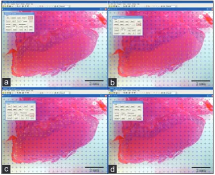
For the analysis of the volume density of trophoblast giant cells and glycogenic cells a “Line System” of equal length of the test
FIGURE 1. Comparison of masses of analyzed placentas on the 16th and the 19th day of gestation. Masses of placentas on the 19th day of gestation are statistically significantly larger than placentas isolated on the 16th day (p<0.05, Student's t test).
FIGURE 3. Changes in proportion of components in the rat pla-centa on gestational day 16 and 19 expressed as perpla-centages of total volume.
FIGURE 2. Presentation of „Point grid system“ with 494 test points and 4 frames. Yellow crosses (+) mark all hits on (A) total rat pla-centa; (B) the labyrinth; (C) the basal plate; and (D) the decidua.
d c
line and the test areas were constructed (Figure 4). On the day 16 of gestation, the volume density of the trophoblast giant cells was 0.174 cm0, and the volume density of glycogenic cells
was 0.379 cm0.
On the day 19 of gestation, as expected, the decline in both volume densities was observed, to 0.107 cm0 for trophoblast
giant cells and 0.236 cm0 for glycogenic cells (Table 2).
On the basis of the obtained results on the volume den-sities, we then calculated the absolute volumes of these cells in the basal layer of the placenta (Table 3). Thus, on the day 19 of gestation, as a consequence of the increase in the absolute volume of the basal layer of the placenta, an increase in the absolute volumes of the trophoblast giant cells was observed as well (Table 3).
DISCUSSION
We obtained the accurate data on absolute volumes of individual components of the placental structure and changes in their values during the last third of gestation. These data
provide an insight into the adaptation of placenta, particularly its labyrinth part, to the greater exchange of matter between mother and fetus as the gestation nears its end. We discov-ered the reduction of volume densities of trophoblast giant cells and trophoblast glycogen cells, which coincides with the reduction of their functions at the end of gestation. At that time, the trophoblast invasion is at its end, and the definitive placenta is formed.
This research also provides results on absolute volumes of the whole placenta and its components during normal pla-cental development in the rat, which might be even a better animal model to study placental function than the mouse [15]. Like humans, rodent placenta is of hemochorial type, i.e. the trophoblast cells originating from the chorion are in direct contact with the erythrocytes of the maternal blood. In hemo-chorial placenta, trophoblast invasion takes place in the uter-ine endometrium.
Similar stereological studies were carried out on the mouse placenta by Coan et al. and they obtained results analogous to our results for rat, wuth respect to the differences in gesta-tional age between the two studies. An increase in the absolute volume of the entire placenta but not in its components [14] was found in the mouse. While the absolute volume of the lab-yrinth increased, the volumes of other placental components (the basal layer and the decidua) exhibited a significant reduc-tion at the end of gestareduc-tion.
We found that the labyrinth of the rat placenta occupies 43% of the placenta on the day 16 of gestation and 57% of the placental volume on the day 19 of gestation, which is similar to values in the mouse [14]. Comparing our results with those of Coan et al. [14]on the absolute volumes of the placental basal layer and the decidua, there is a similarity in the reduction of their volumes at the end of gestation. Volume share of the basal layer in mouse placenta is about 29% on the day 17 and 24% on the day 19 of gestation, while in our analysis of the rat placenta, the basal layer occupies 42% of the placenta on the day 16 and only 36% on the day 19 of pregnancy. The volume share of the decidua in mouse placenta decreased to 23% on the day 17 of gestation and to 18% on the day 19, while in our case of the rat placenta, the reduction in the volume share of the decidua falls to 15% on the day 16 of gestation and to only 7% of placental mass on the day 19 of pregnancy.
The results of our work are important in view of stereolog-ical studies of placenta in normal and in complicated pregnan-cies, where stereology was successfully applied for interpreting the morphology of human and animal placenta [16], in describ-ing the villous trophoblast growth, trophoblast differentiation, vascular morphogenesis and diffusion transport [17,18].
Our results can be of help in future studies if we assume that the basic stereological variables of individual components of the placenta, and of the whole placenta vary depending
TABLE 1. Absolute volumes (cm3) of placentas and their components on the 16th and the 19th day of gestation
Absolute
volume Rat placenta (cm3,±SD) (cmLabyrinth 3,±SD) Basal layer (cm3,±SD) (cmDecidua 3,±SD) Day 16 0.0638±0.0046 0.0274±0.0042 0.0271±0.0017 0.0093±0.0010 Day 19 0.1627±0.0432 0.0922±0.0298 0.0596±0.0177 0.0109±0.0013
TABLE 2. Volume densities (Vv) of trophoblast giant cells (TGC) and glycogen cells on the days 16 and the 19 of gestation
Volume density Vv TGC (cm0,±SD) Vv glycogen cells (cm0,±SD)
Day 16 0.174±0.0318 0.379±0.0350
Day 19 0.107±0.0058 0.236±0.0151
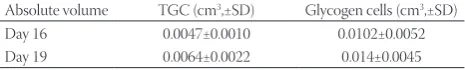
TABLE 3. Absolute volumes (cm3) of trophoblast giant cells (TGC) and glycogen cells on the day 16 and the day 19 of gestation in basal layer
Absolute volume TGC (cm3,±SD) Glycogen cells (cm3,±SD)
Day 16 0.0047±0.0010 0.0102±0.0052
Day 19 0.0064±0.0022 0.014±0.0045
FIGURE 4. „Line system“ was constructed in Ellipse 3D program, where the length of the test line (L/p) was always 0,28 mm and the test area (Ap) was always 0,18 mm2. A – the test system is first adjusted to the calibration on the picture; B – next, the glycogen cells were marked blue and the giant cells were marked orange .
on different harmful environmental influences, or in cases of maternal illness.
The data presented regarding the volume density of TGC and glycogen cells can be very useful in further research of endovascular trophoblast invasion, considering the existing research on human and rat placentas on the effects of pre-eclampsia and other factors that can bring about changes in these processes during pregnancy [19]. Trophoblast invasion in spiral arteries of the uterus is the essential prerequisite for successful development of placental circulation [20], and it is important to understand the dynamics of this process. Crucial roles in this process play trophoblast glycogen cells and tro-phoblast giant cells, for both of which we have obtained data on their volume densities and absolute volumes. Their volume densities were higher on the day 16 of gestation, when the tro-phoblast invasion was very intensive, and they diminished by the day 19 of gestation, when the trophoblast invasion ended.
Taking into account the diversity of application possibili-ties of stereology and the relatively low cost of this technique, stereology should certainly be considered as one of the meth-ods for obtaining quantitative data in both the clinical and basic medical research [21].
CONCLUSION
The results of our study may be used for further research on rodent placentation. The absolute volumes of the labyrinth, decidua and the basal layer, as well as the volume densities and the absolute volumes of the trophoblast giant cells and tropho-blast glycogen cells may be used for characterization of effects of various toxic substances and harmful environmental condi-tions on the components of placenta.
DECLARATION OF INTERESTS
The authors declare no conflict of interest.
REFERENCES
[1] Longo LD, Reynolds LP. Some historical aspects of understanding placental development structure and function. Int J Dev Biol 2010; 54 (2-3): 237-255. http://dx.doi.org/10.1387/ijdb.082774ll.
[2] Serman A, Serman L. Development of placenta in a rodent – model for human placentation. Front Biosci 2011; 3: 233-239. http://dx.doi. org/10.2741/e238.
[3] Sasaki H. Mechanisms of trophoectoderm fate specification in preimplantation mouse development. Dev Growth Differ 2010; 52: 263-273. http://dx.doi.org/10.1111/j.1440-169X.2009.01158.x. [4] Šerman LJ, Šerman A. Uloga glikoproteina u procesima
implant-acije i placentimplant-acije. Gynaecol Perinatol 2006; 15(2):82-88.
[5] Welsh AO, Enders AC. Chorioallantoic formation in the rat: I. Luminal epithelial cell death and extracellular matrix in the mesometrial region of implantation chambers. Am J Anat 1991; 192(3):215-231. http://dx.doi.org/10.1002/aja.1001920302. [6] Enders AC, Blankenship TN. Comparative placental structure.
Adv Drug Deliv Rev 1999; 38(1):3-15. http://dx.doi.org/10.1016/ S0169-409X(99)00003-4.
[7] De Rijk EPCT, Van Esch E, Filk G. Pregnancy dating in the rat: placen-tal morphology and maternal blood parameters. Toxicol Pathol 2002; 30(2):271-282. http://dx.doi.org/10.1080/019262302753559614. [8] Baczyk D, Drewlo S, Proctor L, Dunk C, Lye S, Kingdom J. Glial cell
missing-1 transcription factor is required for the differentiation of the human trophoblast. Cell Death Differ 2009; 16:719-727. http:// dx.doi.org/10.1038/cdd.2009.1.
[9] Carter AM Enders AC. Comparative aspects of trophoblast devel-opment and placentation. Reprod Biol Endocrinol 2004; 2(1):46-61. http://dx.doi.org/10.1186/1477-7827-2-46.
[10] Zybina TG, Kaufmann P, Frank HG, Freed J, Kadyrov M, Biesterfeld S. Genome multiplication of extravillious trophoblast cells in human placenta in the course of differentiation and invasion into endometrium and myometrium I. Dynamics of polyploidiza-tion. Tsitologiia 2002; 44(11):1058-1067.
[11] Huppertz B, Burton G, Cross JC, Kingdom JCP. Placental morphol-ogy. From molecule to mother-A dedication to Peter Kaufmann-A review. Placenta 2006; 27:S3-S8. http://dx.doi.org/10.1016/j. placenta.2006.01.007.
[12] Cross JC, Baczyk D, Dobric NM, Hemberger M, Hughes M, Simmons DG, et al. Genes, development and evolution of the placenta. Placenta 2003; 24:123-130. http://dx.doi.org/10.1053/ plac.2002.0887.
[13] Carter AM. Sources of comparative studies of placentation I. embryological collections. Placenta 2008; 29:95-98. http://dx.doi. org/10.1016/j.placenta.2007.09.008.
[14] Coan PM, Ferguson-Smith AC, Burton GJ. Developmental dynamics of the definitive mouse placenta assessed by stereology. Biol of Reprod 2004; 70: 1806-1813. http://dx.doi.org/10.1095/ biolreprod.103.024166
[15] Pijnenborg R, Vercruysse L. Mathias Duval on placental develop-ment in mice and rats. Placenta 2006; 27: 109-118. http://dx.doi. org/10.1016/j.placenta.2005.01.009.
[16] Serman L, Vlahovic M, Sijan M, Bulic-Jakus F, Serman A, Sincic N, et al. The impact of 5-azacytidine on placental weight, glycopro-tein pattern and proliferating cell nuclear antigen expression in rat placenta. Placenta 2007; 28: 803-811. http://dx.doi.org/10.1016/j. placenta.2007.04.001.
[17] Mayhew TM. A stereological perspective on placental morphol-ogy in normal and complicated pregnancies. Journal of Anatomy 2009 215:77-90. http://dx.doi.org/10.1111/j.1469-7580.2008.00994.x. [18] Veras MM, Damaceno Rodrigues NR, Caldini EG, Maciel
Ribeiro AA, Mayhew TM, Saldiva PH, et al. Particulate urban air pollution affects the functional morphology of mouse pla-centa. Biol Reprod 2008; 79: 578-584. http://dx.doi.org/10.1095/ biolreprod.108.069591.
[19] Geusens N, Verlohren S, Luyten C, Taube M, Hering L, Vercruysse L, et al. Endovascular trophoblast invasion, spiral artery remodelling and uteroplacental haemodynamics in a transgenic rat model of pre-eclampsia. Placenta 2008; 29:614-623. http://dx.doi. org/10.1016/j.placenta.2008.04.005.
[20] Lyall F. Priming and remodelling of human placental bed spiral arteries during pregnancy. Placenta 2005; 26 (Suppl A): S31-36. http://dx.doi.org/10.1016/j.placenta.2005.02.010.