Iran J Public Health, Vol. 45, No.10, Oct 2016, pp.1359-1366
Case Report
A Novel Mutation in the OFD1 Gene in a Family with
Oral-Facial-Digital Syndrome Type 1: A Case Report
Masoud DEHGHAN TEZERJANI
1,2, Reza MAROOFIAN
3,Mohammad Yahya VAHIDI
MEHRJARDI
1,4,
Barry A. CHIOZA
3, Shiva ZAMANINEJAD
5, Seyed Mehdi
KALAN-TAR
2,4, Mahmoud NORI-SHADKAM
6, Hamidreza GHADIMI
7, Emma L. BAPLE
8,
Andrew H. CROSBY
3, *Mohammadreza DEHGHANI
1,21. Medical Genetics Research Center, Shahid Sadoughi University of Medical Sciences, Yazd, Iran 2. Research and Clinical Center for Infertility, Shahid Sadoughi University of Medical Sciences, Yazd, Iran
3. Monogenic Molecular Genetics, University of Exeter Medical School, Exeter EX1 2LU, UK 4. Dept. of Medical Genetics, Shahid Sadoughi University of Medical Sciences, Yazd, Iran
5. Faculty of Dentistry, Golestan Uinversity of Medical Sciences, Gorgan, Iran 6. Dept. of Pediatrics, Shahid Sadoughi University of Medical Sciences, Yazd, Iran
7. Faculty of Medicine, Tehran University of Medical Sciences, Tehran, Iran
8. Human Genetics and Genomic Medicine, Faculty of Medicine, University of Southampton, and Wessex Clinical Genetics Service, Princess Anne Hospital, Southampton, UK
*Corresponding Author: Email: mrezdehghani@gmail.com
(Received 21 Dec 2015; accepted 12 May 2016)
Introduction
Orofaciodigital syndrome (OFDS) (OMIM 311200) is a general term for describing several distinctive developmental genetic diseases that are characterized by malformation of the mouth, face and digits. At least 13 different types of the syndrome have been recognized among which type 1 (OFD1) is the most common form with
an incidence of 1:50000 to 1:250000 live births (1, 2). The cardinal clinical features include cra-niofacial abnormalities (facial asymmetry, hyper-telorism, frontal bossing, microretrognathia, broadened nasal bridge, cleft palate, multi-lobulated tongue with nodules, and abnormal dentition), and digital abnormalities including
Abstract
Oral-facial-digital syndrome as heterogeneous developmental conditions is characterized by abnormalities in the oral cavity, facial features and digits. Furthermore, central nervous system (CNS) abnormalities can also be part of this de-velopmental disorder. At least 13 forms of OFDS based on their pattern of signs and symptoms have been identified so far. Type 1 which is now considered to be a ciliopathy accounts for the majority of cases. It is transmitted in an X-linked dominant pattern and caused by mutations in OFD1 gene, which can result in embryonic male lethality. In this study, we present a family suffering from orofaciodigital syndrome type I who referred to Medical Genetics Research Center, Shahid Sadoughi University of Medical Sciences in 2015. Two female siblings and their mother shared a novel 2-base pair deletion (c.1964-1965delGA) in exon 16 of OFD1 gene. Clinically, the sibling had oral, facial and brain abnormalities, whereas their mother is very mildly affected. She also had history of recurrent miscarriage of male fetus.
brachydactyly, syndactyly, clinodactyly and pre- or post-axial polydactyly. In 60% of cases with OFD1, brain structural abnormalities, develop-mental delay and intellectual disabilities have been identified (3). Majority of cases with onset older than 18 years, suffer from renal cystic dis-ease (4-6). The observation of multiple milia and hypotrichosis is remarkable skin abnormality for OFD1 which are not observed in other types of OFDS (7). Alopecia, deafness and trembling are expressed less frequently in OFD1.
Mutations in the OFD1 gene, which is mapped on chromosome Xp22, are responsible for OFD syndrome type 1. Therefore, OFD syndrome type 1 is inherited in X-linked dominant pattern and has prenatal lethality in the male fetus (5, 8, 9). A protein with 1011 amino acid expressed on the basal body of primary cilia and the centro-some is encoded by OFD1 gene. The protein also has a key role in the biogenesis of cilium and development by modulating Wnt signaling path-ways (10-13). To date, over 130 different muta-tions have been identified in OFD1 with many truncating mutations. The location of mutations causing OFD1 extends to 17 exons of 23 coding exons. About 75% of cases with OFD1 are spo-radic, and there are some reports about correla-tion of genotype with phenotype (12, 14). Muta-tions in OFD1 gene have also been identified in
association with four recessive X-linked pheno-types: Joubert syndrome, intellectual disability, type 2 Simpson-Golabi-Behmel syndrome and retinitis pigmentosa (15-19).
In this case study, we analyzed the sequences of the eight exons of the OFD1 gene in eight indi-viduals from an Iranian family with X-linked do-minant OFD1, and we identified a novel muta-tion. This family had a heterogeneous phenotypic findings ranging from being so mild in the moth-er that she was undiagnosed to sevmoth-ere neurodeve-lopmental delay, distinctive dysmorphic facial features, and multiple oral anomalies in her daughters.
Subjects
The study was approved by Ethics Committee of Yazd University of Medical Sciences and in-formed written consent was obtained from the all subjects participating in the study.
A female sibship was referred to the Medical Ge-netics Department of Yazd University of Medical Sciences due to craniofacial and oral dysmor-phism in addition to neurodevelopmental delay. The patients were thoroughly examined and all their clinical records were reviewed by a physi-cian. A three-generational pedigree for the family was constructed based on their family history ob-tained by a genetic counselor (Fig. 1).
Patient 1 (III-VII)
The first patient was a 9-year-old girl with a birth weight of 3200 g who was born to a family with non-consanguineous parents at 37 wk gestation, because of the fourth pregnancy. At birth, she had ventricular septal defect (VSD) diagnosed with fetal ultrasonography, and deformities of the mouth, jaw, and palate were remarkable. The child could stand and walk without assistance at 29 month. She also started to speak her first words at the age of two. At the time of the study, oral abnormalities included cleft palate, malaligned, abnormal dentition, macroglossia, ankyloglossia, multiple hyperplastic frenulums, and a bifid, lobulated tongue. Facial abnormalities that could be seen in the patient were dolichoce-phaly, macrocephaly (54.3 cm- 88 percentile), saddle nose deformity, low set ears, downslant palpebral fissures, and thin hair and eyebrows (Fig. 2). The girl also had severe psychomotor delay, intellectual disability and seizures. There were no signs of malformation in her feet and hands. Brain magnetic resonance imaging (MRI) revealed heterotopia in right cingulate cortex and brain Computed Tomography (CT) Scan showed ectopic gray matter in right posterior parasagittal (Fig. 3). Abdominal MRI revealed that she had no sign of renal anomaly. A G-band karyotype performed on the patient showed no gross chromosomal aberrations. She had normal vision and hearing and laboratory tests could not detect anything abnormal.
Fig. 2: Clinical features of case 1& 2; A, B, E & F:
Distinctive facial features, C & G: malaligned denti-tion, D & H: bifid tongue & nodules in the lateral border of tongue
Patient 2 (III-III)
The second patient was a 4-yr-old girl born as the result of the seventh pregnancy at 36 wk of gesta-tion with a birth weight of 3300 g. The patient could walk without support at 15 months. She had the following abnormalities: dolichocephaly, macrocephaly (51 cm- 66 percentile), multiple and malaligned dentition, cleft lip and palate, asymmetric, bifid and lobulated tongue, macrog-lossia, multiple hyperplastic frenulum, ankylog-lossia, low set ears, downslant palpebral fissures, and thin hair and eyebrows (Fig. 2). She had moderate psychomotor delay and developmental delay. The patient did not have any symptoms of abnormality in hands and feet. She had normal vision and hearing. Her brain MRI and CT-scan had normal findings and no renal abnormalities such as poly-cystic kidney (PKD) were identified. Her karyotype and routine laboratory test results were also all normal.
Mother of the patients (II-IV)
The mother of patients 1 and 2 was 29 yr old. She had a history of five abortions of malformed male fetuses (I, II, IV, V and III-VI). The abortions of all male fetuses happened during her third month of pregnancy. She was born with bifid tongue that was surgically paired. In addition, her dry and thin hair is re-markable. Although the mother experienced con-secutive abortions with two affected daughters, she did not attend any genetic counseling, which caused delay in diagnosis of the disease.
We extracted genomic DNAs from the peripheral blood samples using the ReliaPrep™ kit (Blood gDNA Miniprep System, Promega). The muta-tional hotspot within 8 exons of OFD1 (includ-ing exons 2, 3, 7, 8, 9, 12, 13 and 16) were ampli-fied based on standard protocols. Primer se-quences are available upon request. Then, the study employed 3730 DNA Analyzer and BigDye Terminator v3.1 cycle sequencing kit (Applied Biosystems) for sequencing of the PCR products in both directions.
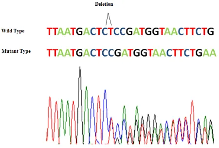
Our genetic studies identified a novel 2-base pair deletion (c.1964-1965delGA) in exon 16 of
two patients and their mother (Fig. 4). The muta-tion has not been previously reported, nor is present in The NHLBI Exome Sequencing Project Exome Variant Server (September 2013), Complete Genomics (February 2012), dbSNP (134–137), 1000 Genomes (May 2012) and Ex-ome Aggregation Consortium (ExAC), Cam-bridge, MA and as it was expected it was absent in the father and the brother of mother.
Fig. 4: Sequence analysis of the exon 16. Direct se-quencing of the PCR products in affected individuals and wildtype. CT (GA in the sense strand) was de-leted.
Discussion
mother, pedigree analysis and finally the genetic screening of the OFD1 gene. Although the find-ings described for OFD1 overlap with those ob-served in other OFDS types, in familial cases, it can be differentiated because of its X-linked do-minant inheritance with multiple male lethality and absence of parental consanguinity from pedi-gree analysis (20).
Unsurprisingly, affected individuals in this family did not display all traditionally key features of OFDS, thus confirming once again the variable phenotypic spectrum of OFDS. For instances, in this family neither of the patients have digital ab-normalities which occur in 88% of Patients with OFDS (12). In addition, polycystic kidney was not observed in the family which is almost present in all the affected individuals with OFDS in their adulthood (21). However, absence of the kidney features in the affected sisters can be due to their young age and there is a very high risk for PKD in the patients. Since PKD leads to renal failure careful morphological assessments and biochemical monitoring of renal function in the family is necessary. Structural brain malformation in patients with OFD1 has often been found in 88.7% among patients displaying neurological symptoms or/and cognitive/behavioral abnor-malities. The most frequent anomalies of brain structure included disorders of neuronal migra-tion and organisamigra-tion, agenesis of the corpus cal-losum, intracerebral cysts, cerebellar malforma-tions and porencephaly (3). Brain MRI analysis of the older sister did not find any abnormalities and younger affected individual only presented mal-formation of cortical development (MCD) in-cluding grey matter heterotopias.
Both affected sister presented mild macrocephaly and dolichocephaly. Although Macrocephaly is a cardinal clinical feature in X-linked recessive OFD1 neurodevelopmental syndromes in males, it has never been reported in OFDS type 1. This is the first report of macrocephaly in female pa-tients with mutation in OFD1.
The mother of the patients was presented with a milder phenotype than that of her daughters (20). Clinical features in the mother of the patients were noticed only after presentation of severe symptoms in her daughters. Milder phenotype in the mother and clinical variability in the affected children, which is common for OFD1, poses challenges for accurate diagnosis and genetic counselling. It can be explained because of the different degree of somatic mosaicism X-inactivation (20). Modifier genes can also be im-portant factor for variability in families with OFD1 patients (22). Male lethality in OFD1 usually occurs in the first or second trimester of pregnancy and only more than a dozen excep-tional male cases with mutation in OFD1 have been reported to date. In this family, the mother had a history of five male miscarriages. There-fore, this mutation can also be responsible for lethality in male fetuses.
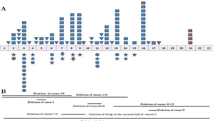
Fig. 5: Schematic representation of the reported OFD1 gene mutations (Last update; main source: Prattichizzo et al. 2008). A: Point mutations. B: Genomic deletions. Square (frame shift ), Triangle (splice site), Star (nonsence), Circle (missense), Diamond (in-frame deletion) and Red symbols indicating mutations found in males with OFD1 X-linked recessive disorder.
Conclusion
We identified a novel truncating mutation in OFD1 in three female members of a family dis-playing variable symptoms and severity of clinical manifestation of OFDS type 1. As observed in previous cases with OFD1, phenotypic variability even within a family is possibly a rule rather than the exception. Hence, this report emphasizes im-portance as well as the challenges of genetic counseling for OFD1 patients and their relatives. In cases of OFDS, thorough physical examina-tion, collecting the family history and genetic screening of the affected individuals and their female relatives, along with monitoring of renal function are mandatory.
Ethical considerations
Ethical issues (Including plagiarism, informed consent, misconduct, data fabrication and/or fal-sification, double publication and/or submission, redundancy, etc.) have been completely observed by the authors.
Acknowledgment
We are thankful to the family members for their willing participation in this study. The authors declare that they have no competing interests.
References
1. Gorlin RJ, Anderson VE, Scott CR (1961). Hypertrophied frenuli, oligophrenia, famflial trembling and anomalies of the hand. Report of four casesin one family and a forme fruste in another. N Engl J Med, 264:486-489. 2. Toriello HV (2009). Are the oral-facial-digital
syndromes ciliopathies? Am J Med Genet A, 149A(5):1089-1095.
3. Del Giudice E, Macca M, Imperati F, D'Amico A, Parent P, Pasquier L, Layet V, Lyonnet S, Stamboul-Darmency V, Thauvin-Robinet C, Franco B, Oral-Facial-Digital Type ICG (2014). CNS involvement in OFD1 syndrome: a clinical, molecular, and neuroimaging study. Orphanet J Rare Dis, 9:74. 4. Feather SA, Winyard PJ, Dodd S, Woolf AS
clinical, radiological and histopathological features of a new kindred. Nephrol Dial Transplant, 12(7):1354-1361.
5. Feather SA, Woolf AS, Donnai D, Malcolm S, Winter RM (1997). The oral-facial-digital syndrome type 1 (OFD1), a cause of polycystic kidney disease and associated malformations, maps to Xp22.2-Xp22.3. Hum Mol Genet, 6(7):1163-1167.
6. Ferrante MI, Giorgio G, Feather SA, Bulfone A, Wright V, Ghiani M, Selicorni A, Gammaro L, Scolari F, Woolf AS, Sylvie O, Bernard L, Malcolm S, Winter R, Ballabio A, Franco B (2001). Identification of the gene for oral-facial-digital type I syndrome. Am J Hum Genet, 68(3):569-576.
7. Habib K, Fraitag S, Couly G, de Prost Y (1992). [Cutaneous lesions in the orofaciodigital syndrome]. Ann Pediatr (Paris), 39(7):449-452. 8. Solomon LM, Fretzin D, Pruzansky S (1970).
Pilosebaceous dysplasia in the oral-facial-digital syndrome. Arch Dermatol, 102(6):598-602.
9. Franco B, Ballabio A (2006). X-inactivation and human disease: X-linked dominant male-lethal disorders. Curr Opin Genet Dev, 16(3):254-259.
10. Romio L, Wright V, Price K, Winyard PJ, Donnai D, Porteous ME, Franco B, Giorgio G, Malcolm S, Woolf AS, Feather SA (2003). OFD1, the gene mutated in oral-facial-digital syndrome type 1, is expressed in the metanephros and in human embryonic renal mesenchymal cells. J Am Soc Nephrol, 14(3):680-689.
11. Giorgio G, Alfieri M, Prattichizzo C, Zullo A, Cairo S, Franco B (2007). Functional characterization of the OFD1 protein reveals a nuclear localization and physical interaction with subunits of a chromatin remodeling complex. Mol Biol Cell, 18(11):4397-4404. 12. Macca M, Franco B (2009). The molecular basis
of oral-facial-digital syndrome, type 1. Am J Med Genet C Semin Med Genet, 15;151C(4):318-325.
13. Hunkapiller J, Singla V, Seol A, Reiter JF (2011). The ciliogenic protein Oral-Facial-Digital 1 regulates the neuronal differentiation of embryonic stem cells. Stem Cells Dev, 20(5):831-841.
14. Bisschoff IJ, Zeschnigk C, Horn D et al. (2013). Novel mutations including deletions of the entire OFD1 gene in 30 families with type 1 orofaciodigital syndrome: a study of the extensive clinical variability. Hum Mutat, 34(1):237-247.
15. Budny B, Chen W, Omran H, Fliegauf M, Tzschach A, Wisniewska M, Jensen LR, Raynaud M, Shoichet SA, Badura M, Lenzner S, Latos-Bielenska A, Ropers HH (2006). A novel X-linked recessive mental retardation syndrome comprising macrocephaly and ciliary dysfunction is allelic to oral-facial-digital type I syndrome. Hum Genet, 120(2):171-178. 16. Coene KL, Roepman R, Doherty D et al. (2009).
OFD1 is mutated in X-linked Joubert syndrome and interacts with LCA5-encoded lebercilin. Am J Hum Genet, 85(4):465-481. 17. Field M, Scheffer IE, Gill D, Wilson M, Christie
L, Shaw M, Gardner A, Glubb G, Hobson L, Corbett M, Friend K, Willis-Owen S, Gecz J (2012). Expanding the molecular basis and phenotypic spectrum of X-linked Joubert syndrome associated with OFD1 mutations.
Eur J Hum Genet, 20(7):806-809.
18. Webb TR, Parfitt DA, Gardner JC et al. (2012). Deep intronic mutation in OFD1, identified by targeted genomic next-generation sequencing, causes a severe form of X-linked retinitis pigmentosa (RP23). Hum Mol Genet, 21(16):3647-3654.
19. Tenorio J, Arias P, Martinez-Glez V, Santos F, Garcia-Minaur S, Nevado J, Lapunzina P (2014). Simpson-Golabi-Behmel syndrome types I and II. Orphanet J Rare Dis, 9:138. 20. Shimojima K, Shimada S, Sugawara M,
Yoshikawa N, Niijima S, Urao M, Yamamoto T (2013). Challenges in genetic counseling because of intra-familial phenotypic variation of oral-facial-digital syndrome type 1. Congenit Anom (Kyoto), 53(4):155-159.
21. Gurrieri F, Franco B, Toriello H, Neri G (2007). Oral-facial-digital syndromes: review and diagnostic guidelines. Am J Med Genet A, 15;143A(24):3314-3323.
23. Rakkolainen A, Ala-Mello S, Kristo P, Orpana A, Jarvela I (2002). Four novel mutations in the OFD1 (Cxorf5) gene in Finnish patients with oral-facial-digital syndrome 1. J Med Genet, 39(4):292-296.
24. Thauvin-Robinet C, Cossee M, Cormier-Daire V et al. (2006). Clinical, molecular, and genotype-phenotype correlation studies from 25 cases of oral-facial-digital syndrome type 1: a French and Belgian collaborative study. J Med Genet, 43(1):54-61.
25. Chetty-John S, Piwnica-Worms K, Bryant J, Bernardini I, Fischer RE, Heller T, Gahl WA, Gunay-Aygun M (2010). Fibrocystic disease of liver and pancreas; under-recognized features of the X-linked ciliopathy oral-facial-digital syndrome type 1 (OFD I). Am J Med Genet A, 152A(10):2640-2645.
26. Diz P, Alvarez-Iglesias V, Feijoo JF, Limeres J, Seoane J, Tomas I, Carracedo A (2011). A novel mutation in the OFD1 (Cxorf5) gene may contribute to oral phenotype in patients with oral-facial-digital syndrome type 1. Oral Dis, 17(6):610-614.
27. Morisawa T, Yagi M, Surono A, Yokoyama N, Ohmori M, Terashi H, Matsuo M (2004). Novel double-deletion mutations of the OFD1 gene creating multiple novel transcripts. Hum Genet, 115(2):97-103. 28. Thauvin-Robinet C, Thomas S, Sinico M et al.
(2013). OFD1 mutations in males: phenotypic spectrum and ciliary basal body docking impairment. Clin Genet, 84(1):86-90. 29. Juric-Sekhar G, Adkins J, Doherty D, Hevner
RF (2012). Joubert syndrome: brain and spinal cord malformations in genotyped cases and implications for neurodevelopmental functions of primary cilia. Acta Neuropathol, 123(5):695-709.