Specific Newborn Individualized Developmental Care and Assessment
Program Movements Are Associated With Acute Pain in Preterm Infants
in the Neonatal Intensive Care Unit
Liisa Holsti, PhD*; Ruth E. Grunau, PhD*‡§; Tim F. Oberlander, MD*‡§; and Michael F. Whitfield, MD‡§
ABSTRACT. Objective. The Newborn Individualized Developmental Care and Assessment Program (NIDCAP) is widely used in neonatal intensive care units and com-prises 85 discrete infant behaviors, some of which may communicate infant distress. The objective of this study was to identify developmentally relevant movements indic-ative of pain in preterm infants.
Methods. Forty-four preterm infants were assessed at 32 weeks’ gestational age (GA) during 3 phases (baseline, lance/squeeze, and recovery) of routine blood collection in the neonatal intensive care unit. The NIDCAP and Neonatal Facial Coding System (NFCS) were coded from separate continuous bedside video recordings; mean heart rate (mHR) was derived from digitally sampled continuous electrographic recordings. Analysis of vari-ance (phase ⴛgender) with Bonferroni corrections was used to compare differences in NIDCAP, NFCS, and mHR. Pearson correlations were used to examine rela-tionships between the NIDCAP and infant background characteristics.
Results. NFCS and mHR increased significantly to lance/squeeze. Eight NIDCAP behaviors also increased significantly to lance/squeeze. Another 5 NIDCAP be-haviors decreased significantly to lance/squeeze. Infants who had lower GA at birth, had been sicker, had expe-rienced more painful procedures, or had greater mor-phine exposure showed increased hand movements in-dicative of increased distress.
Conclusions. Of the 85 NIDCAP behaviors, a subset of 8 NIDCAP movements were associated with pain. Particularly for infants who are born at early GAs, addi-tion of these movements to commonly used measures may improve the accuracy of pain assessment.Pediatrics
2004;114:65–72;preterm infant, pain, NIDCAP, movement, NICU.
ABBREVIATIONS. NIDCAP, Newborn Individualized Develop-mental Care and Assessment Program; NICU, neonatal intensive care unit; GA, gestational age; PCA, postconceptional age; NFCS, Neonatal Facial Coding System; SNAP-II, Score for Neonatal Acute Physiology II.
M
ajor advances in neonatal care now enable a high proportion of preterm infants to sur-vive. During a time when medical concerns focused primarily on stabilizing the physiologic needs of preterm infants, Als, a developmental psy-chologist, developed a theory and systematic method of assessing the developmental needs of preterm newborns, the Newborn Individualized Develop-mental Care and Assessment Program (NIDCAP).1Als hypothesized that early exposure to stress, or the mismatch of the preterm infant brain with the envi-ronment, could be linked to the long-term develop-mental impairments reported in these children at school age.2Since the early 1980s, the NIDCAP
ob-servation system has become widely used in neona-tal intensive care units (NICUs) throughout North America and Europe.3,4With this system, infant
re-sponses, which include motor behaviors, state orga-nizational behaviors, and autonomically related in-dicators (eg, respiratory pattern), are recorded continuously in 2-minute time blocks before, during, and after a procedure.5
Although the NIDCAP is not designed specifically to assess pain, determining whether specific NIDCAP behaviors occur during acute pain might provide more accurate recognition of valid cues that may be useful for clinical pain assessment and pain management. For clinical reasons, accurate interpretation of preterm in-fant pain behaviors is crucial. For example, inin-fants who are in pain or are “stressed” may be given sedatives or analgesics. Analgesics may act differently in the brain according to whether pain is present.6 Therefore,
ap-propriate administration of analgesics only when pain is present may be critical for preventing unwanted long-term side effects of opioid use.7,8Thus, identifying
and treating pain in preterm infants is a high priority for caregivers in the NICU because early pain exposure may alter nociceptive pathways9,10and may also
con-tribute to changes in other areas of development.11,12
Although improved pain assessment and manage-ment is a clinical priority, accurate identification of pain responses is complex for a number of reasons. First, preterm infants respond with facial, motor, and physiologic changes to acute pain, but they differ from term-born infants in that their responses are of smaller magnitude, particularly at younger gesta-tional ages (GAs).13,14Second, preterm infants at
ear-lier GAs may display different pain behaviors than infants at later GAs as a result of neurologic imma-turity. Such behaviors may not be captured in the
From the *Centre for Community Child Health Research, British Columbia Research Institute for Children’s and Women’s Health, Vancouver, British Columbia, Canada; ‡Department of Pediatrics, University of British Colum-bia, Vancouver, British ColumColum-bia, Canada; and §Children’s and Women’s Health Centre of British Columbia, Vancouver, British Columbia, Canada. Received for publication Mar 3, 2003; accepted Jul 7, 2003.
Reprint requests to (L.H.) Centre for Community Child Health Research, Room L-408, 4480 Oak St, Vancouver, British Columbia, V6H 3V4 Canada. E-mail: [email protected]
current pain scales because the behaviors chosen have been based on behaviors seen in term infants. Third, no physiologic or behavioral threshold specif-ically marks the presence of pain. Finally, although using a single pain index is easier for clinicians, the physiologic and behavioral responses of preterm in-fants to painful stimuli are often dissociated15;
there-fore, reliance on current pain indices may not cap-ture the range of responses in this population.16
Currently, changes in facial activity, shifts in infant sleep/waking state, and physiologic indices of heart rate are the most promising biobehavioral pain indi-cators in preterm infants.17–20
In contrast to the research using facial activity and heart rate as pain cues, body movements have not been evaluated thoroughly. Some researchers in-clude knee or leg flexion in their pain index,21–23and
others use a measure of total body movements.13
These measures are problematic because, first, flex-ion and extensflex-ion are not pain specific; and, second, many movements are combined, thus making them more difficult to interpret. Alternatively, using spe-cific behaviors from the NIDCAP to assess pain is appealing because it is an established tool that is developmentally appropriate for preterm infants, and the coding descriptors of each of the facial and body movements are very specific.
Several researchers have used the NIDCAP to as-sess body movements of infants in the NICU, but these studies either did not describe the procedures observed or did not include painful procedures.24–26
Recently, 1 study did examine NIDCAP behaviors in response to events that varied in degree of intrusive-ness (endotracheal suctioning, chest physical ther-apy, diaper change, and nasogastric feed); however, this study used brief observation periods and did not include a pain procedure (eg, skin breaking).27Only
2 studies have used the full NIDCAP to study pain in premature infants. The first of these investigations did not evaluate the system along with other reliable, valid behavioral and physiologic pain measures or take into account GA at assessment, baseline behav-ioral state, or handling before the invasive proce-dure.28 Although the second study measured pain
responses over longer periods of time and compared the NIDCAP with other reliable infant biobehavioral
pain measures, it included only 10 infants, and the time of each handling phase was not controlled.29
The purpose of the present study was to determine whether a subset of NIDCAP movements could be used as valid acute pain cues in preterm infants.
METHODS Study Subjects
The infants were recruited by a NICU-trained research nurse, and written informed consent was obtained from the mother or other legal guardian according to a protocol approved by the Clinical Research Ethics Board of the University of British Colum-bia. The study sample included 44 preterm infants in the level III NICU in the Children’s & Women’s Health Centre of British Columbia (Vancouver, BC, Canada). The infants wereⱕ32 com-pleted weeks’ GA at birth, had no major congenital anomalies, and had no reported illicit maternal drug use during pregnancy. In-fants who had received analgesics or sedatives within 72 hours of the assessment or who had significant intraventricular hemor-rhage (grade III or IV) and/or parenchymal brain injury (periven-tricular leukomalacia) were excluded. All infants were 32 weeks’ postconceptional age (PCA;⫾7 days) at the time of testing. Infant characteristics are presented in Tables 1 to 3. Sample size estimates were calculated as though we were using a between-groups de-sign; this provides a conservative estimate given that we used a repeated measures design. GPOWER30was used to calculate the estimate, and effect sizes entered into the program were based on changes in Neonatal Facial Coding System (NFCS) scores during blood collection at 31 to 33 weeks.13Using this method, 15 infants were needed to detect differences between each phase for a power of .90 with the statistical significance set at .05. For examining gender and associations of clinical variables, the sample size was increased to 44.
Measures
All infants were observed during blood collection that was required for clinical management. The 3 phases of the procedure included in this study were a baseline period of 6 minutes of no handling immediately before the first contact by the laboratory technician (baseline); a blood collection period of 6 minutes from insertion of the lancet into the heel, which included the heel lance and squeezing (lance/squeeze); and an undisturbed recovery pe-riod of 6 minutes from the last contact of the laboratory technician (recovery).
Infant State
Infant sleep/wake state was coded according to the NIDCAP protocol5: 1⫽deep sleep; 2⫽light sleep; 3⫽drowsy; 4⫽quiet awake; 5⫽active awake; 6⫽highly aroused/crying; and 7⫽AA (prolonged respiratory pause⬎8 seconds). The predominant state during each 2-minute period was coded for each phase.
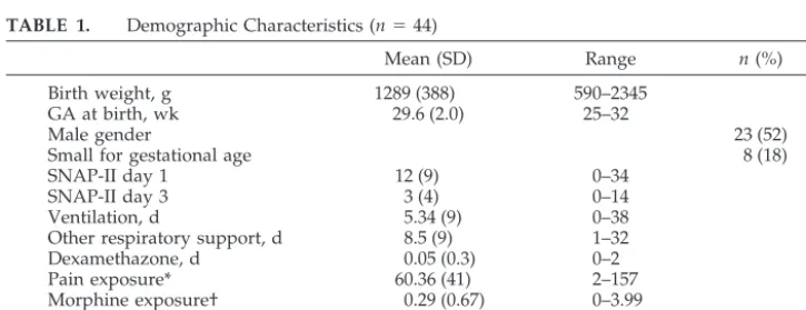
TABLE 1. Demographic Characteristics (n⫽44)
Mean (SD) Range n(%)
Birth weight, g 1289 (388) 590–2345
GA at birth, wk 29.6 (2.0) 25–32
Male gender 23 (52)
Small for gestational age 8 (18)
SNAP-II day 1 12 (9) 0–34
SNAP-II day 3 3 (4) 0–14
Ventilation, d 5.34 (9) 0–38
Other respiratory support, d 8.5 (9) 1–32
Dexamethazone, d 0.05 (0.3) 0–2
Pain exposure* 60.36 (41) 2–157
Morphine exposure† 0.29 (0.67) 0–3.99
Ethnicity (white) 34 (77)
Maternal age, y 32.1 (5.7) 19–47
* Number of invasive (skin-breaking) procedures from birth to the study day.
† Morphine exposure⫽(daily average/kg per os dose/3⫹daily average intravenous mg/kg)⫻
Facial Activity (NFCS)
The NFCS is a reliable, well-validated behavioral pain measure that is used widely in studies of term-born19,31 and preterm in-fants.13,14,32–34Traditionally, the full NFCS has been applied for brief periods (eg, 20 seconds per phase) to capture the acute pain response. However, for this study, the frequency of NFCS brow bulge was coded continuously for 18 minutes using the Noldus Observer system35(throughout 6 minutes of baseline, 6 minutes of lance/squeeze, and 6 minutes of recovery) to match the NIDCAP coding. Brow bulge was selected as a proxy for upper facial actions because it has been shown to correlate highly with the other upper facial actions of the NFCS.36Lower facial actions were not used in that they are sometimes obscured in preterm infants. Videotapes were edited for coding in random order of events, and coders were blind to all clinical information about the infants and to events. To establish reliability, both the primary NFCS coder (L.H.) and the reliability coder were trained on the entire tool to achieve a reliability coefficient of .87.19 In addition, reliability coding was conducted on 20% of the sample. For data analysis, the frequency of NFCS brow bulge was summed across all infants for each 6-minute phase.
Full Body (NIDCAP)
The NIDCAP behaviors were coded continuously, from video recordings of each infant, for the 3 phases (baseline, lance/ squeeze, and recovery), and coding was conducted blind to all clinical information. Following published NIDCAP procedures, the frequency of each infant’s movements was recorded system-atically in 2-minute time blocks.5The primary coder (L.H.) was an occupational therapist, and the reliability coder was a physiother-apist, both of whom were NIDCAP certified. Reliability for the NIDCAP was initially established during the certification pro-cess.37In addition, a randomly selected sample of 5% of NIDCAP video segments from the study (baseline segment, lance/squeeze segment, or recovery segment) was coded to evaluate reliability. NIDCAP reliability was calculated by determining the percentage of agreement of occurrence (both coders indicating the presence or absence of a behavior) within every 2-minute time segment during each 6-minute phase for each infant. Interrater agreement was
87%. Physiologic measures (heart rate) were recorded by custom computer software and so were not scored using the NIDCAP observation record.
Heart Rate
Continuous electrocardiographic activity was recorded from a single lead of surface electrocardiogram (lead II) and was digitally sampled at 360 Hz off-line using a specially adapted computer acquisition system. Custom physiologic signal processing soft-ware38 was used to acquire, process, and analyze heart rate. R waves were detected from the sampled electrocardiogram and were used to form a smoothed instantaneous 4-Hz time series as described previously.39Mean heart rate was calculated for each 2-minute segment of each study period to correspond to the 2-minute NIDCAP time blocks and averaged over 6 minutes of each of the 3 phases (baseline, lance/squeeze, and recovery). Before statistical analysis, 25 (6%) of the 2-minute HR segments were dropped because of poor signal and were replaced by the group mHR for that phase.
Background Data
A NICU-trained research nurse completed the prospective clin-ical chart review and obtained information from birth to day of testing, including but not limited to the following: birth weight, GA at birth, Apgar score at 1 minute, illness severity using the Score for Neonatal Acute Physiology II (SNAP-II),40 amount of opioid and other analgesic and sedative exposure, numbers and types of invasive skin-breaking procedures, respiratory support, and type and time of last handling just before blood collection. Invasive procedures were defined as those involving skin break-ing, such as heel lance, venipuncture, insertion of arterial and venous lines, lumbar puncture, and chest-tube insertion. In addi-tion, number of endotracheal intubations was collected (Table 1). Study day characteristics of the infants are presented in Table 2.
Procedures
Each infant was lying in the incubator undisturbed for a period of at least 30 minutes before recording. Heart rate data were collected by attaching the leads from the bedside monitor to a custom-designed computer data acquisition system. Two cameras (one positioned for close-up on the face, the other on the full body) were attached to a custom-made recording set-up on a moveable cart, including two 9-in video monitors. The signals were fed directly to 2 VCRs, and a time code was imprinted automatically. Each study phase was marked with an inaudible event cue signal recorded simultaneously on the videotape and physiologic acqui-sition systems. During recording, the incubator was partially cov-ered with a blanket, and the infant’s position was supported (nested) using a continuous roll around both sides and feet. At the time the infants were studied, 10 were supine, 27 were prone, and 7 were side lying with the face and full body in view for video coding. The infant’s position was not altered before or during the procedure, because handling to alter the position may affect the infant pain response.41For the blood-collection procedure, the research nurse applied a foot-warming pack 5 minutes before the laboratory technician drew the blood. The research nurse deter-mined which foot would be used for the blood collection accord-ing to which foot would be most easily accessed by the laboratory technician to minimize extreme stretching of the leg and foot TABLE 2. Infant Characteristics on the Study Day (n⫽44)
Mean (SD) Range n
(%)
PCA on study day, wk 32.3 (0.7) 31–33 Postnatal age on study
day, d
18.2 (13) 3–51
Mechanical ventilation on study day
4 (9)
Time since last feed, min*
58.8 (4) 0–116
No. of painful procedures in 24 h before study day
1.5 (1) 0–11
Time since last invasive procedure, min
1242.2 (1187) 40–6690
* Four infants were not on oral feeds.
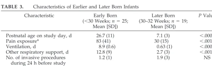
TABLE 3. Characteristics of Earlier and Later Born Infants
Characteristic Early Born (⬍30 Weeks;n⫽25;
Mean [SD])
Later Born (30–32 Weeks;n⫽19;
Mean [SD])
PValue
Postnatal age on study day, d 26.7 (11) 7.1 (3) ⬍.0001
Pain exposure* 83 (41) 30 (15) ⬍.001
Ventilation, d 8.9 (0.6) 0.63 (1) ⬍.0001
Other respiratory support, d 12.8 (9) 2.7 (3) ⬍.001
No. of invasive procedures during 24 h before study day
1.2 (1) 1.9 (3) NS
Time since last handling 86.6 (55) 96.4 (47) NS
NS indicates not significant.
during the procedure. Fifteen different laboratory technicians con-ducted the blood collection on the 44 infants. The laboratory technician’s standard protocol involved checking the infant’s identification band on the incubator, removing the warming pack from the foot, swabbing the heel with a small gauze pad with disinfectant, lancing the heel, and then gently squeezing the heel intermittently until the amount of blood that was required for clinical care was collected. A research technician set up the video camera and the VCR machines, operated the cardiac data acqui-sition computer, and marked each phase during the procedure.
Data Analysis
The frequencies of each NIDCAP behavior were reviewed, and the 31 movements that occurred in ⬍25% of the infants were excluded from statistical analysis (Fig 1).27Total frequencies of each NIDCAP movement were summed for each 6-minute phase to reveal in a clinically meaningful way the amount of infant movement exhibited throughout the procedure. Repeated mea-sures analysis of variance was conducted across the 3 phases of blood collection, with gender examined as a between-subjects variable. Bonferroni corrections were used to correct for overall error. Statistically significant analysis of variance was followed by planned t tests for paired comparisons to identify differences between specific phases. Pearson product-moment correlations were used to examine associations between perinatal variables and to describe relationships between the NIDCAP and infant background characteristics during lance/squeeze.
RESULTS Infant State
Most infants were in “sleep” (75%) or “drowsy” (25%) states during baseline, and then shifted signif-icantly from baseline to lance/squeeze (t⫽ ⫺7.40,P ⬍.0001). Seven percent of infants remained in “deep sleep” during lance/squeeze, no infants were in “ac-tive sleep,” 56% were “drowsy,” and 36% were cry-ing. Sleep/wake state also shifted significantly from lance/squeeze to recovery (t⫽6.43,P⬍.0001); 70%
of infants returned to sleep, and only 2% remained highly aroused.
NFCS and NIDCAP
There were no statistically significant differences in gender with any of the behavioral measures. The frequency of NFCS brow bulge increased signifi-cantly across the 3 phases (F[1,43] ⫽ 49.43; P ⬍ .0001). Of the 25 NIDCAP behaviors included in the statistical analyses, the frequency of a subset of 8 NIDCAP behaviors—flex arms, flex legs, extend arms, extend legs, hand on face, finger splay, fisting, and frown—increased significantly across the 3 phases. The frequency of a second subset of 5 NIDCAP behaviors—twitch face, twitch body, twitch extremities, mouthing, and foot clasping— decreased significantly across the 3 phases. In addition, the frequency of 2 NIDCAP behaviors— diffuse squirms (F[1,43]⫽5.22;P⬍.008) and arching (F[1,43]⫽4.22; P ⬍ .03)— decreased significantly from lance/ squeeze to recovery. Finally, during recovery, all but 1 of the NIDCAP behaviors returned to baseline frequencies; the frequency of fisting (t⫽ ⫺2.5,P⬍0. 02) remained increased. The total frequencies of the NIDCAP behaviors across the 3 phases of blood col-lection are shown in Table 4.
Heart Rate
Heart rate increased significantly across the 3 phases (F[1,43]⫽40.67;P⬍.0001). Heart rate (mean ⫾standard deviation) increased from baseline (157.5 ⫾12) to lance/squeeze (174.0⫾ 20;t⫽ ⫺6.74,P⬍ .0001) and decreased during recovery (156.3 ⫾ 16; t⫽9.48,P⬍ .0001).
Infant Background Characteristics
GA at birth and postnatal age (days) at testing were highly correlated (r⫽ ⫺0.91,P⬍.0001); GA at birth was used to examine the relationships between infant characteristics and the NIDCAP behaviors. Perinatal and study day characteristics of the earlier born and later born infants are shown in Table 3.
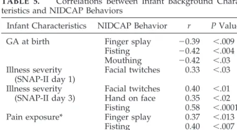
During blood collection at 32 weeks, infants who were born at lower GAs (⬍30 weeks) showed signif-icantly higher frequencies of finger splay, fisting, and mouthing during lance/squeeze than infants who were born at later GAs. Infants who had been ex-posed to greater numbers of invasive procedures since birth showed significantly more finger splays and fisting. Moreover, infants who had greater opi-oid exposure displayed significantly more fisting and hand on face. Finally, infants who were sicker earlier in the neonatal course (SNAP-II day 1) dis-played more facial twitches. Finally, infants who continued to be physiologically unstable on day 3 (SNAP-II day 3) continued to show more facial twitches, greater numbers of hand on face, and fist-ing than infants who became medically more stable (Table 5).
DISCUSSION
Although the NIDCAP has been used widely in NICUs throughout North America and Europe since
the 1980s, this study is the first to examine whether the NIDCAP catalog behaviors can be used to iden-tify acute pain in preterm infants under well-con-trolled conditions using a relatively large sample. The infants in this study showed facial, behavioral state, and heart rate responses similar to responses documented in other studies of responses of preterm infants to acute pain.13,42 Although we found that a
significant proportion (37%) of the NIDCAP move-ments either did not occur or occurred in ⬍25% of the infants, this finding is consistent with another study that recently reported that more than one third of the movements described by the NIDCAP were not observed in infants⬍30 weeks’ GA in the NICU even when observed sequentially over a 7-week pe-riod. However, these investigators did not specify the procedures that they observed.26
The most important finding of this study was that we found a subset of 8 NIDCAP movements that seem to be associated with acute pain in preterm infants. Although some of the 8 NIDCAP movements that we identified are included in other pain mea-sures, others have not been described as behavioral pain cues before this study. For example, the in-creased frequency of flexing and extending the ex-tremities has been reported in other studies that de-scribed and assessed pain responses in both term-born23,43 and preterm infants.22,28,44 In particular,
extending the legs seems to be a consistently ob-served distress cue.27,29 The combined flexing and
extending of the legs observed in these infants may be reflexive in nature (ie, the flexor withdrawal re-sponse),45,46 but because the flexor withdrawal
re-sponse is not pain specific, the use of these move-ments for pain assessment is unlikely to improve the identification of pain if facial activity, sleep/wake state, and change in heart rate are also used.
As in previous studies, we found that finger splay increased during the lance phase. Moreover, finger splay may be a developmentally specific distress cue, because the infants who were born before 30 weeks’ GA had a higher frequency of finger splays to lance than those who were bornⱖ30 weeks. These earlier born infants show greater finger splays not only during lance/squeeze but also during the baseline phase. This suggests that these infants may be rela-tively more stressed. This finding is consistent with previous studies that examined procedural stress and pain responses in this population27,29 and may
be indicative of “sensitization” that results from greater early pain exposure.47,48
Fisting has been described in 1 other study that examined preterm infants’ responses to pain.49
Fur-thermore, a recent study showed that fisting is con-sidered by a majority of nurses to be a pain indica-tor.50Like finger splay, fisting seems to be a sensitive
distress cue in infants who are born at earlier gesta-tional ages (⬍30 weeks) and may be useful in iden-tification of pain in infants who are sicker, who have been exposed to greater numbers of invasive proce-dures, and who require opioids during their care. This finding is important because preterm infants’ behavioral pain expression is influenced by their pre-vious experience in the NICU; that is, infants who are TABLE 4. Changes in Frequency of NIDCAP Behaviors
Across the Phases
NIDCAP Behavior Total Frequencies Across Phases
Baseline Lance/Squeeze Recovery PValue
Flex arms 38 88 49 ⬍.01
Flex legs 118 310 117 ⬍.0001
Extend arms 26 52 27 ⬍.02
Extend legs 103 216 79 ⬍.01
Hand on face 14 45 11 ⬍.001
Finger splay 42 86 51 ⬍.048
Fisting 0 12 7 ⬍.004
Frown 12 51 16 ⬍.0001
Twitch face 153 19 127 ⬍.0001
Twitch body 74 12 60 ⬍.001
Twitch extremities 371 39 287 ⬍.0001
Mouthing 22 8 36 ⬍.02
Foot clasping 18 1 16 ⬍.01
TABLE 5. Correlations Between Infant Background Charac-teristics and NIDCAP Behaviors
Infant Characteristics NIDCAP Behavior r PValue
GA at birth Finger splay ⫺0.39 ⬍.009 Fisting ⫺0.42 ⬍.004 Mouthing ⫺0.42 ⬍.03 Illness severity
(SNAP-II day 1)
Facial twitches 0.33 ⬍.03
Illness severity Facial twitches 0.40 ⬍.01 (SNAP-II day 3) Hand on face 0.35 ⬍.02
Fisting 0.58 ⬍.0001
Pain exposure* Finger splay 0.37 ⬍.013
Fisting 0.40 ⬍.007
Morphine exposure† Fisting 0.57 ⬍.0001 Hand on face 0.34 ⬍.02
* Number of invasive (skin-breaking) procedures from birth to study day.
born at earlier gestational ages and who have expe-rienced greater numbers of painful procedures may show diminished facial responses to acute pain.14,42
In addition to fisting and finger splaying, which have been previously associated with painful experiences in the NICU, the movement of hand on face (which involves a defensive-like action with the infant plac-ing a hand on its face) may represent an additional pain cue in the preterm population.
The only NIDCAP facial movement that increased significantly during lance/squeeze was frowning (brow lowering). Frowning is a flexor motion that involves knitting of the eyebrows or darkening of the eyes, a motion that is consistent with brow lowering shown in facial responses to pain.13,19
Whereas we found a subset of 8 NIDCAP behav-iors associated with pain, we also found 5 behavbehav-iors that decreased significantly during the lance/ squeeze phase. As in our previous study of stress during endotracheal suctioning, twitches decreased and startles did not change significantly during a more invasive procedure. Although in some infant pain scales, jitteriness and startles have been in-cluded with other behavioral indicators of pain,44,51
we conclude that, in preterm infants, these are not specific pain cues.27,29Indeed, many fetal ultrasound
studies show that twitches and startles are behaviors associated with sleep states in the normal fetus.52–54
Rather than indicators of pain, twitches are necessary movements for normal infant development that in-fluence neuron cell death, synapse elimination, mus-cle fiber differentiation, and formation of topo-graphic maps.55 Although we concluded that
twitches may not be specific pain cues, infants who were sicker on day 1 and who remained sicker on day 3 had more facial twitches associated with the lance/squeeze phase at 32 weeks’ PCA, a finding that has not been reported in previous studies. We cannot attribute this finding to the infants’ being in active sleep during the lance, because none of our infants remained in this sleep state during blood collection.
Another NIDCAP behavior that decreased to lance/squeeze was foot clasping. This finding was not unexpected, because infants who are rapidly flexing and extending their legs are less likely to clasp their feet together. Similarly, the frequency of mouthing (more than 1 opening and closing of the mouth) decreased during lance/squeeze. Indeed, fe-tal ultrasound studies have shown that mouthing is a regularly observed movement in utero (ie, under optimal, nonstressed conditions).56,57
Like finger splay, the frequency of 2 behaviors— diffuse squirms and arching— occurred at high fre-quencies during baseline. However, they differed from finger splay in that they did not increase to lance/squeeze and furthermore dropped signifi-cantly during recovery. The high frequency of these movements during baseline may indicate a higher basal arousal in earlier born preterm infants; how-ever, even infants who were born atⱖ30 weeks and had little previous pain exposure showed these be-haviors. It is possible that their diminished frequency
during recovery was an indication of fatigue caused by the length of the procedure.
Before this study, most of the literature describing the responses of preterm infants to acute pain used very short periods of observation. Our findings dem-onstrated that many preterm infants remain in a higher state of physiologic and behavioral arousal not only during the tissue-damaging portion but also during the entire blood collection. It may be that this higher and sustained level of stress contributes to altered reactivity and self-regulation observed later in these children.11,58
Another important finding of this study is that we found increased body movements associated with acute pain in infants who were born at earlier gesta-tional ages. This finding is contrary to the dampened facial activity that has been associated with infants of lower GAs36and those who experience greater
num-bers of painful procedures.14,42 Increased body
movements during pain may be indicative of in-creased pain sensitivity as a result of sensitization, which is then followed by the “wind-up” phenome-non, both of which are spinal cord–mediated ef-fects.48
Although our study carefully controlled for age at assessment, length of assessment time, and proce-dure order, there remain some limitations. First, it was not possible to be blinded to events when coding body movements. However, with the use of a second video camera, facial coding was conducted blinded to events. Second, the infants’ position during the lance was not controlled; more than half of the in-fants were positioned in prone during the assess-ment. It is standard practice in our nursery to pro-mote prone positioning to support physiologic stability, particularly for infants with respiratory dif-ficulties.59We deliberately did not alter the position
of the infant for the study because change of position would likely alter biobehavioral reactions. Although prone position does not seem to affect the facial responses during blood work,41it may have altered
the frequency of 1 NIDCAP body movement, sitting on air, an action whereby the legs are flexed at the hips and extended at the knees. This movement would be very unlikely to occur in prone.
CONCLUSION
The NIDCAP is a developmentally sensitive model of care that provides a detailed behavioral catalog of infant behaviors. We found a subset of 8 NIDCAP movements that were associated with acute pain in preterm infants at 32 weeks’ PCA. By adding a few discrete body movements to the assessment of pain, particularly hand movements such as finger splay, fisting, and hand on face, we can use some elements of the NIDCAP assessment to provide additional behavioral cues that may make identifying pain in preterm infants more accurate, particularly for in-fants who are born at early GAs.
ACKNOWLEDGMENTS
Oc-cupational Therapy Foundation Post-Doctoral Fellowship (Dr Holsti), and a Senior Scholar Award from the Michael Smith Foundation for Health Research (Dr Grunau).
We thank the staff and families of the Special Care Nursery at B.C.’s Children’s Hospital for participation in this study; Colleen Fitzgerald, Study Co-ordinator; Gisella Gosse and Adi Amir for data collection; and Colleen Jantzen for carrying out reliability NFCS coding, all of whom are staff of the Biobehavioral Research Unit of Centre for Community Health and Health Evaluation Research, B.C. Research Institute for Children’s & Women’s Health. We also thank Linda Williams, Clinical Supervisor, Phys-iotherapy Department, B.C.’s Children’s Hospital, for carrying out NIDCAP reliability coding.
REFERENCES
1. Als H. Toward a synactive theory of development: promise for the assessment and support of infant individuality.Infant Ment Health J.
1982;3:229 –243
2. Als H, Gilkerson L. The role of relationship-based developmentally supportive newborn intensive care in strengthening outcome of pre-term infants.Semin Perinatol.1997;21:178 –189
3. Symington A, Pinelli J. Developmental care for promoting development and preventing morbidity in preterm infants.Cochrane Database Syst Rev.2003;(4):CD001814
4. Jacobs SE, Sokol J, Ohlsson A. The Newborn Individualized Develop-mental Care and Assessment Program is not supported by meta-analysis of the data.J Pediatr.2002;140:699 –706
5. Als H.Manual for the Naturalistic Observation of Newborn Behavior (Pre-term and Full(Pre-term). Boston, MA: The Children’s Hospital; 1984 6. Rahman W, Fitzgerald M, Aynsley-Green A, Dickenson A. The effects of
neonatal exposure to inflammation and/or morphine on neuronal re-sponses and morphine analgesia in adult rats. In: Jensen TS, Turner JA, Weisenfeld-Halling Z, eds.Progress in Pain Research and Management. Vol 8. Seattle, WA: IASP Press; 1997:738 –794
7. Ng E, Taddio A, Ohlsson A. Intravenous midazolam infusion for seda-tion of infants in the neonatal intensive care unit.Cochrane Database Syst Rev.2003;(1):CD002052
8. Mao J. Opioid-induced abnormal pain sensitivity: implications in clin-ical opioid therapy.Pain.2002;100:213–217
9. Anand KJ, Coskun V, Thrivikraman KV, Nemeroff CB, Plotsky PM. Long-term behavioral effects of repetitive pain in neonatal rat pups.
Physiol Behav.1999;66:627– 637
10. Anand KJS. Effects of perinatal pain and stress.Prog Brain Res.2000; 122:117–129
11. Grunau RE. Long-term consequences of pain in human neonates. In: Anand KJS, Stevens BJ, McGrath PJ, eds.Pain in Neonates. 2nd Revised and Enlarged Edition, Pain Research and Clinical Management.Vol 10. New York, NY: Elsevier Science; 2000:55–76
12. Grunau R. Early pain in preterm infants. A model of long-term effects.
Clin Perinatol.2002;29:373–394
13. Craig KD, Whitfield MF, Grunau RV, Linton RV, Hadjistavropoulos HD. Pain in the preterm neonate: behavioral and physiological indices.
Pain.1993;52:287–299
14. Johnston CC, Stevens BJ. Experience in a neonatal intensive care unit affects pain response.Pediatrics.1996;98:925–903
15. Morison SJ, Grunau RE, Oberlander TF, Whitfield MF. Relations be-tween behavioral and cardiac autonomic reactivity to acute pain in preterm neonates.Clin J Pain.2001;17:350 –358
16. Porter FL, Wolf CM, Miller JP. Procedural pain in newborn infants: the influence of intensity and development.Pediatrics.1999;104(1). Avail-able at: www.pediatrics/org/cgi/content/full/104/1/e13
17. Stevens BJ, Johnston CC, Grunau RV. Issues of assessment of pain and discomfort in neonates.J Obstet Gynecol Neonatal Nurs.1995;24:849 – 855 18. Stevens B, Johnston CC, Petryshen P, Taddio A. The premature infant
pain profile.Clin J Pain.1996;12:13–22
19. Grunau RE, Craig KD. Pain expression in neonates: facial action and cry.Pain.1987;28:395– 410
20. Franck LS, Greenburg CS, Stevens B. Pain assessment in infants and children. Acute pain in children.Pediatr Clin North Am.2000;47:487–512 21. Evans JC, Vogelpohl DG, Bourguignon CM, Morcott CS. Pain behaviors in LBW infants accompany some “non-painful” caregiving procedures.
Neonatal Netw.1997;16:33– 40
22. Lawrence J, Alcock D, McGrath PJ, Kay, MacMurray SB, Dulberg C. The development of a tool to assess neonatal pain.Neonatal Netw.1993;12: 59 – 66
23. Franck LS. A new method to quantitatively describe pain behavior in infants.Nurs Res.1986;35:28 –31
24. Cheng CM, Chapman JS. Assessment of reliability and validity for the behavioral observation record for developmental care.Nurs Res.1997; 46:40 – 45
25. Peters KL. Bathing premature infants: physiological and behavioral consequences.Am J Crit Care.1998;7:90 –100
26. Pressler JL, Helm JM, Hepworth JT, Wells NL. Behaviors of very pre-term neonates as documented using NIDCAP observations.Neonatal Netw.2001;20:15–24
27. Grunau RE, Holsti L, Whitfield MF, Ling E. Are twitches, startles and body movements pain indicators in extremely low birth weight infants?
Clin J Pain.2000;16:37– 45
28. Van Cleve L, Andrews S. Pain responses of hospitalized neonates to venipuncture.MCN Am J Matern Child Nurs.1995;20:148 –152 29. Morison SJ, Holsti L, Grunau RE, et al. Are there developmentally
distinct motor indicators of pain in preterm infants?Early Hum Dev.
2003;72:131–146
30. Faul F, Erdfelder E.GPOWER: A priori-, post hoc-, and compromise power analyses for MS-DOS (Computer program). Bonn, Germany: Bonn University; 1998
31. Grunau RE, Johnston CC, Craig KD. Neonatal facial and cry re-sponses to invasive and non-invasive procedures. Pain. 1990;42: 295–305
32. Lindh V, Wiklund U, Sandman P-O, Hakansson S. Assessment of acute pain in preterm infants by evaluation of facial expression and frequency domain analysis of heart rate variability. Early Hum Dev. 1997;48: 131–142
33. Stevens BJ, Johnston CC, Horton L. Factors that influence the behavioral pain responses of premature infants.Pain.1994;59:101–109
34. Johnston CC, Stevens B, Craig KD, Grunau RE. Developmental changes in pain expression in premature, full-term, two-and four-month-old infants.Pain.1993;52:201–208
35.The Observer, Base Package for Windows Reference Manual.Wageningen, The Netherlands: Noldus Information Technology; 1995
36. Johnston CC, Stevens BJ, Yang F, Horton L. Differential response to pain by very premature neonates.Pain.1995;61:471– 479
37. Pressler JL, Hepworth JT. A quantitative use of the NIDCAP tool.Clin Nurs Res.2002;11:89 –102
38.HR View Software.Brighton, MA: Boston Medical Technologies; 1996 39. Berger RD, Saul P, Cohen RJ. Transfer function analysis of autonomic
regulation: canine atrial rate response. Am J Physiol. 1989;256: H142–H152
40. Lee SK, Ohlsson A, Synnes AR, et al. Mortality variations and illness severity (SNAP II) in Canadian NICUs.Pediatr Res.1999;45:248A 41. Grunau RE, Linhares MBM, Holsti L, Oberlander TF, Whitfield MF.
Does prone or supine position influence pain responses in preterm infants at 32 weeks gestational age?Clin J Pain.2004;20:76 – 82 42. Grunau RE, Oberlander TF, Whitfield MF, Fitzgerald C, Lee SK.
Demo-graphic and therapeutic determinants of pain reactivity in very low birth weight neonates at 32 weeks’ postconceptional age.Pediatrics.
2001;107:105–112
43. Rich EC, Marshal RE, Volpe JJ. The normal neonatal response to pin-prick.Dev Med Child Neurol.1974;16:432– 434
44. Sparshott MM. The development of a clinical distress scale for venti-lated newborn infants: identification of pain and distress based on validated behavioural scores.J Neonat Nurs.1996;5–10
45. Fitzgerald M, Shaw A, McIntosh N. Postnatal development of the cutaneous flexor reflex: comparative study of preterm infants and new-born rat pups.Dev Med Child Neurol.1988;30:520 –526
46. Andrews K, Fitzgerald M. Cutaneous flexion reflex in human neo-nates: a quantitative study of threshold and stimulus-response charac-teristics after single and repeated stimuli.Dev Med Child Neurol.1999; 41:696 –703
47. Andrews K, Fitzgerald M. The cutaneous withdrawal reflex in human neonates: sensitization, receptive fields, and the effects of contralateral stimulation.Pain.1994;56:95–101
48. Fitzgerald M, Millard C, McIntosh N. Cutaneous hypersensitivity fol-lowing peripheral tissue damage in newborn infants and its reversal with topical anaesthesia.Pain.1989;39:36
49. Bozzette M. Observation of pain behavior in the NICU: an exploratory study.J Perinat Neonat Nurs.1993;7:76 – 87
50. Howard VA, Thurber FW. The interpretation of infant pain: physiolog-ical and behavioral indicators used by NICU nurses.J Pediatr Nurs.
1998;13:164 –174
assess-ment and manageassess-ment of pain related to nursing procedures used with premature babies: questionnaire study for caregivers.Int J Nurs Pract.
2001;7:422– 430
52. Kisilevsky BS, Low JA. Human fetal behavior: 100 years of study.Dev Rev.1998;18:1–29
53. DiPietro JA, Hodgson DM, Costigan KA, Hilton SC. Fetal neurobehav-ioral development.Child Dev.1996;67:2553–2567
54. Visser GHA. The second trimester. In: Nijhuis JG, ed.Fetal Behavior. Developmental and Perinatal Aspects.. Oxford, UK: Oxford Medical Pub-lications, Oxford University Press; 1992:17–25
55. Blumberg MS, Lucas DE. A developmental and component analysis of active sleep.Dev Psychobiol.1996;29:1–22
56. Roodenburg PJ, Wladimiroff JW, Prechtl HFR. Classification and quan-titative aspects of fetal movements during the second half of normal pregnancy.Early Hum Dev.1991;25:19 –35
57. D’Elia A, Pighetti M, Moccia G, Santangelo N. Spontaneous motor activity in normal fetuses.Early Hum Dev.2001;65:139 –147
58. Grunau RE, Oberlander TF, Whitfield MF, Fitzgerald C, Morison SJ, Saul JP. Pain reactivity in former extremely low birth weight infants at corrected age 8 months compared with term born controls.Infant Behav Dev.2001;24:41–55
59. Hutchinson AA, Ross KR, Russel G. The effect of posture on ventilation and lung mechanisms in preterm and light-for-date infants.Pediatrics.
1979;64:429 – 432
OUR INCREASINGLY MULTILINGUAL COUNTRY
“According to the US Bureau of the Census, 10 years ago about 1 in 7 people in this country spoke a language other than English at home, and since then the proportion of immigrants in the population has grown and grown. Ever-wider swaths of Florida, California, and the Southwest are heavily Spanish-speaking. Hispanic people make up 30 percent of the population of New York City, and a television station there that is affiliated with a Spanish-language network has been known to draw a larger daily audience than at least one of the city’s English-language network affiliates. Even Sioux City, Iowa, now has a Spanish-English-language newspaper. According to the census, from 1980 to 1990 the number of Spanish-speakers in the United States grew by 50 percent. Over the same decade, the number of speakers of Chinese in the United States grew by 98 percent. Today approximately 2.4 million Chinese-speakers live in America, and more than 4 out of 5 of them prefer to speak Chinese at home. The rate of growth of certain other languages in the United States has been higher still. From 1980 to 1990, the number of speakers of Korean increased by 127 percent and of speakers of Vietnamese by 150 percent. Small American towns from Huntsville, Alabama, to Meriden, Con-necticut, to Wausau, Wisconsin, to El Cenizo, Texas—all sites of linguistic contro-versy in recent years— have been alarmed to find that many new arrivals do not speak English well and some may not even see the point of going to the trouble of learning it.”
Wallraff B. What global language?Atlantic Monthly. November 2000
DOI: 10.1542/peds.114.1.65
2004;114;65
Pediatrics
Liisa Holsti, Ruth E. Grunau, Tim F. Oberlander and Michael F. Whitfield
Intensive Care Unit
Movements Are Associated With Acute Pain in Preterm Infants in the Neonatal
Specific Newborn Individualized Developmental Care and Assessment Program
Services
Updated Information &
http://pediatrics.aappublications.org/content/114/1/65
including high resolution figures, can be found at:
References
http://pediatrics.aappublications.org/content/114/1/65#BIBL
This article cites 48 articles, 4 of which you can access for free at:
Subspecialty Collections
http://www.aappublications.org/cgi/collection/neonatology_sub
Neonatology
following collection(s):
This article, along with others on similar topics, appears in the
Permissions & Licensing
http://www.aappublications.org/site/misc/Permissions.xhtml
in its entirety can be found online at:
Information about reproducing this article in parts (figures, tables) or
Reprints
http://www.aappublications.org/site/misc/reprints.xhtml
DOI: 10.1542/peds.114.1.65
2004;114;65
Pediatrics
Liisa Holsti, Ruth E. Grunau, Tim F. Oberlander and Michael F. Whitfield
Intensive Care Unit
Movements Are Associated With Acute Pain in Preterm Infants in the Neonatal
Specific Newborn Individualized Developmental Care and Assessment Program
http://pediatrics.aappublications.org/content/114/1/65
located on the World Wide Web at:
The online version of this article, along with updated information and services, is
by the American Academy of Pediatrics. All rights reserved. Print ISSN: 1073-0397.