Stimulus-Induced Drop Episodes in Coffin-Lowry Syndrome
□VGregg B. Nelson, MD, and Jin S. Hahn, MD
ABSTRACT. Objective. Coffin-Lowry syndrome (CLS) is a rare disorder characterized by moderate to severe mental retardation, facial dysmorphism, tapering digits, and skeletal deformity. Paroxysmal drop attacks occur in patients with CLS, characterized by sudden loss of mus-cle tone induced by unexpected tactile or auditory stim-uli. Our objective is to characterize these attacks better using neurophysiologic studies.
Methods. We report 2 teenage boys with CLS and stimulus-induced drop episodes (SIDEs). Simultaneous surface electromyogram (EMG) and video electroenceph-alogram were performed during SIDEs on our 2 patients.
Results. Both patients had SIDEs stimulated by a loud noise, unexpected light touch stimulation, or visual threat that were characterized by abrupt episodes of com-plete or partial loss of lower extremity tone. These events were not associated with impairment of consciousness, and immediate recovery was noted. Simultaneous sur-face EMG and video electroencephalogram revealed no epileptiform discharges in either patient. In the first pa-tient, after unexpected tactile or auditory stimulation, tonic EMG activity in paraspinal muscles was lost briefly, similar to that seen in cataplexy. In the second patient, at 6 years of age, sudden nonepileptic drop epi-sodes were induced by an unexpected tactile, auditory, or visual stimulation. At 11 years of age, his episodes had changed to brief myoclonic jerk and tonic spasm that were triggered by unexpected tactile and auditory stim-uli. An increase in tonic EMG activity occurred during the attacks, consistent with hyperekplexia.
Conclusions. Our data suggest that SIDEs in CLS are a heterogeneous group of nonepileptic events that may manifest features of both cataplexy and hyperekplexia, even in the same patient.Pediatrics 2003;111:e197–e202. URL: http://www.pediatrics.org/cgi/content/full/111/3/ e197;Coffin-Lowry syndrome, drop episodes, nonepileptic events, hyperekplexia, cataplexy.
ABBREVIATIONS. CLS, Coffin-Lowry syndrome; SIDE, stimulus-induced drop episode; sEMG, surface electromyogram; VEEG, video electroencephalogram.
C
offin-Lowry syndrome (CLS) was indepen-dently reported by Coffin et al1 in 1966 and Lowry et al2in 1971 as a phenotype charac-terized by moderate to severe mental retardation,facial dysmorphism, tapering digits, and skeletal de-formity. The condition is transmitted by X-linked semidominant inheritance. The gene locus has been mapped to Xp22.2, and mutations have been identi-fied in affected patients in the RSK-2 gene, a growth factor–regulated protein kinase.3
Stimulus-induced drop episodes (SIDEs) have been recently recognized in patients with CLS.4 –7 These episodes are characterized by a sudden falling that is induced by unexpected tactile or auditory stimuli.4 –7These events have been labeled by various names, including cataplexy, nonepileptic collapses with atonia, exaggerated startle responses, hyperek-plexia, and drop episodes. The pathophysiology of SIDEs is not well understood. It does not seem to be an epileptic phenomenon, as epileptiform activity was absent during the episodes in all previously reported cases.4,5,7 We report our experience in 2 patients with SIDEs in CLS. On the basis of our cases and review of the literature, we suggest that there are diverse mechanisms of SIDEs in CLS.
METHODS
Two patients who had CLS and were referred to the Neurology Clinic at Packard Children’s Hospital at Stanford for evaluation of drop episodes constitute our cohort. Event descriptions for both patients were obtained from detailed medical history and a vali-dated cataplexy questionnaire.8With the use of the latter, patients
were scored to determine level of cataplexy risk.
Surface electromyogram (sEMG) and video electroencephalo-gram (VEEG) were obtained simultaneously during SIDEs. The sEMG electrodes were placed over the left lower limb in the quadriceps, hamstring, tibialis anterior, and gastrocnemius mus-cle. A left lumbar paraspinal muscles electrode was also applied. VEEG was recorded simultaneously using conventional electrodes fixed with collodion, following the International 10 –20 system, and recording a total of 14 EEG channels.
Case 1
The patient was born at term via uncomplicated normal spon-taneous vaginal delivery to a 33-year-old gravida 1 para 0 woman. During infancy, he was noted to have delay in gross motor mile-stones and hypotonia. At 23 months, the patient received a diag-nosis of CLS on the basis of typical facial and digital features. He continued to have delayed development and hypotonia through-out childhood and at 10 years developed thoracolumbar kypho-scoliosis.
At 13 years of age, he developed SIDEs and sought neurologic evaluation for this problem at 16 years. These events were stim-ulated by loud noises or unexpected light tactile stimulation that led to a sudden loss of tone with an abrupt drop to the ground usually when standing or walking. They could also be provoked by suddenly waving a hand across his visual field. There was no history of startle or myoclonic jerks. No loss of consciousness was noted, and he was able to recover immediately. SIDEs were not provoked by emotional content or sudden laughter. The events initially had a frequency of 3 to 5 times per year but became more frequent— up to several times per week. Rarely, he injured his face and teeth during episodes. Neurologic evaluation was remarkable From the Department of Neurology and Neurological Sciences, Stanford
University School of Medicine, Stanford, California; and Lucile Packard Children’s Hospital at Stanford, Stanford, California.
□VOnline version of this article contains video material for Figs 1 and 2.
Online version available at: www.pediatrics.org.
Received for publication Aug 12, 2002; accepted Oct 9, 2002.
Reprint requests to (J.S.H.) Department of Neurology, 300 Pasteur Dr, A343, Stanford, CA 94305-5235. E-mail: jhahn@stanford.edu
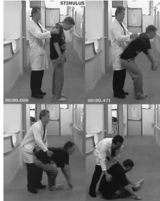
only for low limb tone and a wide-based gait. A SIDE was induced with an unexpected tactile stimulation of the shoulders (Fig 1).
At 17 years of age, a trial of tiagabine at 12 mg/d led to a decrease in severity of SIDEs. His events were described as occur-ring in “slow motion,” which allowed him to generate protective reflexes with his hands.
Case 2
The patient was born to a 23-year-old gravida 1 para 0 woman at term via cesarean section as a result of breech presentation. During infancy, he had hypotonia and delay in motor milestones. Chromosomes were normal. A head magnetic resonance imaging scan was consistent with benign extra-axial fluid collections of infancy. At 5 years of age, he received a diagnosis of CLS on the basis of typical facial and digital features.
At 5 years of age, he developed SIDEs that occurred several times a day. He was noted to lose postural tone in the lower extremities and suddenly drop to the floor after either loud noise or light tactile stimulation. Specific auditory triggers included loud noises such as toilet flushes, yelling, clapping behind his head, opening a door unexpectedly, or the sound of the doorbell. Specific tactile triggers included someone unexpectedly brushing against the patient or touching him from behind. Episodes were also triggered by visual stimuli (eg, by moving a hand across his visual field, moving a hand toward his face). These events oc-curred while sitting, standing, or walking. No loss of conscious-ness was noted, and he was able to recover immediately. SIDEs were not provoked by emotion or sudden laughter. Protective reflexes were present during observed episodes but were incom-plete. He sustained recurrent injury to his face and chin and knocked out 2 incisors because of the episodes. Neurologic
eval-uation was remarkable for generalized hypotonia and a wide-based gait. EEG was unremarkable.
By 6.5 years, the frequency of SIDEs had increased to 10 to 20 episodes per day and he was placed in a wheelchair for safety. Felbamate was started and titrated to 36 mg/kg/d with a reduc-tion in frequency to 3 to 4 episodes per day. Between 8 and 10 years of age, he was treated with valproate followed by clonaz-epam, without any significant reduction of SIDEs. Off medication, SIDEs occurred at 5 to 6 episodes per day.
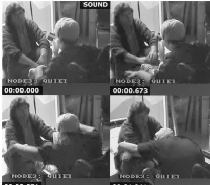
At 11 years of age, the character of the SIDEs changed. They were described as a sudden drop of his head with his arms spreading out laterally as the body lurched forward. These epi-sodes were induced by unexpected auditory and tactile stimuli. A trial of tiagabine at 12 mg/d led to improvement with a reduction in the frequency of events to 3 to 4 events per day (Fig 2). Although he was still able to ambulate, he preferred to use a wheelchair because of SIDEs.
RESULTS Cataplexy Questionnaire
The validated cataplexy questionnaire8was com-pleted by the patients with the assistance of the parents. The parents were able to provide responses for critical questions, including onset of SIDEs after telling or hearing a joke, laughing, or experiencing anger. Neither patient had an affirmative response to these 3 highly predictive questions. Using a decision
tree based on the questionnaire,8 both patients fell into the low-risk category for cataplexy (risk: 0.6%).
Neurophysiologic Studies
Case 1
At 16 years of age, simultaneous sEMG/VEEG studies during SIDEs did not show epileptiform dis-charges but demonstrated a brief transient decrease in sEMG activity in the lumbar paraspinal muscles shortly after the stimulus. Decreases in the sEMG activity were also seen in the quadriceps and ham-string muscles. The average latency was 224 msec, and the average duration was 159 msec, followed by an increase in sEMG activity to prestimulus levels.
Case 2
At 6 years, a VEEG captured 3 SIDEs unaccompa-nied by epileptiform activity. SIDEs were induced by lightly bumping into furniture or objects as he walked backward and consisted of sudden flexion of the hips with arms following in an extended posture, and rapid falling to the ground. Partial protective reflexes were present, and immediate recovery was noted. At 13 years of age, simultaneous sEMG/
VEEG studies during SIDEs did not show epilepti-form discharges. Events that were triggered by a loud noise were captured while seated and consisted of sudden forward flexion of the head and upper torso with symmetric abduction of the shoulders and flexion at the elbows. A generalized increase in sEMG activity was present during events consistent with myoclonic jerks with brief tonic spasms during events (Fig 2).
DISCUSSION
A characteristic paroxysmal disorder has been de-scribed in patients with CLS, characterized by sud-den loss of muscle tone induced by unexpected tac-tile or auditory stimuli.4 –7 These events have been given several names, indicating a poor understand-ing of their pathophysiology. These names include cataplexy, nonepileptic collapses with atonia, exag-gerated startle responses, hyperekplexia, and drop episodes.
One possibility is that SIDEs in CLS represent ep-ileptic events. In startle-induced epilepsy, seizures are precipitated by sudden stimuli and are usually tonic or myoclonic in nature.9,10 Ictal EEG usually
shows paroxysmal epileptiform activity. However, SIDEs in CLS do not seem to be epileptic in nature, because epileptiform activity is absent during epi-sodes in all reported cases,4,5,7including our 2 cases. Furthermore, startle-induced seizures are generally more prolonged or elaborate than SIDEs.
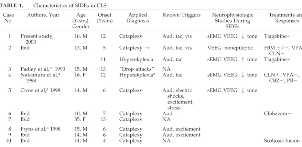
We reviewed the English literature and found 9 reported cases of “drop episodes” in CLS.4 –7,11 A comparison of 9 previously reported cases with our cases is presented in Table 1. The mean age of drop episodes onset was 8.6 years. Nine of 11 patients were male. All patients were described as having atonic collapse. In all cases in which details of SIDE was available, the patients experienced sudden giv-ing away of legs from a standgiv-ing position or durgiv-ing walking with return of normal tone after several seconds. There was no apparent lapse of conscious-ness during SIDEs. Four reported increasing fre-quency of episodes with time. Four reported some form of injury to self during events, and 2 of these cases were confined to a wheelchair by the age of 10 years.
Triggers for SIDEs are also presented in Table 1. Auditory triggers were most common, reported in all 8 patients in whom triggers were noted. Tactile trig-gers and “excitement” were reported in 3 cases each. Visual triggers have not been reported previously but were noted with both of our patients. No cases noted SIDEs induced by laughter or anger or after telling or hearing a joke, common triggers for cata-plexy.
Changes in tone with SIDEs were described as instantaneous in all cases. Most of the patients had sudden loss of tone in the lower limbs, rather than myoclonic jerks. Duration of episodes was very short, usually lasting a few seconds in all reported cases. Recovery was immediate in these cases, and
no episodes of more prolonged paralysis were re-ported (Table 1).
Three of the previously reported cases4,5,7 and both of our cases used combined sEMG and EEG studies during SIDEs. None of the 5 patients showed epileptic activity on EEG during SIDEs. Muscles sampled by sEMG included abnormal, paraspinal or quadriceps. Cases 1 and 5 were thought to be con-sistent with “cataplexy-like” episodes, and cases 2, 4, and 11 were thought to represent exaggerated startle responses or hyperekplexia. Four of these cases had a sudden transient decrease in sEMG activity after stimulus (Table 1). The latency after the stimuli ranged from 60 to 224 ms. The duration of this de-creased muscle activity varied from 4 to 159 ms. The latency and duration of decreased activity were somewhat longer in our case 1 than those cases pre-viously reported. This may be attributable to differ-ences in stimuli, recording technique, and patient variability.
Our case 2 was unusual in that during the second decade, the SIDEs evolved from sudden loss of tone (“cataplexy-like” episodes) to sudden increased tone with myoclonic jerks (“hyperekplexia-like” epi-sodes). Neurophysiologic studies during SIDEs at this later stage revealed a generalized increase in sEMG activity in muscles corresponding to a myo-clonic jerk. There was no significant latency after stimulus or transient loss of sEMG activity.
SIDEs do not conform well to either hyperekplexia or cataplexy. Table 2 provides a summary of the features differentiating these 2 disorders. Hyperek-plexia is defined as an exaggerated motor response to auditory, somesthetic, or visual stimuli that is resistant to habituation and may be characterized by either a brief pathologic startle reflex or a sustained tonic spasm.12 An appropriate stimulus produces a
TABLE 1. Characteristics of SIDEs in CLS Case
No.
Authors, Year Age (Years), Gender Onset (Years) Applied Diagnosis
Known Triggers Neurophysiologic Studies During
SIDEs
Treatments and Responses
1 Present study, 2003
16, M 12 Cataplexy Aud, tac, vis sEMG VEEG:2tone Tiagabine⫹
2 Ibid 13, M 5 Cataplexy3 Aud, tac, vis VEEG: nonepileptic FBM⫹/⫺, VPA⫺, CLN⫺
11 Hyperekplexia Aud, tac sEMG VEEG:1tone Tiagabine⫹
3 Padley et al,111990 15, M ⬃13 “Drop attacks” NA
4 Nakamura et al,4
1998
16, F 12 Hyperekplexia* Aud, tac sEMG VEEG:2tone CLN⫹, VPA⫺, CBZ⫺, PB⫺ 5 Crow et al,51998 14, M 6 Cataplexy Aud, electric
shocks, excitement, stress
sEMG VEEG:2tone
6 Ibid 10, M 7 Cataplexy Aud Clobazam⫺
7 Ibid 35, F 13 Cataplexy NA
8 Fryns et al,61998 15, M 6 Cataplexy Aud, excitement
9 Ibid 14, M 6 Cataplexy Aud, excitement
10 Ibid 14, M 4 Cataplexy NA Scoliosis fusion⫹
11 Caraballo et al,7
2000
16, M 11 Hyperekplexia* Aud sEMG VEEG:2tone CLN and LMT⫺, clomipramine⫹
Aud indicates auditory; tac, tactile; vis, visual; NA, not available or mentioned; FBM, felbamate; VPA, valproic acid; CLN, clonazepam; CBZ, carbamazepine; PB, phenobarbital; LMT, lamotrigine;⫹, reduction or elimination of SIDEs; ⫹/⫺, transient response;⫺, no significant response.
sudden stereotyped shock-like movement usually in-volving facial grimacing; abduction of the shoulders; and flexion of the neck, trunk, elbows, and knees. Consciousness remains intact during episodes, and most patients are able to rise immediately. Patients with sustained tonic spasm are reported to fall rig-idly to the floor, unable to take protective action, resulting in self-injury.13 Cataplexy, an ancillary symptom of narcolepsy, is defined as the sudden loss of bilateral muscle tone that is more pronounced in the antigravity muscles and is provoked by a strong emotion with preservation of consciousness lasting less than a few minutes.14 The most common emo-tional expression that provokes cataplexy is laugh-ing, which is reported to be the trigger in⬎80% of cases. Other common precipitants include anger, fear, surprise, and excitement. In most patients, cat-aplexy lasts for 1 minute or less. Closure of the eyes, sagging of the jaw, slurred speech, nodding of the head, dropping of the arms, and giving way of the knees are characteristic. Loss of muscle tone may be partial and is not always instantaneous but can progress over a few seconds and spread from cranial muscles to upper and lower limb muscles. In these cases, patients may be able to mount a protective response. Only approximately one third of patients with cataplexy experience sudden and severe loss of muscle tone causing collapse to the ground.14During these episodes, the patient cannot move the extrem-ities. Muscle hypotonia and decreased or abolished muscle stretch reflexes are noted, whereas eye move-ments and sometimes verbal communication are possible.8
The typical triggers that induce hyperekplexia (sudden unexpected auditory or tactile stimuli) were also found in the majority of our patients with SIDEs. However, in hyperekplexia, the stimuli are followed by a shock-like jerk (increased tone) rather than a loss of muscle tone. Hence, the sudden loss of tone in SIDEs more closely resembles the motor response in cataplexy. Only 1 patient (case 2) had sEMG-docu-mented myoclonic or shock-like jerks after stimuli. Episodes in cases 4 and 11 were considered as hy-perekplexia-like but in fact were accompanied by loss of tone rather than increased tone on sEMG. The typical triggers of cataplexy (elation, laughter, and anger) that are the most sensitive indicators for identifying cataplexy8were not reported in any case of CLS.
The duration of the SIDEs in CLS is much shorter than typical cataplexy. All cases reported SIDEs that were of no more than several seconds in duration. In
cataplexy, the majority of cases are less than 1 minute in duration but usually longer than a few seconds. Approximately 27% of cases of cataplexy are of a longer duration (⬎1 minute).14
Various therapies were used for SIDEs for 5 of 11 patients (Table 1).4 –7Rarely, near-complete improve-ment of symptoms has been reported with the use of clonazepam (case 4) and clomipramine (case 11). It is interesting that complete resolution of SIDEs was noted after scoliosis fusion surgery in 1 patient (case 10). We treated both of our patients with tiagabine because it is a potent GABA-uptake inhibitor that enhances neuronal inhibition. In case 1, a reduction in the severity of SIDEs was observed. In case 2, a reduction of SIDE frequency was noted. However, in neither case was the response to tiagabine dramatic. Conventional antiepileptic medications including valproic acid, carbamazepine, phenobarbital, and lamotrigine have been generally ineffective in the treatment of SIDEs.
Case 2 was the only one that seems to manifest signs and symptoms of true hyperekplexia with con-firmation on sEMG. Episodes in other cases (4 and 11) labeled as “hyperekplexia-like” demonstrated loss of tone during SIDEs. However, case 2 had also previously experienced documented atonic SIDEs 8 years earlier. Although no case fulfilled the criteria for cataplexy, case 6 was considered to have “cata-plexy-like” episodes because of the abrupt loss of tone from the head downward and successful volun-tary attempts to overcome attacks.
Proposed neurotransmitter mechanisms differ in hyperekplexia and cataplexy. Hyperekplexia is be-lieved to be mediated by reduced glycine receptor sensitivity leading to reduced glycinergic inhibition in the spinal cord and subsequent neuronal hyper-excitability.15 In cataplexy, hypersensitivity of the muscarinic cholinergic system has been postulated to be the mechanism.14
In regard to structural localization, it has been suggested that hyperekplexia is mediated by path-ways concentrated in the pontomedullary reticular formation,15whereas cataplexy has been proposed to be mediated by pathways concentrated in the me-diodorsal pontine tegmentum.14Given the proximity of suspected localization of neuronal circuitry in these 2 conditions and the unusual characteristics of case 2 in which a change in tonal characteristics of SIDEs occurred over time, the possibility exists that these conditions may involve parallel neuronal path-ways allowing shifting expression of tonal control through pontomedullary reticular neurons.
TABLE 2. Features Differentiating Hyperekplexia, Cataplexy, and SIDEs
Hyperekplexia Cataplexy SIDEs
Trigger Physical Emotional Auditory, tactile, visual
Tonal change during attack Increased Decreased Usually decreased
Partial tone loss No Possible No
Brief paralysis No Possible No
Protective reflexes No Possible Possible
Duration of event Seconds Seconds to minutes Seconds
Recovery Immediate May be delayed Immediate
Treatment VPA, CLN TCA, SSRI CLN, tiagabine
CONCLUSION
SIDEs in CLS are nonepileptic events usually char-acterized by atonic collapses triggered by sudden stimuli with immediate and complete recovery. Al-though there are similarities to hyperekplexia and cataplexy, SIDEs have a distinctive pattern of fea-tures (Table 2). Given the lack of characteristic emo-tional triggers, the reliably short duration of events, and the presence of immediate recovery in all cases, SIDEs should not be classified as cataplexy. Com-bined VEEG and sEMG recordings of SIDEs demon-strate that a brief loss of tone in the paraspinal or quadriceps muscles is characteristic in the majority of cases. Finally, we report a single case that demon-strates evolution of tonal characteristics over time and not only suggests that SIDEs may have the po-tential for evolution over time but also provides a possible explanation for the broad range of previ-ously reported presentations.
ACKNOWLEDGMENTS
We thank Dr Christian Guilleminault and Dr Emmanuel J. Mignot for helpful discussions regarding cataplexy and narco-lepsy.
REFERENCES
1. Coffin GS, Siris E, Wegienka LC. Mental retardation with osteocartilag-inous anomalies.Am J Dis Child.1966;112:205–213
2. Lowry B, Miller JR, Frazer FC. A new dominant gene mental retardation syndrome.Am J Dis Child.1971;121:496 –500
3. Trivier E, De Cesare D, Jacquot S, et al. Mutations in the kinase Rsk-2 associated with Coffin-Lowry syndrome.Nature.1996;384:567–570 4. Nakamura M, Yamagata T, Momoi MY, Yamazaki T. Drop episodes in
Coffin-Lowry syndrome: exaggerated startle responses treated with clonazepam.Pediatr Neurol.1998;19:148 –150
5. Crow YJ, Zuberi SM, McWilliam R, et al. “Cataplexy” and muscle ultrasound abnormalities in Coffin-Lowry syndrome.J Med Genet.1998; 35:94 –98
6. Fryns JP, Smeets E. “Cataplexy” in Coffin-Lowry syndrome. J Med Genet.1998;35:702
7. Caraballo R, Tesi Rocha A, Medina C, Fejerman N. Drop episodes in Coffin-Lowry syndrome: an unusual type of startle response.Epileptic Disord.2000;2:173–176
8. Anic-Labat S, Guilleminault C, Kraemer H, Meehan J, Arrigoni J, Mi-gnot E. Validation of a cataplexy questionnaire in 983 sleep-disorders patients.Sleep.1999;22:77– 87
9. Manford MRA, Fish DR, Shorvon SD. Startle provoked epileptic seizures: features in 19 patients.J Neurol Neurosurg Psychiatry.1996;61: 151–156
10. Ricci S, Cusmai R, Fusco L, Vigevano F. Reflex myoclonic epilepsy in infancy: a new age-dependent idiopathic epileptic syndrome related to startle reaction.Epilepsia.1995;36:342–348
11. Padley S, Hodgson SV, Sherwood T. The radiology of Coffin-Lowry syndrome.Br J Radiol.1990;63:72–75
12. Brown P, Rothwell JC, Thompson PD, Britton TC, Day BL, Marsden CD. The hyperekplexias and their relationship to the normal startle reflex. Brain.1991;114:1903–1928
13. Saenz-Lope E, Herranz-Tanarro FJ, Masdeu JC, Charcon Pena JR. Hyperekplexia: a syndrome of pathological startle responses.Ann Neu-rol.1984;15:36 – 41
14. Guilleminault C, Gelb M. Clinical aspects and features of cataplexy.Adv Neurol.1995;67:65–77
DOI: 10.1542/peds.111.3.e197
2003;111;e197
Pediatrics
Gregg B. Nelson and Jin S. Hahn
Stimulus-Induced Drop Episodes in Coffin-Lowry Syndrome
Services
Updated Information &
http://pediatrics.aappublications.org/content/111/3/e197
including high resolution figures, can be found at:
References
http://pediatrics.aappublications.org/content/111/3/e197#BIBL
This article cites 15 articles, 4 of which you can access for free at:
Subspecialty Collections
http://www.aappublications.org/cgi/collection/therapeutics_sub Therapeutics
http://www.aappublications.org/cgi/collection/pharmacology_sub Pharmacology
following collection(s):
This article, along with others on similar topics, appears in the
Permissions & Licensing
http://www.aappublications.org/site/misc/Permissions.xhtml
in its entirety can be found online at:
Information about reproducing this article in parts (figures, tables) or
Reprints
http://www.aappublications.org/site/misc/reprints.xhtml
DOI: 10.1542/peds.111.3.e197
2003;111;e197
Pediatrics
Gregg B. Nelson and Jin S. Hahn
Stimulus-Induced Drop Episodes in Coffin-Lowry Syndrome
http://pediatrics.aappublications.org/content/111/3/e197
located on the World Wide Web at:
The online version of this article, along with updated information and services, is
http://pediatrics.aappublications.org/content/suppl/2003/02/28/111.3.e197.DC1
Data Supplement at:
by the American Academy of Pediatrics. All rights reserved. Print ISSN: 1073-0397.